Your browser does not fully support modern features. Please upgrade for a smoother experience.
Please note this is a comparison between Version 1 by Pooja Chawla and Version 2 by Catherine Yang.
The family of nuclear peroxisome proliferator-activated receptors (PPARα, PPARβ/δ, and PPARγ) is a set of ligand-activated transcription factors that regulate different functions in the body. Whereas activation of PPARα is known to reduce the levels of circulating triglycerides and regulate energy homeostasis, the activation of PPARγ brings about insulin sensitization and increases the metabolism of glucose. On the other hand, PPARβ when activated increases the metabolism of fatty acids. Further, these PPARs have been claimed to be utilized in various metabolic, neurological, and inflammatory diseases, neurodegenerative disorders, fertility or reproduction, pain, and obesity.
- peroxisome proliferator-activated receptors
- ligands
- a thiazolidinedione
1. Introduction
Nuclear receptors are a type of protein that recognize steroid and thyroid hormones in the body [1]. Nuclear receptors tend to influence the growth, homeostasis, and metabolism of organisms by binding to DNA and regulating the expression of specific genes. As a result of their ability to modulate transcription, these are known as transcription factors [2][3][2,3]. The nuclear receptor superfamily’s peroxisome proliferator-activated receptor (PPAR) is a ligand-dependent transcription factor [4]. To build a heterodimer, all PPARs interact with the retinoid X receptor [5][6][7][5,6,7]. In 1990, PPARs were first observed in rodents [8]. PPARs are receptors associated with a superfamily of nuclear receptors that also contain steroids, retinoid receptors [9], thyroid hormone receptors, and Vit. D receptors [10]. They are multiple and persuasive regulators of various cellular functions and metabolic functions such as glucose and lipid homeostasis, cholesterol, and energy balance [11][12][11,12]. Peroxisome proliferator-activated receptors are a subgroup of the ligand-dependent transcription factor that contains peroxisome proliferator response elements as transcription factors to regulate transcriptional activity [13][14][15][13,14,15].
2. Structure of PPAR
The three-dimensional structure of PPARs includes the N-terminus and C-terminus where DNA binding domain and ligand-binding domain are attached, respectively, [16]. They are translocated to the nucleus and heterodimerized with retinoid X receptors (RXR) [17]. In targeted genes, the PPREs (peroxisome proliferator hormone response elements) are particular DNA areas that interact with PPARs [18][19][18,19]. They are connate in the fatty acid-binding protein like PPAR-responsive gene promoters that activate transcription of multiple genes involved in various physiological processes shown in Figure 1 [20][21][20,21]. The protein structure of various PPARs is fetched from the protein data bank using three PDB ids such as 2P54 (PPARα), 2A5G (PPA, Rβ,), and 3VI8 (PPARγ), shown in Figure 2 [22][23][24][25][26][22,23,24,25,26].
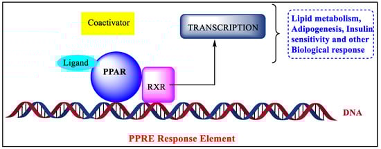
Figure 1.
Mechanism of biological responses through PPAR.
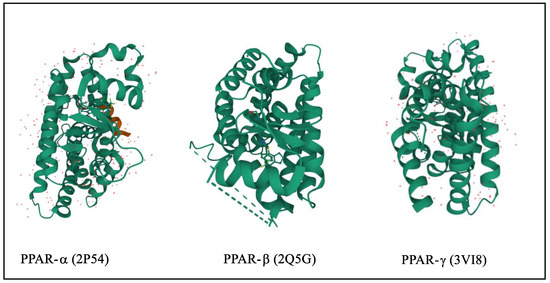
Figure 2.
3D structure of PPARα/β/γ.
3. Types and Expressions of PPARs
The transcriptional factors of peroxisome proliferator-activated receptors (PPARs) contain three different isoforms, namely nuclear receptor subfamily PPARα or NR1C1, PPARβ/δ or NR1C2, and PPARγ or NR1C3 [27][28][29][27,28,29]. These isoforms are expressed in multiple tissue and organs with identical character and ligand specificity [30]. The PPAR-α is expressed mainly in the liver but is also present in muscle, bone, and heart. The PPAR-δ is expressed in most parts of the body and regulates energy expenditure [31][32][31,32]. The PPAR-γ is expressed in vascular smooth muscle cells, and endothelial cells [33][34][33,34]. The expression of various types of PPARs is shown in Figure 3 [35][36][37][35,36,37].
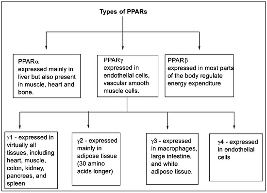
Figure 3.
Types and expression of PPARs.
4. Functions of PPARs
In the human body, the three forms of PPARs are responsible for the activation of most of the enzymes that are required for fatty acid oxidation, glucose metabolism, and lipid metabolism [38]. These PPARs perform specific or distinct functions such as cellular energy balancing, and maintenance of energy homeostasis on their own or by helping each other [39]. Energy metabolism at a molecular level through PPARs is still not explained but when ligands are attached to these receptors, they transcript the genes involved in regulatory energy functions [40]. The PPARα is involved in the activation of gene-encoding enzymes such as carnitine palmityl transferase 1 (CPT1), fatty acid transport protein, acyl-CoA dehydrogenase or oxidase, or synthase required in the fatty acid oxidation pathway [41]. It also reduces plasma triglyceride levels and increases high-density lipoprotein, which gives benefits such as ketogenesis [42]. The PPARβ controls fatty acid metabolism, increases insulin sensitivity, obesity resistance, suppression of macrophage-derived inflammation, and also the formation of oxidative muscle fibers. The PPARγ promotes fatty acid uptake, and adipokine production raises insulin sensitivity, also in adipogenesis [43][44][45][46][43,44,45,46]. Other functions associated with PPARs are described in Figure 4 [47][48][49][47,48,49].
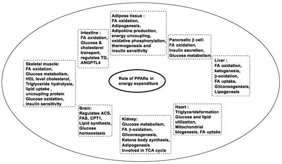
Figure 4.
Functions of PPARs.
In several areas of medicinal chemistry, heterocyclic molecules are indispensable [50][51][50,51]. These compounds constitute nitrogen, sulfur, and oxygen atoms with various positional combinations in the cyclic ring [52]. Medicinal chemists and researchers always explore heterocycles due to their vital contribution to drug discovery and design by boosting the novel lead moieties with potential biological action in medicinal chemistry [53]. Based on statistics, more than 85% of biologically potential chemical moieties contain heterocyclic motifs [54]. Some of the most common heterocycles are pyrimidine, imidazole, oxazole, tetrazole, triazine, triazle, thiazole, indole, pyridine, thiophene, pyrazole, coumarin, oxindole, furan, etc. [55][56][57][58][55,56,57,58]. These heterocyclic nuclei possess a broad spectrum of medicinal potential such as an anti-microbial [59], antimalarial [60], anti-anxiety [61], anti-cancer [62], anti-depressant [63], anti-tubercular [64], anti-virus [65], anti-protozoal [66], and anti-convulsant, etc. Several heterocyclic analogs have been reported to affect PPAR in different diseases [67][68][69][70][71][72][67,68,69,70,71,72]. Apart from commercially available marketed drugs, Table 1. depicts the patented drugs acting on PPARs.
Table 1.
Various patents on Peroxisome proliferator-activated receptor.
| Sr No | Date | Patent Number | Description | Ref. | ||||
|---|---|---|---|---|---|---|---|---|
| 1. | Thiazolidinediones19 August 2021 | WO2021161218 | Sulfinic acid and sulfonic acid compounds for use in modulating peroxisome proliferator-activated receptors | [73] | ||||
| Allyl derivative ( | 1a) | EC50 of −4.95 μM in the human PPAR-γ transactivation assay | [83] | 2. | 23 December 2020 | US20210238125 | Diabetes and Metabolic Syndrome Treatment with a Novel Dual Modulator of Soluble Epoxide Hydrolase and Peroxisome Proliferator-Activated Receptors | [ |
| Amino derivative (3b) | 1.7 times more than reference compounds in glucose uptake assay | [84] | 74] | |||||
| 3. | 18 July 2018 | US20190077774A1 | Peroxisome proliferator-activated receptor agonists | [75] | ||||
| Chloro derivative (8a) | 63.15%, PPAR-γ transactivation | [85] | 4. | 28 July 2018 | US20180305403A1 | PPAR agonists and methods of use thereof | ||
| Dichloro derivative (9a) | [ | 76 | ] | |||||
| (−11.6930) | Docking score | [86] | 5. | 01 July 2018 | US20190000790A1 | PPAR-γ activators and their therapeutical usage | ||
| Nitro and carboxylic acid derivative (10d) | [ | 77 | ] | |||||
| (−17.44) Docking score | [ | 87 | ] | 6. | 13 December 2017 | US20190060269A1 | Brain-Derived PPARα Ligands | [78 |
| ] | ||||||||
| Flouro derivative ( | 12e) | Reduced 0.09-fold the expression gene of PPARγ in cultured adipocytes compared to the control group | [88] | 7. | ||||
| Oxadiazole | 07 April 2017 | No substitution (13dUS20180015062A1 | ) | ECPPAR-α Activator, Pharmaceutical Composition, Food and Drink, Food Additive, Supplement, and Method of Manufacturing the Same | 50 = −0.15 for PPARα [79] |
|||
| EC | 8. | 13 April 2017 | WO2017180818A1 | PPAR agonists, compounds, pharmaceutical compositions, and methods of use thereof | [80] | |||
| 50 | = −0.29 for PPARδ | [ | 89] | |||||
| ADAM (14) | 2.5-fold greater efficacy in activating PPARα | [90] | 9. | 07 April 2017 | US20170304255A1 | |||
| Bromo derivative (15a) | Potency and selectivity towards PPARα/δ receptors with PPARα/δ/γ EC50, EC50 | PPAR agonists, compounds, pharmaceutical compositions, and methods of use thereof | [81] | |||||
| 10. | 30 January 2017 | US20170210711A1 | Competitive PPAR-α antagonists | [82] |
The SAR of these derivatives will be discussed to pave the way for the development of new, safe, and economical ligands shortly. Various heterocyclic moieties that act on the PPAR receptors have been summarized and displayed in Table 2.
Table 2. Summary of various heterocycles with the different substitutions for Peroxisome proliferator-activated receptor.
Summary of various heterocycles with the different substitutions for Peroxisome proliferator-activated receptor.
| Heterocycles | Most Potent Substitution | Activity | Reference |
|---|---|---|---|
| γ/α ratio, and EC | |||
| 50 | |||
| γ/δ ratio value was 8/5/2939 nM, 367, 588 respectively | |||
| [ | |||
| 91 | |||
| ] | |||
| Flouro derivative (16d) | PPARα −0.06 ± 0.0005, PPARγ −0.07 ± 0.0006 | [92] | |
| Bemzoimidazole | Chloro derivative | EC50 = −0.19 ± 0.01 | [93] |
| Flouro and chloro derivative | −68 ± 28j | [94] | |
| (20c) | −10.6 ± 0.4 | [95] | |
| Chloro and iodo derivative | Ki = 1023 µM for PPARγ and Ki = 0.106 µM for PPARδ | [96] | |
| Chloro derivative (22b) | PPARα 307 PPARγ 2052 PPARδ 214 | [97] | |
| (23) | Kd value = −2.8 ± 0.8 nM | [98] | |
| Methoxy derivative (24a) | [99] | ||
| Thiazole | - | - | [100] |
| (26) | EC50 > 10 µM | [101] | |
| Phenyl derivative (27b) | PPARα Imax% 22 ± 16 |
[102] | |
| p-halogenated phenyl substituted thiazole derivative (28f) | Increased PPARγ activity by almost 4- to 5-fold while rosiglitazone exhibited approximately 10-fold activation | [103] | |
| fluoro and nitro derivative (29b) | - | [104] | |
| indole | 2,4 dimethoxy derivative 30d | - | [105] |
| Chloro and phenyl derivative (31a) | PPARα/δ/γ profile at potency and efficacy level.(31a, Emax = 50%) | [106] | |
| Cyclohexyl derivative | 24.43% Reduction in blood glucose level | [107] | |
| Furan | phenyl derivative 33h | hPPARα (LBD)-GAL4 EC50 (µM) −7.31 hPPARγ (LBD)-GAL4 EC50 (µM) −2.97 hPPARδ (LBD)-GAL4 EC50 (µM) −1.98 |
[108] |
| Triflouro carbon phenyl derivative (34a) | 16.2-fold and 8.4-fold more PPAR-α/δ agonistic activity than PPARγ | [109] | |
| Triflouro carbon phenyl derivative (35a) | High potency toward PPAR- α/δ (0.26 ± 0.08 µM 0.50 ± 0.10 µM) and higher selectivity against PPARγ (4.22 ± 0.18 M) than that of GFT505 | [110] | |
| Benzopyran | Nitro derivative (36b) | EC50 −0.91 µM | [111] |
| Butane derivative (37c) | hPPARα and γ with % values of 91% and 88% using a transactivation assay | [112] | |
| Di-hydroxyl deritive (38b) | Ki value 1.41 μM | [113] | |
| 39 | hPPARα with high selectivity (123 % and 38% for α and γ, respectively). | [114] | |
| Bavachinin | (40) | PPARα agonistic activity EC50 −0.43 | [115] |
| 41 | 21 (EC50 = −22.28 μM) | [116] | |
| Miscellaneous | 42 | - | [117] |
| Cyanophenyl (43a) | IC50 6.5 µM time-resolved FRET technique | [118] | |
| 44 | EC50 (nM) = −170 ± 10 | [119] | |
| 45a | HepG2 and A549 cell growth with IC50 values of −0.54 and −0.47 μM, respectively, CCK-8 assay | [120] | |
| 46 | - | [121] | |
| Methoxy and cyclohexyl derivative | IC50 values −0.350 fluorescence polarization assay |
[122] | |
| 50a | [123] | ||
| o-Tolyl (51a1–5) | glucose uptake activity in vivo evaluation, with −37.05 ± 0.44 |
[124] | |
| 52 | - | [125] | |
| 53a | - | [126] | |
| Ethylcyclopropane derivative (54a) | - | [127] |