1. Introduction
Mastitis is the most frequently occurring infectious disease in dairy cattle, with worldwide economic losses estimated to be more than USD 40 billion USD per year
[1][7]. Udder health is of paramount importance for sustainable milk production. Mastitis is more than just a medical condition; it impacts milk quality, cattle performance, and farm antimicrobial use
[2][8].
The diagnosis of subclinical mastitis in dairy cows, as well as the early detection of clinically expressed mastitis that induces macroscopic changes in milk secretion, is an essential component of any program established to improve the health parameters of a dairy farm. This goal is of tremendous economic interest, both for farmers and manufacturers, given that a large number of somatic cells in milk and the presence of bacteria have a severe negative influence on milk production and quality
[3][4][9,10].
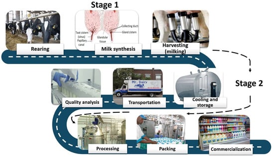
From its origin to the point of consumption, milk moves through two separate stages
[5][93]. The first stage extends from the mammary alveolar tissue to the papillary orifice of the galactophore (papillary) canal. In contrast, the second stage covers the way from the milking machine to the consumer (
Figure 1).
Figure 1. Milk way—from the rearing to the consumer.
The essential thing to mention in this respect is that the current set of mandatory tests for milk quality control pertains only to the second stage of the milk journey; thus, only the existing hygienic-sanitary circumstances in this phase after milking are shown. Thereby, the two main parameters, somatic cell count (SCC), respectively total bacteria count (TBC), may offer different types of information regarding udder health and milk quality
[6][94]. In general, the presence of inflammation and subclinical mastitis is indicated by SCC levels of more than 200,000 cells/mL of milk. In most European nations, the limit for farm milk commercialization was set at 400,000 cells/mL, while in the United States, the limit is set at 750,000 cells/mL, and the price of milk decreases as the amount of SCC cells gets closer to the legal limit
[7][8][9][54,92,95].
According to Regulation (EC) No 853:2004 of the European Parliament and the Council, raw milk must originate from animals that do not exhibit any evidence of contagious diseases that may be transmitted to people, as well as any indicators of illnesses of the mammary or genital tract that might potentially contaminate milk. The total bacteria count and the bulk milk somatic cells are the two primary health regulatory standards taken into consideration for assessing milk quality. Somatic cell count is defined just for raw cow milk, and it corresponds to a cell count of fewer than 400,000 cells per milliliter. In contrast, the plate count at 30 degrees Celsius must be less than 100,000 forming colony-forming units per milliliter. These two regulatory standards are each stated as a rolling geometric average over two or three months, with at least one sample collected per month
[10][96]. The requirements for testing total bacteria and somatic cell count are outlined in Regulation (EC) No. 2074/2005, revised by Regulation (EC) No. 1664/2006 regarding the implementation of specific measures for original animal products intended for human consumption. The requirements for testing total bacteria and somatic cell count are outlined in Regulation (EC) No. 2074/2005, revised by Regulation (EC) No. 1664/2006 regarding the implementation of specific measures for original animal products intended for human consumption. The reference methods are EN/ISO 4833 for the plate count at 30 degrees Celsius, respectively ISO 13366-1 for the somatic cell count
[11][97].
Increased SCC always indicates the presence of infection in some mammary quarters, whereas TBC mostly indicates milking hygiene or sanitation problems
[12][13][14][98,99,100]. The presence of elevated SCC in the second phase of the milk route suggests the existence of an undesirable scenario in the herd, most likely owing to bovine mastitis. Increased SCC has a negative impact on the organoleptic characteristics of milk and its appropriateness for producing quality products
[15][16][17][18][101,102,103,104], whereas increased TBC in raw milk represents, among other things, the risk of food-borne disease for consumers
[19][20][21][22][105,106,107,108]. If both limits are surpassed, the milk is entirely inadequate in terms of quality, both nutritionally and hygienically, economically, and technologically. Screening the herd somatic cells count levels weekly, as an integrating part of the milk quality monitoring procedure, may provide farmers with useful information regarding the potential going ongoing herd-level disease, as well as the effectiveness of the implemented barn and milking hygiene procedures.
2. Conventional Methods for Monitoring Udder Health
The effectiveness of the milking routine and the performance of the milk collection equipment has a crucial impact on milk quality and udder health in dairy cattle
[23][109]. The teat canal acts as the main physical barrier that prevents bacteria from entering the udder via the teat canal. Between milkings, the smooth muscles surrounding the teat canal should be constricted, and the teat canal should be securely closed to prevent infections from entering the teat canal and, from there, the udder
[24][110]. This defense mechanism is reinforced by the presence of keratin cells, rich in lipids, present inside the teat canal. When the skin is elastic and smooth, without any lesions, the teat is in the best position to provide a natural barrier against the invasion of pathogens that cause mastitis. This is because the teat’s skin is more likely to withstand the pressure of the pathogens.
Any stress applied to the teats, even for a very short period, might affect their inherent capability to withstand a pathogen invasion. While the majority of attention is focused on teat-end hyperkeratosis, other short-term teat disorders, such as discoloring, sores, edema, and congestion, indicate poor milking performance
[25][26][111,112].
Teat scoring, more accurately known as teat-end scoring, is a helpful technique to assess the amount of teat-end hyperkeratosis and other teat lesions in a dairy herd (
Table 1). This method may be a useful management tool for farmers, providing information regarding the efficiency of the milking equipment and the milking procedure
[27][113]. Research has shown that a circulatory impairment of any kind may be connected with an increased risk of a mastitis infection that is not yet clinically apparent
[27][113]. Mastitis has been linked to teat hyperkeratosis, which is thought to cause alterations in the surrounding tissue of the teat canal, enabling bacteria to enter easily into the mammary gland. For this reason, it is essential that the assessment of teat-end scoring be carried out at regular intervals on at least 20% of the herd so that changes may be monitored over time
[28][114] before the quality of milk is hindered
[23][109].
Table 1. Summary of primary conventional methods, based on physicochemical milk modifications, used for monitoring udder health.
Several tests have been developed to identify some of the changes that take place in milk yield throughout the progression of a mammary gland infection
[29][115]. The majority of the tests pursued to reveal certain physicochemical modifications, such as an increase in the number of somatic cells (SCC) by direct or indirect counting (tests based on organic detergents: California Mastitis Test and similar tests such as the Wisconsin Mastitis Test, R-mastitest), the accumulation of chlorides, an increase in pH, electroconductivity (EC), viscosity, or catalase, an increase in udder skin surface temperature, the presence of grains with a diameter 0.1 mm.
Mastitis causes alterations in the real ionic dynamics of vascular components due to excessive cellular destruction and weakened milk–blood barrier. Loss of intracellular potassium results in a rise in the amounts of sodium, potassium, calcium, magnesium, and chloride ions in the blood, while the concentration of potassium ions declines. The electro-conductivity (EC) of milk is altered, and the pH is elevated due to these processes. These variations serve as a diagnostic sign for distinguishing milk with unusual qualities. Due to its simplicity and rapidity, with a cost/sample almost equal to the cost of the equipment, the electrical conductivity (EC) of milk has been studied extensively for the detection of clinical mastitis in the past
[30][116]. The research conducted by Khatun et al. (2022)
[31][117] highlighted that mastitis detection systems that rely only on EC are unlikely to accomplish the appropriate sensitivity and specificity criteria, but improvements are possible if several measures are performed (
Table 1).
On the other hand, a study carried out by Kandeel et al. (2019)
[32][55] showed that milk sodium, potassium, and calcium concentrations, as well as EC, were not sufficiently accurate to diagnose subclinical mastitis (SCM) and intramammary infections (IMI) in cattle, therefore they cannot serve as routinely udder health monitoring tools. Milk pH testing has also been proposed as a simple, inexpensive, and useful on-farm approach for identifying SCM and IMI in cattle. However, different authors concluded that milk pH does not offer a clinically effective technique for identifying SCM or IMI in dairy cattle
[33][34][118,119].
Multiple kinds of predictive variables were proposed by Kamphuis et al. (2008)
[35][120] as a means to enhance mastitis detection performance. Modifications in milk yield, milk temperature, milk color, cow activity, and other milk components are further markers used in the diagnosis of mastitis
[34][36][119,121]. Using numerous criteria allows for a more accurate prediction of mastitis status, as shown in research by Khatun et al. (2018)
[37][122]. It is also expected that their usefulness in farms will increase if more precise detection technologies are developed that use various measurements.
Currently, the California Mastitis test (CMT) and similar tests are routinely used by small and large-size farms to assess udder health. However, due to the subjectivity of reading and interpreting the results, they give less reliable results than the direct counting of somatic cells but more correct than other methods. The advantage of this method is that it is relatively fast, less expensive, and within reach of any farmer
[38][123]. The favorable reviews enjoyed by CMT and similar tests are probably also because they were the first in the category of those simultaneously assessing two changes from two different categories: the number of cells and the pH
[4][39][10,124] (
Table 1).
Infections of the udder may also be identified by examining many additional biomarkers, such as secreted enzymes that indicate tissue damage. Colorimetric and fluorometric assays may be used to determine the activity of lysosomal N-acetyl-β-d-glucosaminidase (NAGase) or lactate dehydrogenase (LDH) in milk. A significant fraction of the enzyme NAGase is generated by epithelial cells of the udder that have been injured; such is the case of mastitis. According to a study conducted by Hovinen et al. (2016)
[40][125], NAGase activity may be a reliable indication of both subclinical and clinical mastitis. Due to the breakdown of the blood–milk barrier that occurs after an intramammary infection, there is a rise in the amount of immunoglobulin G (IgG) found in the milk. Both lactate dehydrogenase (LDH) and serum albumin (SA) can cross the aforementioned barrier; hence, both may be utilized as indicators to predict the IgG transfer into milk and, subsequently, the presence of an intramammary infection
[31][117]. One of the commercially available methods for assessing the LDH activity is the UdderCheck
TM from PortaCheck, which utilizes paper-based test strips and evaluates the color changes in the presence of an LDH-specific substrate. The severity of the disease is determined by making a qualitative comparison of the results using a color chart (
www.portacheck.com). However, a comparative test showed that this diagnostic tool’s applicability is limited, and its accuracy is lower compared to other methods, such as the California mastitis test
[31][41][42][117,126,127]. Other possible biomarkers for mastitis diagnosis, such as procalcitonin (PCT), neopterin (NPT), haptoglobin (HP), serum amyloid A (SAA), proinflammatory cytokines (IL-1β, IL-8, TNF-α, IF-γ)
[43][44][45][46][47][48][128,129,130,131,132,133], as well as lactose
[49][70] are now the subject of research and analysis.
The most widely used method for detecting mastitis, particularly in its subclinical forms, is monitoring the SCC content in milk
[15][101]. When the values of SCC go above the limit, the value of the milk significantly declines. For this reason, researchers consider SCC level to be essential criteria for udder health evaluation
[8][50][51][52][53][92,134,135,136,137]. Although the direct measurement of SCC level offers great accuracy and reliable information regarding udder health status, this approach may, in some cases, be inaccessible for some dairy farmers and dairy associations due to its high costs (
Table 1).
In the past, direct microscopy assessment of the somatic cells was seen as a time-consuming process, whether performed on a single sample or a collection of samples, with uncertain results due to subjective interpretation. Nowadays, due to cutting-edge diagnostic tools such as the DeLaval cell counter, Fossomatic cell counter, PortaCheck
®, and Somaticell
®, SCC levels may be evaluated quickly and automatically on many samples
[4][54][10,138]. Cell counters with high capacity, based on the concept of flow cytometry (fluorooptoelectronic method), such as the Fossomatic cell counter or SomaScope, are often used for measuring SCC in large numbers of samples at once (400–600 samples per hour)
[39][55][124,139] (
Table 1).
3. Methods Based on the Detection of the Pathogen Agent Causing Mastitis
The diagnostic approaches mentioned in Table 1 provide information regarding the udder’s health status, and some may even indicate the degree to which mastitis has progressed. However, none of them can pinpoint the pathogen agent that is causing the problem. Early and precise detection of the pathogen implicated is associated with a number of benefits, such as appropriate therapy options, including the choice of adequate antibiotics and improved management measures to restrict the spread of disease and antibiotic resistance.
Environmental pathogens that spread predominantly outside the milking parlor account for about 90% of pathogens responsible for udder infections. The most predominant species are
Escherichia coli,
Streptococcus uberis,
Streptococcus dysgalactiae, and
Proteus spp.
[56][140]. Contagious mastitis is usually caused by pathogens such as
Staphylococcus aureus,
Streptococcus agalactiae, and
Mycoplasma spp., which occur mainly in the cow’s udder, their presence in bulk milk indicating the existence of intramammary infections in the herd
[57][58][141,142]. Fungi are a less common cause of mastitis, with fewer documented cases, and are most often seen on farms with poor environmental and sanitary conditions
[59][60][61][62][143,144,145,146]. Contamination with microalgae from
Prototheca spp, frequently related to poor milking conditions and extended antibiotic medication, has also been documented
[63][64][65][147,148,149].
The culture-based technique has long been the gold standard for identifying mastitis pathogens. To stimulate growth, a known amount of milk, from either bulk tank or udder quarter, is incubated on culture plates for about 18 h at set temperatures. After the growth phase is over, colony-forming units (CFU) are counted, and the colony phenotype is analyzed to identify the pathogens. Additional biochemical testing may be performed if required. Most pathogens grow well on conventional culture medium, either under aerobe (the vast majority) or under anaerobe conditions (e.g.,
Mycoplasma spp.). The principal disadvantages of bacterial culture are associated with the need for sterile conditions to prevent the development of bovine mastitis non-related microorganisms, the requirement for special equipment, and the need for competent operators to accurately conduct the microbiological procedures and interpret the phenotypic findings. Furthermore, the approach often requires lengthy growth periods (up to 48 h) and is prone to false negatives, with a reported probability of 20–50%
[66][150].
Over the past years, several types of on-farm culture plates were specially designed for farmers and veterinarians, providing a rapid, simple, and low-cost method for determining the probable bacterial etiology of mastitis. While some of the on-farm culturing systems can distinguish only between the main two types of pathogens, Gram-negative and Gram-positive, others may stimulate the development of specific microorganisms and reduce the incubation time using a selective culture medium. For instance, the Accumast
TM system separates staphylococci, streptococcus, and Gram-negative bacteria using a tri-plate containing three different chromogenic media. A noticeable color shift is produced when particular bacterial enzymes break chromogens contained in the culture medium
[39][124]. The Minnesota Easy
® Culture System uses three different kinds of culture media, Factor™, MacConkey, and Focus™, to differentiate between Gram-positive, and Gram-negative, respectively,
Streptococcus and
Streptococcus-like bacteria
[67][151]. Likewise, ClearMilk Test culturing systems enable specialists to identify the pathogen in roughly 22 h using a tri-plate-based culturing system designed to distinguish between
Staphylococcus spp.,
Streptococcus spp., Gram-negative, as well as yeast
[68][152].
However, although these on-farm culturing systems have become commercially available at reasonable prices, some of the studies have pointed out that the commercial on-farm culturing systems differed significantly in their ability to classify bacterial colonies by genus and species
[69][153] and training beyond the instruction manual is required for untrained observers to make this type of systems effective for pathogen-based mastitis control
[70][154].
Furthermore, given the frequency of false negatives with culture-based methods, the development of molecular diagnostic tests with high test sensitivity and specificity, as well as the necessity to detect non-viable bacteria, has been approached by different researchers that demonstrated effective PCR-based amplification and identification of mastitis pathogens
[71][72][73][74][75][76][155,156,157,158,159,160]. Polymerase chain reaction (PCR) is known to be highly sensitive and specific for detecting mastitis pathogens, providing accurate pathogen identification, including those that do not grow using conventional culturing techniques. Although the results when using PCR may be obtained in a matter of hours, a study conducted by Hiitiö et al. (2015)
[72][156] concluded that when low DNA levels have been identified in milk samples, the clinical importance of the data should be carefully reviewed before making any further judgments.
Due to sterility standards, the requirement for sophisticated equipment, and skilled staff, PCR is challenging to deploy on-farm. Furthermore, the presence of recognized PCR inhibitors such as calcium, fat, or protein in milk necessitates using specific DNA extraction techniques to ensure high-quality findings. Alternative to regular PCR and quantitative (qPCR) procedures, loop-mediated isothermal amplification (LAMP) has been described as a promising molecular tool for quick on-farm diagnostics
[77][78][161,162] and food pathogen detection
[79][80][81][82][83][163,164,165,166,167]. This approach is quicker than PCR, less costly, highly selective for the target sequence, and requires less template quality and complicated apparatus. Finally, as an isothermal amplification approach, it might be used in the field, needing just a water bath or heat block for the reaction to take place
[84][85][168,169]. LAMP tests for common mastitis pathogens such as
Staphylococcus aureus,
Streptococcus agalactiae, or
Streptococcus uberis have been developed and validated
[86][87][88][170,171,172].
As next-generation sequencing (NGS) is becoming more accessible and less expensive, a new opportunity for developing novel genotyping tools to detect mastitis infectious pathogens arises. Studies have shown that target-specific primers for PCR-mediated amplification with NGS technology to enrich and accurately sequence pathogen genomic areas of interest may contribute to identifying pathogens that were overlooked by other methods
[89][90][91][173,174,175]. This outcome indicates the NGS’s practicability and suggests that it is possible to integrate this technique as a diagnostic tool into a veterinary diagnostic laboratory in a cost-effective manner and that in the near future, NGS sequencing can be used as a tool in routine identification of mastitis-related microorganisms
[39][124].
4. Emergent Methods for Monitoring Udder Health: Infrared Thermography, Biosensors, and Lab-on-Chip Devices
The main barrier to adopting new diagnosis tools is the challenge related to their implementation without disrupting the technological flow in large and medium-sized herds. The even more significant challenge is their incorporation into the technical flow of intensive, free-stall farms. This is why, despite its advantages, the usage of “cow side” tests has decreased in practice as intensive dairy farming has progressed
[4][92][10,176]. In the context of intensive dairy farming, the traditional method of hand-milking has been mostly phased out in favor of either automated or machine milking. Subsequently, automatic detection techniques for bovine mastitis based on biosensors and employing appropriate sensing technology, such as in-line monitoring of somatic cell count (ISCC) along with quarter-based electrical conductivity (EC) of milk, were developed for the assessment of udder health and early detection of mastitis in large-scale farms
[93][94][95][177,178,179]. Precision livestock farming, which makes use of a broad range of technologies, but also incorporates increasingly cutting-edge technologies such as microfluidics, sound analyzers, image-detection, sweat, and salivary sensing, pH and temperature determinations, or serodiagnosis, is becoming one of the most influential and practically applicable in the animal health sector. Biosensors and wearable technologies are now considered state-of-the-art in dairy health management
[96][180].
Biosensors are devices that combine a biological component known as a bioreceptor with a physical transducer known as a sensor. These devices are at the junction of biology and microsystems technology
[97][181]. When a biological recognition element interacts with a target analyte, a quantifiable signal is generated due to the interaction. This signal may then be translated into data by an integrated transducer. There are many different kinds of transducing principles, but the ones that are most frequently researched and used for biomarker and pathogen detection are electrochemical
[98][99][182,183], optical
[100][101][184,185], surface plasmon resonance (SPR)
[102][103][104][186,187,188], and piezoelectric
[105][189]. Other sensors include acoustic, magnetic, calorimetric, and gravimetric measurement devices
[106][107][190,191].
Recent developments in microtechnology and nanotechnology have paved the way for improving analytical systems. According to Pérez-López and Merkoci (2011)
[108][192], the foundation of more integrated biosensors for in situ food analysis may be found in improved microfabrication techniques and novel nanomaterials with enhanced sensing capabilities or coupled to biomolecules to work as reporters or signal amplification systems. It has been demonstrated that the incorporation of nanostructures such as carbon materials (for example, nanotubes and graphene sheets), metal nanoparticles (for example, gold, silver, and metal oxides) in various shapes (for example, beads, rods, wires, and discs), and many other structures may promote better signal transduction, assist in biorecognition, and enhance signal amplification.
Identifying the pathogen agent that causes the disease is a paramount step for the successful management of bovine mastitis because it enables veterinarians to lower the risk of developing chronic infections and plan accordingly the antibiotic treatment that will be provided to the animals. For this reason, researchers have orientated their attention to developing fast and user-friendly diagnosis tools for molecular detection, based on either nanotechnology or microfluidics, which may be used “cow-side” and offer an accurate result in a very short amount of time without the milk sample requiring complex processing. For instance, Duarte et al. (2016)
[109][193] designed a magnetic counter that may detect the presence of
Streptococcus agalactiae (Group B Streptococci) in raw milk. An integrated microfluidic platform was used for the detection process. On this platform, magnetoresistive sensors were employed to dynamically detect magnetic beads of 50 nm in diameter connected to
Streptococcus agalactiae. Deb et al. (2022)
[110][194] developed an amplification-free visual assay for rapid and sensitive detection of
E coli. based on numerous gold nanoparticles (AuNPs) trapped on a magnetic microbead surface. The assay was performed without expensive equipment and could detect bacterial DNA as small as 10
2 CFU/μL
[110][194].
Coatrini-Soares et al. (2022)
[111][195] on the other hand, used machine learning with decision tree models in the development of a low-cost microfluidic-based electronic tongue for the detection of bovine mastitis. The electronic tongue was manufactured using biocompatible molecular architecture and could identify
Staphylococcus aureus in milk samples with 100% accuracy. Over the past years, different point-of-care (POC) tests were developed for the diagnosis of bovine mastitis
[112][113][196,197] and further on, research on this topic is currently being carried out in different EU-funded projects
[114][115][198,199].
Additionally, novel diagnostic methods such as infrared thermography (IRT) have proven to be effective in evaluating udder health and identifying quarters with subclinical mastitis
[116][200]. IRT is an easy-to-use, efficient, cow-side, and noninvasive diagnostic tool that uses infrared imaging and a measurement camera to assess the invisible infrared energy (radiation) emitted by skin or udder surface by converting it to thermal images or thermograms
[117][201]. The very sensitive thermal camera of the IRT can detect even minute shifts in surface temperature or inflammation of the udder. When combined with the mobile-based application, the IRT may transform into a diagnostic tool that is both easy and portable
[118][202]. In their 2018 study, Zaninelli et al.
[119][203] assessed the potential of IRT in the diagnosis of mastitis and found that it correlates very well with the somatic cell count.
This method has been reported to have diagnostic sensitivity and specificity comparable to CMT, distinguishing between clinical and subclinical mastitis in large and small ruminants
[120][204]. Thereby, with further refinements and developments, the IRT has the potential to become a beneficial and practical tool for use on farms in the future
[121][122][123][205,206,207] since it is both farmer-friendly and non-invasive. It may enable farmers to assess the milk quality in the first phase of its way (intramammary). Determinations may be made for each mammary compartment separately, with increased local temperature indicating inflammation. Thereby, mixing regular milk with mastitic milk and overall quality deterioration and potential food-borne diseases may be avoided.
5. Discussions
On a farm, it is essential that the variation in milk SCC from all of the animals that are housed in natural conditions be collected and processed efficiently. Any variation from these changes should be closely analyzed, and the appropriate procedures should be performed to keep the milk quality at its optimal level. SCC may be an effective management instrument for increasing herd immunity, boosting milk production and quality, and enhancing cow health and welfare.