Loss of Homeostatic Microglia Signature in Prion Diseases: Comparison
Please note this is a comparison between Version 1 by Yue Wang and Version 4 by Lindsay Dong.

Prion diseases are neurodegenerative diseases that affect humans and animals. They are always fatal and, to date, no treatment exists. The hallmark of prion disease pathophysiology is the misfolding of an endogenous protein, the cellular prion protein (PrPC), into its disease-associated isoform PrPSc. Besides the aggregation and deposition of misfolded PrPSc, prion diseases are characterized by spongiform lesions and the activation of astrocytes and microglia. Microglia are the innate immune cells of the brain. Activated microglia and astrocytes represent a common pathological feature in neurodegenerative disorders. Microglia are dysregulated in neurodegenerative diseases. However, the loss of the microglia homeostatic signature might contribute to disease progression. 

  • prion diseases
  • microglia
  • homeostatic microglia
  • prion protein
  • astrocytes

1. Introduction

Prion diseases, also known as transmissible spongiform encephalopathies, are neurodegenerative, progressive disorders. They are always fatal and, to date, no therapeutic or disease-modifying strategies exist. Prion diseases affect humans and animals alike and, occasionally, may even be transmitted between different species. The animal prion disorders include scrapie in sheep and goats, bovine spongiform encephalopathy (BSE; also known as “mad cow disease”) in cattle, and chronic wasting disease in deer and elk [1][2][3][1,2,3]. The human diseases include Creutzfeldt–Jakob disease (CJD), Gerstmann–Sträussler–Scheinker Syndrome, Kuru, and Fatal Familial Insomnia [4][5][6][7][8][9][10][4,5,6,7,8,9,10]. The most common human prion disease is sporadic CJD (sCJD) with an incidence of 1–2 per million people per year [11]. Several sCJD subtypes have been defined, which may differ in clinical presentation. The molecular basis for this phenotypic variability is defined by the distinct biochemical properties of the prion protein and an allelic variation in codon 129 (methionine or valine) of the prion protein gene (PRNP) [12][13][12,13]. This polymorphism also modulates the degree of disease susceptibility, e.g., for BSE, since BSE has been transmitted to humans in a number of cases, mainly in 129 MM carriers during the BSE crisis, and gave rise to a subtype of CJD, which was termed variant CJD [14][15][14,15]. However, variants of PRNP were also identified as a key risk factor for sCJD, besides the recent identification of novel risk loci [16][17][16,17]. Upon adaptive passaging, prion diseases can also be transmitted to small animal models such as hamsters and mice, and such adapted prion strains and animal models have been intensively used to study the pathophysiology of the disease. Prion diseases are characterized by the conformational conversion of the endogenous cellular prion protein PrPC into the disease-associated protein isoform PrPSc, which is key to prion formation and disease progression [18] (Figure 1A). PrPC is a glycosylphosphatidylinositol (GPI)-anchored protein that is attached to the outer leaflet of the plasma membrane [19]. Misfolding of this cellular isoform leads to the β-sheet-enriched and protease-resistant disease conformer PrPSc [20][21][20,21]. Since PrPC is the essential substrate for the disease-associated PrPSc-templated misfolding cascade, PrP-knockout mice are resistant to prion infection and disease [22]. Besides the accumulation of aggregated PrPSc in the brain, prion diseases are characterized by neuronal loss, spongiform lesions, and the widespread activation of astrocytes and microglia (Figure 1B).
Figure 1. The cellular prion protein is misfolded in prion diseases. (A) A glycosylphosphatidylinositol (GPI) anchor tethers the cellular prion protein (PrPC; schematic in green) to the outer leaflet of the plasma membrane. In prion disease, PrPC is conformationally converted into its disease-associated isoform PrPSc (schematic in red). Misfolded PrPSc is more stable against proteolytic digestion and can be detected in prion disease but not in healthy brain tissue after proteinase K (PK) digestion by Western blotting. (B) Prion disease histopathological hallmarks include spongiform changes (hematoxylin and eosin (H&E) staining, see vacuolation), deposition of aggregated protein (misfolded PrPSc), astrogliosis (GFAP), and microglial activation (IBA1).


2. Loss of Homeostatic Microglia Signature in Prion Diseases

2.1. Microglia Are Dysregulated and May Have a Dual Role in Prion Diseases

Small rodents can be infected with prions from various natural sources (i.e., isolated from animals such as sheep that succumbed to prion disease and then serially passaged in mice). These experiments led to the generation of different mouse-adapted prion variants, so-called strains (such as 22L, RML, ME7, etc.) with distinct neuropathological features and disease kinetics [23][24][29,30]. Hamster and mouse models faithfully recapitulate all aspects of prion disease with generation and deposition of misfolded PrPSc, astrogliosis, and microgliosis, and also including behavioral changes and a shortened life span. Therefore, prion-infected mice represent a versatile tool to study and understand prion biology [25][31]. The use of these rodent models also led to the early discovery that microglia are activated in prion diseases with the mounting of an atypical inflammatory response [26][27][28][32,33,34].
The clinical course of prion disease in mice is accurately predictable, suggesting that prion pathogenesis is driven by precisely timed molecular events [29][35]. In the RML infection mouse model of prion diseases, the incubation time to terminal disease is about 150 days ± 5 days (Figure 2) [30][36]. While infectious prions can be detected in the brains of intracerebrally infected animals at about 30 dpi, misfolded PrPSc can be detected by Western blotting only after about 60 dpi. Symptomatic disease starts at around day 90, with mice displaying a stiff tail and a gradual loss of nest-building behavior. The latter deteriorates from the building of a proper nest via the unorganized stacking of nesting material to the complete absence of a nest, with the material tramped to the floor and soiled by excrement close to terminal disease. Progressive dysregulation of glia cells including microglia could also be demonstrated on the gene expression level using novel techniques [31][32][37,38]. Microglia are the innate immune cells of the brain, yet in contrast to their counterparts in peripheral organs, the macrophages, they arise from primitive yolk sack macrophages that colonize the brain early during development [33][34][39,40]. In the adult brain, microglia replenish by a self-renewal process and local proliferation upon stimulation [35][36][37][38][41,42,43,44]. Different groups have independently shown that the appearance of reactive microglia starts before the onset of symptoms, can be detected at around day 70–80 in a region-dependent manner, and is visible mainly in the thalamus at this time point (Figure 2) [39][40][41][45,46,47]. In terminal prion disease, two changes in microglia are apparent: First, microglia morphology is changed from the ramified presentation typical in homeostasis to a bushy/reactive or even amoeboid morphology in a region-dependent manner (Figure 3A,B) [39][40][41][45,46,47]. The number of activated microglia is highest in the thalamus, a region where reactive microglia can already be detected at a preclinical time point (see Figure 2) [39][40][41][45,46,47]. Second, microglia proliferation leads to a significant increase in microglia numbers (Figure 3B). Although microglia numbers were significantly increased in all four brain regions at the terminal stage, the increase was highest in the thalamus [39][40][41][45,46,47]. The region-specific increase in microglia numbers during the course of a prion disease had already been shown with several mouse adapted prion strains [39][40][41][45,46,47] and may be aggravated by systemic infection [42][43][48,49]. Moreover, it has been shown that suppression of microglia proliferation in the clinical disease phase significantly prolonged survival in the mouse model [39][45], suggesting a detrimental role for this cell type at a late disease stage. In contrast, reducing microglia numbers by knocking out interleukin 34 (IL-34), an essential signaling molecule, led to augmented PrPSc deposition and shortened survival in a prion mouse model [44][45][50,51], while the inhibition of IL-34 resulted in reduced microglia proliferation [46][52].
/media/item_content/202303/64115fe29f0d2cells-11-02948-g002.png


Figure 2. Prion disease course in the mouse model. Prion diseases are neurodegenerative diseases that can be easily experimentally transmitted by the inoculation of mouse-adapted prions (such as the Rocky Mountain Laboratory strain; RML inoculum) to the brains of mice. Disease onset and clinical course are relatively accurately predictable, eventually leading to terminal disease usually within 150 ± 5 days after high-dose intracerebral inoculation. RML 5.0 is a specific and very well-characterized passage of RML that was and is used in several laboratories [29][35]. After a latency period, in this model, prion infectivity is already detectable in the brain at 30 days post infection (dpi), and misfolded PrPSc can be detected by Western blot (WB) at around 60 dpi. Symptoms appear around 90–100 dpi. Lower panel: The pan-microglia/monocyte marker ionized calcium-binding adaptor molecule 1 (IBA1) is shown at 80 dpi in different brain regions in comparison to the brain of a mock-infected control mouse (healthy brain). Note the region-specific change towards a reactive microglia phenotype in the thalamus in prion disease during the preclinical stage before the onset of symptoms, including an increase in microglia numbers [39][40][41][45,46,47]. Scale bar: 100 µm.
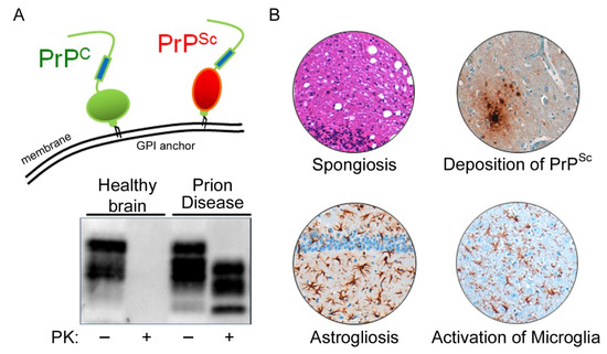
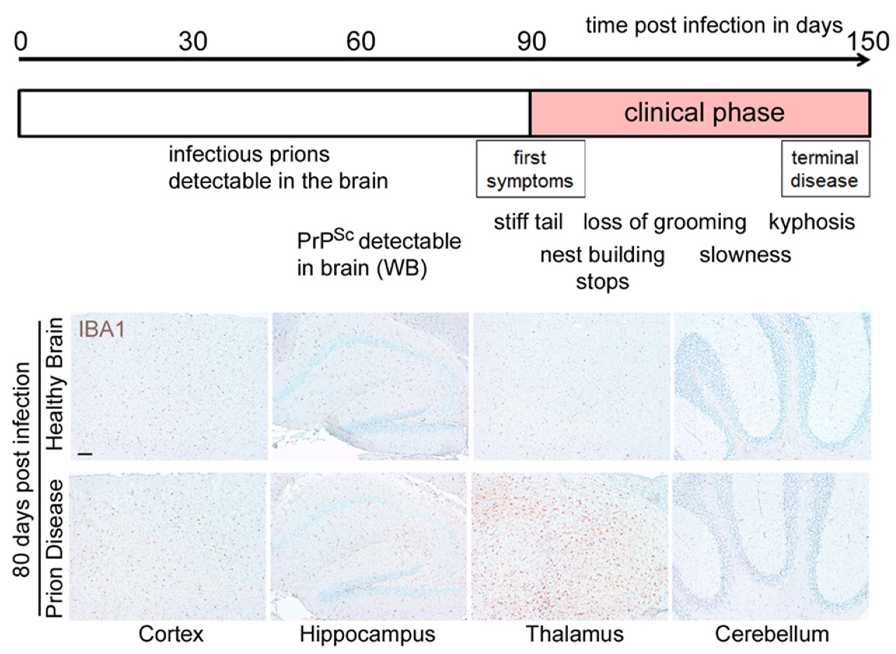
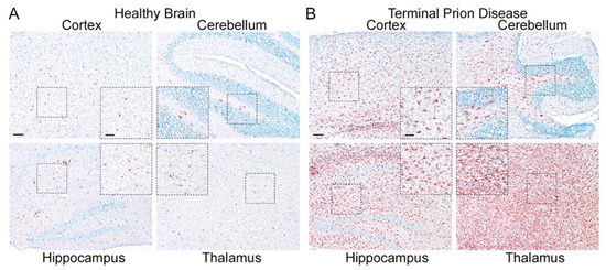
Figure 3. Microglia are highly activated in terminal prion disease. The pan-microglia/macrophage marker IBA1 is shown in mouse brain sections of (A) mock-infected healthy mice and (B) RML 5.0 prion-infected mice at a terminal disease state. (A) Microglia in the healthy brain show a ramified phenotype with a small soma and thin processes in all four regions displayed here (see magnified close-up). (B) During the terminal prion disease stage, microglia massively proliferate (see overview) and change their morphology towards bigger cell bodies and thicker arms (see close-ups) with a bushy appearance in the hippocampus and an amoeboid phenotype in the thalamus [39][40][41][45,46,47]. Scale bar: 100 µm; close-up: 50 µm.
Microglia in the healthy brain display a homeostatic morphology characterized by a small cell body and long branching processes which are constantly surveying their environment [47][61]. Moreover, microglia are involved in several mechanisms regulating brain development and plasticity. They actively shape neuronal connectivity by the removal of excess/neglected synapses, which are formed during development [48][49][50][62,63,64]. For this, microglia have been shown to directly contact tagged pre- and postsynaptic structures and remove them by phagocytosis. The targeting of unwanted elements for removal involves complement receptors and adaptor proteins, among other signals [48][50][62,64].
Microglia can react to alterations in the brain environment or the presence of threats to its cellular integrity e.g., invading pathogens and aggregated proteins, but also apoptotic cells or cell debris, by mounting an inflammatory response to restore homeostasis. The latter enables microglia to increase their phagocytic capacity to remove unwanted structures and to release proinflammatory mediators including cytokines and chemokines [51][65]. Activated microglia represent a common feature of neurodegenerative diseases [52][53][54][54,66,67]. Activation can be identified by a combination of morphological and immuno-phenotypic changes [55][68]. Several proinflammatory cytokines are upregulated in the brain during the prion disease course including TNF α, IL 1α, and C1qa [40][56][57][58][59][60][46,69,70,71,72,73].
Since pro-inflammatory mediators are upregulated slightly before the onset of symptoms, several studies were conducted to investigate the impact of manipulating microglia receptor abundance or cytokine release, and yielded a somewhat mixed outcome. In line with the proposed capacity of microglia to phagocytose PrPSc, the knockout of C-X-C chemokine receptor type 3 (CXCR3) led to accelerated PrPSc accumulation and increased prion infectivity titers, but prolonged survival in the mouse model [61][76]. Moreover, these mice developed excessive astrocytosis. The knockout of the cluster of differentiation CD14 also led to a prolonged survival with enhanced microglia activation in two different mouse models [62][77]. The NLRP3 inflammasome is a multi-molecular complex which can sense heterogeneous pathogen-associated molecular patterns (PAMPs) culminating in the activation of caspase 1 and the release of interleukin IL 1β. It has been shown to play a fundamental role in Alzheimer’s disease (AD) pathophysiology [63][64][78,79]. In contrast to AD, the knockout of NLRP3 or the adaptor protein ASC does not influence the prion disease course [65][80]. Interestingly, retroviral infection preceding prion infection in mice led to a stimulation of microglia at early disease time points with a reduction in infectious prions [41][47], while another stimulator of inflammation, the bacterial lipopolysaccharide (LPS), could not further stimulate their PrPSc-degrading function during disease progression [66][81]. In addition to the upregulation of PAMPs in prion diseases, the transcription of several genes encoding damage-associated molecular pattern (DAMP) proteins and receptors such as Toll-like receptors or proteins of the complement cascade are also increased in the brains of prion-infected mice [57][67][70,82]. Interestingly, depletion of any of the DAMP receptor genes Tlr2, C3ar1, and C5ar1 in a prion mouse model only led to a slightly increased survival in the Tlr2-model, while C3ar1, and C5ar1 did not influence prion disease course [67][82].

2.2. Homeostatic Microglia: Guardians of the Physiological State

For decades, microglia research was focused on the inflammatory context and profile. Only recent research has brought into the focus the homeostatic functions of microglia, which—if lost—might be a driver of neurodegeneration in disease [68][69][70][71][72][55,88,89,90,91]. However, the identification of microglia-specific profiles was complicated by the lack of understanding of whether and how brain-resident microglia functionally differ from peripheral myeloid cells which might enter the brain in certain instances [73][92]. Only recently were the molecular and functional characteristics of murine bona fide homeostatic microglia identified by different groups using a set of novel methodologies such as quantitative proteomics or RNA sequencing [69][74][75][88,93,94]. Thus, a unique transcriptional expression signature of homeostatic microglia was identified which allowed for their differentiation from peripheral myeloid cells. Homeostatic microglia are characterized by the expression of specific markers such as Tmem119, Hexb, Gpr34, P2ry12, Olfml3, and Tgfbr1. Several of these genes are also expressed on human microglia, including the P2Y purinoceptor 12 (P2RY12) [76][95] and the transmembrane protein TMEM119 [77][96]. The development of robust tools including microglia-specific antibodies followed soon after.
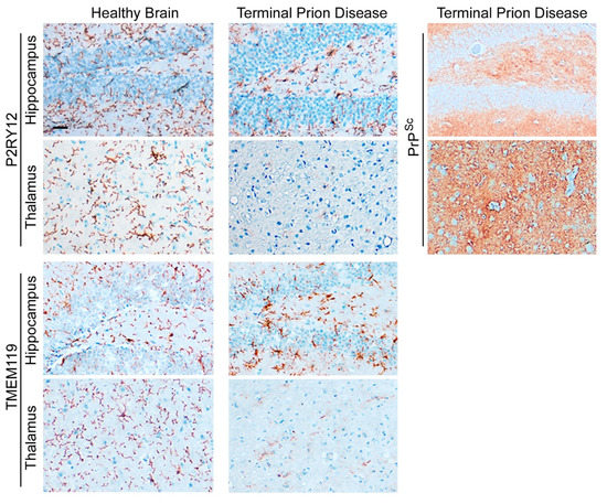
The abundance of homeostatic microglia proteins such as TMEM119 and P2RY12 is significantly reduced in terminal prion disease (Figure 4) [41][60][78][47,73,97]. In the healthy brain, microglia show a ramified morphology with a small cell body and thin processes that are especially visible after staining with P2RY12 [68][69][70][71][72][55,88,89,90,91]. In contrast, both markers are affected in terminal prion disease. While the homeostatic markers are still abundant in the hippocampus, this signature is almost completely lost in the thalamus, an effect which is especially severe for P2RY12 (Figure 4) [41][60][78][47,73,97].
Figure 4. Loss of microglial homeostatic phenotype in terminal prion disease. The microglia-specific homeostasis markers P2RY12 and TMEM119 show a ramified morphology of microglia in the healthy brain [41][60][78][47,73,97]. In terminal prion disease, this homeostatic microglia signature is lost in a region-dependent manner [41][60][78][47,73,97]. While the activated microglia in the dentate gyrus of the hippocampus display a bushy morphology with thick and retracted processes and still express both markers, microglia in the thalamus (posterior complex) have almost completely lost the expression of the markers, especially P2RY12. In the RML-prion mouse model, the deposition of PrPSc-specific staining is stronger in the thalamus than in the hippocampus in terminal prion disease. Scale bar: 25 µm.

2.3. Human Microglia and Their Regional Heterogeneity

For decades it has been noted, mainly based on morphological data from immunohistochemical staining, that the microglia population within the brain is not uniform. Over the last ten years, microglia heterogeneity has been studied in murine models, determining that microglia have distinct region-dependent transcriptional identities and that they age in a regionally variable manner [79][80][109,110]. Recent advances in single-cell sequencing have shown the diversity and regional heterogeneity of murine brain microglia in more detail [81][111]. Although a variety of new molecular tools, such as nuclear sequencing, are now available to study expression profiles on a single-cell basis in the human brain in health and disease [82][113], the transmissible nature of the fatal yet untreatable prion disorders and their respective biosafety issues limit their use in the study of human prion disease. However, bulk expression analysis in two brain regions in sCJD displayed regionally distinct inflammatory profiles [83][114]. Prion disease-associated astrogliosis is also prominent in human prion diseases as shown by the upregulation of GFAP and YKL-40 (or Chitinase-3-like protein 1; CHI3L1) [60][84][73,107]. However, while a GFAP increase is also found in other neurodegenerative diseases such as AD [78][97], YKL-40 upregulation is more specific for human CJD and might serve as a diagnostic marker [84][107]. The microglia signature of sCJD patients differ from those of rapid AD, with significant downregulation of the microglia homeostatic marker TMEM119 [78][97].  The regional differences in inflammation-associated expression changes are also beginning to be investigated and described in the frontal cortex and cerebellum of patients with sCJD [83][114]. Neuronal loss shows considerable variation between various regions of the brain within a CJD-diseased individual and between CJD patients. However, the cortex areas and cerebellum are often severely affected in sCJD, with a relative sparing of the hippocampus and subcortical grey matter [85][86][117,118]. Thus, it will be of the highest interest to investigate, if other, less-affected brain regions will show distinct inflammatory profiles in human prion diseases. Although the deposition patterns of misfolded PrPSc are diverse in murine models of prion disease, the heterogeneity of PrPSc deposition patterns is higher in human prion diseases and comprises synaptic, perivacuolar, plaque-like, kuru-plaque, florid-plaque, punctuate, perineuronal, and intraneuronal patterns, which might occur side-by-side or even overlapping in the same patient [87][88][119,120]. If and how these protein deposits directly influence the inflammatory profiles in individual brain regions in human prion diseases is currently not clear.
ScholarVision Creations