CSCs have been defined ancer Stem Cells (CSCs) is a subset of cancer cells with the ability to self-renew and to differentiate into non-CSC cancer cells within the tumor mass. The CSC field was shaped by great research done on hematopoietic stem cells (HSCs). HSCs are hierarchically arranged with HSCs being the founder cells that undergo asymmetric cell division giving rise to differentiated daughter cells and one quiescent stem cell with self-renewal abilities. CSCs are a subpopulation of cancer cells known to be resistant to therapy and cause metastasis. CSCs have been characterized in many cancers with data illustrating that CSCs display great abilities to self-renew, resist therapies due to enhanced epithelial to mesenchymal (EMT) properties, enhanced expression of ATP-binding cassette (ABC) membrane drug transporters, activation of several survival signaling pathways and increased immune evasion as well as DNA repair mechanisms. CSCs also display great heterogeneity with the consequential lack of specific CSC markers presenting a great challenge to their targeting.
- Cancer stem cells
- Drug response
- Chemotherapy
- Radiotherapy
- Drug efflux pumps
- Tumor Microenvironment
- CSC Niche
- EMT
- Metabolism
- Immunotherapy
1. Introduction
The dividing daughter cells will over time become restricted in terms of lineages it can form. The studies on HSCs ignited research on mammalian tissue and cell renewal as well as in cancer. In addition, cancer patients with chronic myeloid leukemia (CML) were shown to have rare quiescent cells also referred to as Philadelphia chromosome-positive and BCR-ABL-positive cells and these cells were able to resist drug treatment [10,11]. The above-mentioned studies and revelations allowed further research on self-renewal and eventually gave birth to the CSC field as it is today. CSCs are able to reproduce primary tumor heterogeneity as well as metastases in distant tissues and organs [12]. As postulated by Paget, cancer cells can escape the primary tumor site and spread to other tissues and organs where they can proliferate and therefore act as “seeds” for the growth of secondary tumors [12]. It is possible that cancer cells can detach from the primary tumor and enter circulation, however, they are likely not to survive the arduous journey to other organs and cannot “seed” metastases at secondary sites. With their demonstrable survival abilities, enhanced expression of transmembrane transporters and tumorigenic abilities, CSCs on the other hand are likely to survive in circulation and be able to “seed” new tumors at secondary sites [13,14]. CSCs are also responsible for the development of therapy resistance, with many studies demonstrating that CSCs are able to withstand conventional therapies such as chemotherapy and radiotherapy [15]. The ability to resist conventional therapies has been attributed to many properties including increased expression of drug transporters, maintenance of a slow dividing state (quiescence) as well as efficient DNA repair mechanisms [16,17,18]. To overcome CSC resistance, new therapies are under development including epigenetic therapies, immunotherapy as well as drugs targeting angiogenesis [19].
From the early days of their discovery, many studies have shown that CSCs are undifferentiated tumor cells able to generate tumors [20,21,22]. To date, several studies have been able to prove the existence of CSCs in cancers such as CML, ovarian, lung and breast cancer [23,24]. Methods used to identify CSCs range from antibody-based isolation, enzyme activity of ALDH, tumorsphere formation, use of dyes such as PKH26 and side population sorting [25,26]. Side population cells display enhanced abilities to efflux dyes and drugs at a higher rate than the main cell population due to increased expression of ATP-binding cassette (ABC) transporter proteins. These methods are all not specific, and in most cases, scientists combine these methods to get a cell population with high numbers of CSCs. The gold standard method to study whether cancer cells have tumor-initiating capabilities is the use of limiting dilution in xenograft animals. A detailed review of CSCs definition and terminology is provided by Valent and colleagues [15]. Recently introduced “humanized” animal models are better models than traditional animal models as they can recapitulate some human cancers better [27,28].
Due to their ability to resist therapy, CSCs can travel to distant sites and form new tumors. Whilst the process of metastasis appears disorganized, metastatic lesions are the main cause of cancer deaths and therapy resistance [29]. Signaling pathways upregulated and dysregulated in CSCs and CSC-cell interactions are therefore some of the targets of new drugs under development. Conventional cancer treatment strategies mainly target rapidly proliferating cancer cells and can reduce tumor mass, tumor relapse can result from a few remaining cancer cells including CSCs (Figure 1) [30]. Our ability to target CSCs largely depends on new evidence and in-depth characterization of these cells. It is plausible to postulate that long-lasting cancer treatment efficacy can only come from both shrinkage of the primary tumor as well as the prevention of cells such as CSCs from metastasizing to new sites throughout the body.
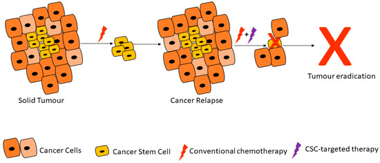
Figure 1. Cancer stem cells are able to resist conventional therapies and form new tumors, unless targeted by cancer stem cell (CSC)-specific therapy. Adapted from Dzobo et al. [30].
2. Properties of Cancer Stem Cells
2.1. Cancer Stem Cell Markers and Therapy Resistance
Current therapies are unable to eliminate cancer partly due to CSCs’ enhanced ability to withstand treatment regimens [15,30]. CSCs are thought to account for a small percentage of the total number of cancer cells within a tumor but have self-renewal and differentiation capabilities [21]. A major hurdle faced by scientists working with CSCs has been the isolation and characterization of these cells. Antibodies against several CSC markers have been used to isolate CSCs from solid tumors [26]. Commonly used CSC markers and methods for isolation and characterization include CD24, CD44, CD133 and ALDH enzymatic assay (Figure 2; Table 1) [31]. These CSC markers are either used alone or in different combinations in different cancers. For example, gastric CSCs display high CD44, CD133 as well as Lgr5 [32]. Lung CSCs express several markers including CD133+, ALDH1+ and CD44+ [33]. Whilst the same CSC markers can be found in different cancers, some cancers have distinct markers for example melanoma CSCs are ABCB5+ whilst medulloblastoma CSCs are CD15+ (Table 1).
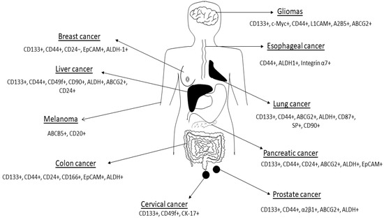
Figure 2. Cancer stem cell markers expressed in some human cancers are shown in the figure. Figure adapted from Dzobo et al. [34]. See Table 1 for references. The list of CSC markers is not exhaustive. The CSC markers continue to be refined based on new data.
Table 1. CSC markers expressed in different human cancers *.
| Cancer | CSC Markers | References |
|---|---|---|
| Cervical | CD133+, CD49f+, CK-17+ | [35,36,37] |
| Esophageal | CD44+, ALDH1+, Integrin α7+ | [38,39] |
| Kidney | CD24-, CD44+, CD105+, CD133+ | [40,41,42] |
| Lung cancer | CD44+, CD90+, CD133+, ABCG2+, ALDH+ | [24,33] |
| Colon cancer | CD24+, CD44+, CD133+, EpCAM+, ALDH+ | [43,44,45,46] |
| Liver cancer | CD24+, CD44+, CD90+, CD133+, ALDH+, ABCG2+ | [47,48] |
| Breast cancer | CD24-, CD44+, CD133+, ALDH-1+ | [5,49,50] |
| Gastric | CD44+, CD133+ | [51,52,53] |
| Glioma | CD44+, CD133+, A2B5+, BCRP1+, SSEA-1+ | [54,55] |
| Leukemia (AML) | CD34+, CD38−, CD123+ | [56,57,58] |
| Leukemia (CML) | CD25+, CD26+, CD44+, CD93+, IL1RAP+ | [59,60] |
| Ovarian | CD44+, CD117+, CD133+, ALDH1+ | [61,62] |
| Prostate cancer | CD44+, CD133+, α2β1+, ALDH+ | [63,64,65] |
| Pancreatic cancer | CD44+, CD133+, ABCG2+, ALDH+, EpCAM+ | [66,67,68] |
| Melanoma | ABCB5+, CD20+ | [69,70] |
| Head and neck cancer | CD44+, CD133+ | [71,72] |
| Sarcoma | CD29+, CD117+, CD133+, Nestin+, Stro-1+ | [73,74] |
* The list of CSC markers is not exhaustive. The CSC markers continue to be refined based on new data.
Several studies demonstrated an increase in CSCs in tumors after cancer treatment, clearly illustrating their persistence during treatment [75,76,77]. CSCs are able to resist therapeutic interventions due to several reasons including their cellular plasticity, enhanced expression of ABC drug transporters, ability to detoxify of drugs and compounds, increased adaptation to stressful conditions such as hypoxia, attaining quiescence and activation of survival pathways [77,78,79].
CSCs ability to resist therapy is widespread and referred to as multidrug resistance. This capability stems from the ability of CSCs to express increased detoxifying enzymes, increased activation of survival signaling pathways, DNA repair mechanisms as well as drug efflux pumps [30,78]. In addition, CSCs have been noted for their immune evasion capabilities, their ability to undergo epithelial to EMT as well as to adapt their metabolism to survive low nutrient conditions [77,78]. Thus, the hallmarks of CSCs include quiescence, increased expression of drug metabolizing and detoxifying enzymes, enhanced DNA reparability, the ability to undergo EMT and overexpression of ABC membrane transporters. Lately, CSCs have also been shown to undergo epigenetic reprogramming, making them very difficult to eradicate in cancers [80].
The ALDH superfamily is a large family of proteins and several members including aldehyde dehydrogenase 1 (ALDH1) have been implicated in drug detoxifying activities [81,82]. In its entirety, the ALDH superfamily is composed of 19 enzymes with ALDH1 being the main isoform [81,82,83]. This family of detoxifying enzymes is involved in the oxidation of aldehydes to carboxylic acids as well as retinol to retinoic acid [84,85]. Besides being expressed by normal cells, ALDH1 is expressed highly in CSCs [86,87]. As a result, ALDH1 expression and activity can be used reliably to identify CSCs in some cancers. Vogler and colleagues demonstrated that ALDH1 expression can be used as an independent prognostic marker for low survival in colorectal patients [88]. In addition, van den Hoogen and coworkers also showed that enhanced ALDH1 activity can be used to identify tumor-forming cells as well as cells with the propensity to form prostate cancer metastases [89]. Ueda and colleagues also showed that ALDH1 activity can be used to identify cancer cells with CSC-like properties in human renal cell carcinoma cell line [90]. Ginestier and colleagues demonstrated that ALDH1 is highly expressed in breast CSCs and is a predictor of poor clinical outcome [91]. In addition, ALDH1-expressing cells were able to form xenograft tumors easily [91]. Several other studies demonstrated the successful transplantation of ALDH1-expressing cells into mice [92,93]. The expression of ALDH1 by normal stem cells may explain the aberrant expression of this enzyme in CSCs as normal stem cells are a potential source of CSCs, among other cells [94]. Furthermore, ALDH1 expression has been shown to allow CSCs to resist conventional therapy including commonly used drugs such as paclitaxel, gemcitabine and cisplatin [95,96]. In agreement with the above, several studies demonstrated that inhibition of ALDH1 activity in CSCs sensitizes these cells to several drugs, linking ALDH1 with therapy resistance [97,98].
In addition, CSCs demonstrate increased expression of drug effluxing proteins such as the ABC transporters (Figure 3) [99,100,101]. The ABC family of transporters consists of 49 molecules using ATP as an energy source during the trafficking of proteins across the cell membrane. Many studies have been performed on the characterization of members of this family including ABCB1 (multidrug resistance 1 (MDR1)), ABCG2, ABCC1 and ABCB5 [102,103]. Through elaborate experiments, several research groups demonstrated that CSCs aberrantly express ABC transporters and are able to withstand toxic levels of drugs and other toxins [104,105]. In elaborate experiments performed by Wright and colleagues, the researchers demonstrated that ABCB1 was aberrantly overexpressed in breast CSCs causing resistance to conventional chemotherapy such as paclitaxel and doxorubicin [106]. Frank and coworkers demonstrated that ABCB5 was overexpressed and caused resistance to doxorubicin in CD133+ circulating melanoma cells [107]. Through the use of a monoclonal antibody against ABCB5, the authors were able to induce cancer cell sensitivity to drugs such as doxorubicin [107]. Shi and colleagues demonstrated that ABCG2-expressing CSCs isolated from hepatocellular carcinoma cell lines via the side population technique are able to resist cisplatin and 5-fluorouracil [108]. The above studies and others demonstrated that inhibition of ABC transporters is a potential mechanism of overcoming CSC chemoresistance [109,110]. Several studies have been performed on the inhibition of ABC transporters and have shown remarkable success in sensitizing both cancer cells and CSCs to several drugs [111,112]. For example, Marcelletti and colleagues utilized zosuquidar, an inhibitor of P-gp (ABCB1) to sensitize cancer cells in acute myeloid leukemia [111,112].
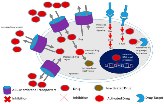
Figure 3. Hallmarks of cancer stem cells include increased expression of ATP-binding cassette (ABC) membrane transporters, enhanced survival signaling, increased drug in activation as well as increased DNA repair processes compared to cancer cells. This allows CSCs to survive conventional therapy and thus contribute to chemoresistance for example. Adapted from Senthebane et al. [3].
Several other proteins associated with apoptosis are also involved in the survival of cancer cells and CSCs [113,114]. For example, several pro-survival proteins including BCL-2, B-cell lymphoma extra-large (Bcl-xL) and BCL-2-like-2 (BCL-W) have been found to be overexpressed in several cancer types including lymphoid cancer [115,116]. The overexpression of these pro-survival proteins has also been linked with carcinogenesis, with the blocking of these proteins and their associated pathways resulting in reduced tumor growth and enhanced response to chemotherapy [116,117,118].
Cytotoxic drugs target rapidly growing cancer cells, making them ineffective against slow dividing or dormant CSCs [30]. Viale and colleagues demonstrated that leukemia CSCs proliferate at a much lower rate than other cancer cells [119]. Therapies that target cancer cell cycling would therefore be ineffective against CSCs. Therapeutic agents such as paclitaxel would be unable to be less effective against slow dividing CSCs [120]. In addition, several studies demonstrate that CSCs show enhanced DNA damage repair capacity, with phosphorylation of repair enzymes observed in cancers such as breast and gliomas [121,122]. CSCs including glioma stem cells demonstrate great abilities at ROS scavenging thus protecting themselves against oxidative DNA damage [123,124,125]. Therapy itself has been shown to selectively increase CSCs in tumors. For example, Rizzo and colleagues demonstrated that CSCs are enriched in ovarian tumors after chemotherapy [126]. In addition, Levina and coworkers showed that chemotherapy can lead to the propagation of CSCs in lung cancer [127]. Thus, chemotherapy only targets the rapidly proliferating cancer cells leaving the CSCs to propagate the tumor after therapy. Chen and colleagues demonstrated that the drug temozolomide (TMZ) activates CSCs to produce cancer cells after therapy [128]. Qiu and colleagues demonstrated that elevated O6-methylguanine DNA methyltransferase (MGMT) expression and activity in glioma stem-like cells were responsible for temozolomide resistance [129]. Kurtova and coworkers also demonstrated that blockage of tumor repopulation by CSCs is effective at attenuating therapy resistance in bladder cancer [130]. Saito and colleagues demonstrated that inducing cell cycle re-entry through treatment with granulocyte colony-stimulating factor (G-CSF) allows normal chemotherapy to eliminate cancer cells effectively [131]. In addition, the induction of CSCs differentiation has been used successfully to increase CSCs sensitivity to commonly used cancer drugs. Lombardo and coworkers induced colorectal CSCs terminal differentiation via the use of bone morphogenic protein 4 (BMP4) and observed increased CSCs sensitization to standard chemotherapy [132]. Wang and coworkers used silibinin, which blocks colon CSCs self-renewal, resulting in reduced CSC population leading to reduced cancer cell proliferation [133]. Whilst several strategies have been developed to induce CSCs differentiation, all-trans retinoic acid (ATRA) is one of the common drugs used for this purpose [134,135].
2.2. Cancer Stem Cells and Angiogenesis
Many biological processes are dependent on the formation of new blood vessels, a process referred to as angiogenesis. Normal development and tissue repair and regeneration are especially dependent on new blood vessels for the supply of nutrients as well as the removal of toxic material [136,137]. Besides normal biological activities, angiogenesis is a requirement for tumor formation beyond a certain diameter [138,139]. During tumor formation, the usual delicate balance between pro-angiogenesis and anti-angiogenesis is altered, with pro-angiogenesis factors dominating [138]. New blood vessels sprout from pre-existing vessels within and around the tumor, fueling the rapid growth of the tumor [140,141]. The rapid growth of a tumor results in hypoxic conditions within the tumor. CSCs are known to release factors such as hypoxia-inducible factor 1 which induces the release of proangiogenic factors (Figure 4) [142,143]. Hypoxia has also been shown to fuel CSCs [144]. Hypoxia promotes CSC growth in several cancers via the upregulation of adaptive transcriptional programs, allowing CSCs to survive, invade and metastasize [144,145]. Soeda and colleagues demonstrated that hypoxia promotes the self-renewal capacity of CD133-positive human glioma-derived cancer stem cells [144]. Heddleston and colleagues showed that hypoxia promotes CSC self-renewal capabilities and stem-like phenotype even in non-stem cancer cell populations [146]. As reviewed by Heddleston and colleagues, CSC plasticity is influenced by hypoxia and new drugs must target such microenvironmental conditions for durable cancer treatment [147].
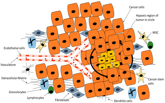
Figure 4. Cancer stem cells are able to reside deep within the tumor in hypoxic regions that are normally toxic to normal cells, whilst CSCs are able to release factors such as hypoxia-inducible factor 1 which induces the release of proangiogenic factors, this position means CSCs are inaccessible to drugs or are exposed to reduced drug doses. Adapted from Senthebane et al. [3].
Endothelial cells are also recruited to the tumor site. Endothelial cells express VEGFR and the binding of VEGF-A results in the activation of several signaling cascades involved in migration and ECM remodeling [143,148]. Survival pathways including the PI3K-Akt and the MEK-ERK cascades are activated and play key roles in the activation of endothelial cells to form new blood vessels [148]. Several cytokines are also known to be secreted by CSCs within the TME and these include IL-6 and TNF-α [149,150]. The secreted cytokines are involved in the recruitment of immune cells such as myeloid cells to further promote tumorigenesis [149,150]. Dysregulation of the Notch pathway has also been associated with tumor growth in general and survival of CSCs [151,152,153]. Several reports demonstrated that cells showing high expression of Notch signaling have elevated tumor-forming abilities and self-renewal capacity than those with less Notch activation [154,155,156]. Activation of the Notch pathway has also been associated with proangiogenic activity, with Notch ligand Jagged-1 promoting blood vessel formation [157,158,159].
In addition, matrix metalloproteases (MMPs) secreted by both cancer cells and stromal cells remodel the TME, allowing the creation of space for blood vessels formation as well as recruitment of different cells [160,161]. Due to the plasticity of CSCs, suggestions have been made to the effect that CSCs can give rise to endothelial cells and pericytes within and around the tumor [162,163]. Blood vessels within the TME are convoluted and “leaky”, resulting in fewer drugs able to reach cancer cells and CSCs deep within the TME. Tumor-derived cells are also able to intravasate and travel to distance sites, promoting metastasis in the process. Several studies have demonstrated the presence of circulating tumor-derived cells that are able to act as “seeds” for new tumors in distant sites [164,165]. Once the circulating cancer cells reach distant sites, they are able to extravasate and form new tumors in favorable microenvironments [19,166]. The formation of new tumors is dependent on CSCs successfully inducing angiogenesis to allow the exchange of nutrients and metabolic byproducts. Whilst it has been shown that stromal cells play a key role in inducing angiogenesis within tumors, CSCs are also involved in releasing angiogenic factors [167]. For example, Bao and colleagues demonstrated that glioma CSCs release VEGF resulting in increased microvascular density in malignant glioma [168]. Monzani and colleagues also showed that melanoma CSCs co-expressed CD133 and VEGF [169]. Maeda and colleagues also showed that pancreatic CSCs co-express CD133 and VEGF-C resulting in increased microvascular density [170]. It is also possible that cancer cells may enter a state of dormancy in which they remain until induced to proliferate and form new tumors [171,172].
2.3. Cancer Stem Cells and Epithelial to Mesenchymal Transition
Besides the influence of genetic and epigenetic mechanisms on the CSC phenotype, the TME within which CSCs are located plays a huge role in the CSC behavior [30]. As more data emerges the CSC field continues to change and be refined [117,173]. Overall, the CSC phenotype is dynamic and never constant. When CSCs undergo EMT, they acquire characteristics allowing them to migrate, invade surrounding tissues and metastasize [174]. EMT and CSC characteristics appear to share similar molecular pathways that are involved in invasion and migration of cancer cells from the primary tumor. In addition, transcriptional analysis of EMT and those associated with CSCs reveal significant overlap in gene expression including TGF-β, Hedgehog signaling and microRNAs [26]. EMT has been associated with poor prognosis in several cancers including esophageal and colon cancers [175,176]. Several signaling pathways have been identified to be key in modulating CSCs behavior including invasiveness and metastatic ability [177,178]. In addition, several markers identifying CSCs with invasive and metastatic abilities have been revealed including CD44v6 [179,180]. CD44 is specifically expressed by breast epithelial cells undergoing EMT [181]. EMT is characterized by the loss of cell to cell adhesion with cells becoming mesenchymal and markers such as E-cadherin lacking in such cells [182,183]. The loss of E-cadherin from the cell surface is accompanied by the expression of N-cadherin [184]. Histone deacetylation of the CDH1 promoter through the actions of DNMT and HDACs leads to gene silencing [185,186]. Histone methylation within the CDH1 promoter via the EZH2 and PRC2 complex is known to silence its expression [187].
EMT is influenced by several protein factors as well as microRNAs. For example, TGF-β has been regarded as a master regulator of EMT in certain cancers including breast and colorectal cancers [188]. Besides influencing cancer cells, TGF-β can also regulate CAFs with a net effect of promoting metastasis [189]. Furthermore, microRNA-200 family members have been shown to suppress EMT via binding to two transcription factors, zinc finger E-box-binding homeobox 1 (ZEB1) and ZEB2 [190,191]. Tellez and colleagues demonstrated that EMT can be induced by epigenetic mechanisms including chromatin remodeling through H3K27me3 enrichment as well as DNA methylation to sustain silencing of tumor-suppressive microRNAs, microRNA-200b, microRNA-200c and microRNA-205 [192]. Thus, silencing these microRNAs through tri-methylation of DNMT and H3K27 can induce EMT-like and CSC characteristics [192].
2.4. Cancer Stem Cells and Metabolic Activity
Recently, metabolic alterations have been identified to cause cells to acquire stem-cell-like characteristics [193]. These alterations and the subsequent acquisition of stem-cell-like characteristics are thought to be caused by epigenetic changes in adult stem cells as well as cancer cells. Based on the CSC theory, acquisition of stem-cell-like characteristics makes these cells achieve a higher status within the hierarchy through the expression of self-renewal and pluripotent genes [30,78]. According to Menendez and Alarcon, products of mutated metabolic enzymes can behave as oncometabolites, inducing epigenetic changes in genetic material and thus drive tumor initiation and progression [193]. This and more pieces of evidence point to the need for a full view of tumor initiation and progression and not just focus on cancer cells. Metabolic processes can thus be targeted to stop tumor initiation and progression. Specifically, the TME is characterized by low oxygen and glucose levels and thus tends to favor oxidative phosphorylation as the main supplier of energy [194]. Hypoxia has been shown to induce metabolic alterations resulting in acidosis in several cancers [195,196]. Lee and colleagues demonstrated that chemoresistance and enhanced oxidative phosphorylation are correlated [194]. Recent studies demonstrated that indeed, the targeting of oxidative phosphorylation has shown some success in inhibiting CSCs metabolic processes and proliferation in some cancers [197,198].
Inhibition of the mitochondrial complex III resulted in decreased breast CSCs [199]. When relapse occurs, CSCs have been shown to increase oxidative phosphorylation levels to pretreatment levels, demonstrating the importance of oxidative phosphorylation in chemoresistance [200]. The adipose tissue and adipose-derived cells are able to interact with CSCs and have been shown to promote fatty acid oxidation in CSCs and chemoresistance [201]. The mitochondria are also known to play a role in CSC chemoresistance [202]. This is unsurprising as the mitochondria are central to many cellular processes such as metabolism, signaling and apoptosis. Mitochondria have recently been shown to play key roles in CSC behavior [203]. Sancho and colleagues concluded that the removal of CSCs through targeting mitochondrial function might prevent cancer disease from recurring and thus prevent fatal disease [204]. In colon CSCs, tumorigenic ability was associated with enhanced mitochondrial functions [205]. Atovaquone has been used to inhibit the mitochondrial complex II resulting in decreased breast CSCs [199]. Isayev and colleagues demonstrated that inhibition of glucose metabolism through the use of 3-bromopyruvate inhibited pancreatic CSCs growth and resistance to gemcitabine [206]. Several other studies also showed that inhibition of mitochondrial function affect CSC proliferation and self-renewal capabilities [207,208].
2.5. Cancer Stem Cells and Epigenetic Reprogramming
A contributing factor to the complex intra- and inter-tumor heterogeneity and the resulting failure of many anticancer therapies comes from CSC epigenetic alterations. The heritable non-genetic changes to CSCs phenotypes are what are called epigenetic reprogramming of CSCs [209,210]. Most of the proteins and enzymes involved in epigenetic reprogramming of cells including histone modifications and DNA methylations have been well-characterized [211,212]. For example, histone methyltransferases (HMTs) are responsible for methylation of histones whilst histone acetyltransferases are responsible for the acetylation of histones [213]. Demethylation and deacetylation of histones are carried out by histone demethylases (HDMs) and histone deacetylases (HDACs) respectively [213]. When acetylated, histones are more loosely packed and can be accessed by RNA polymerases, allowing transcription of genes around a specific location. On the other hand, methylation can activate or repress gene transcription. For example, the acetylation of histone H3/H4 is linked to the transcription of genes [214,215]. In addition, H3 lysine 4 methylation is also linked to transcription of several genes [216,217]. In contrast, the methylation of H3 lysine 9 and 27 is linked to gene repression [218,219,220]. It has been observed that different patterns of histone modification produce variable transcriptional outcomes, with some giving rise to activation of genes and others to repression [221,222]. Various mechanisms are known to be involved in epigenetic gene regulation, from modifications of cytosines in DNA, covalent modifications of histones, the involvement of noncoding RNAs to chromatin remodeling [223,224,225].
CpG islands are regions of the genome containing a large number of CpG dinucleotide repeats and usually extend for 300–3000 base pairs [226]. In most cases, CpG islands are located close to gene promoters in humans [227]. DNMT in addition to histone modification determines whether transcription occurs or not. When CpG islands are unmethylated, transcription can take place. When CpG islands are methylated the chromatin becomes transcription-suppressive. Methylation of CpG islands is catalyzed by DNMT1, DNMT3A and DNMT3B. Several tumor suppressor genes are silenced via CpG island methylation [228]. Transcription can also be repressed via the Polycomb repressive complexes 1 and 2 (PRC1 and PRC2) [229,230]. Polycomb repressors are able to catalyze the trimethylation of histone 3 lysine 27 (H3K27me3) giving rise to repression of genes associated with many cellular processes such as differentiation, development and choice of lineage [229,231]. Collinson and colleagues demonstrated that Polycomb complex PRC2 mediates H3K27me3 via the histone methyltransferase EZH2, leading to transcriptional repression of several genes [232].
CSCs and their subsets display epigenetic alterations including histone modifications and this eventually contributes to the intratumor heterogeneity observed in many tumors [233]. Several epigenetic regulators have mutations leading to tumor formation and progression as a result of epigenetic dysregulation [234,235]. Several CSC markers including CD133 are known to be regulated by epigenetic alterations [236]. Tabu and colleagues demonstrated that the hypomethylation of the CD133 promoter influences its expression in gliomas [237]. Yi and colleagues observed abnormal DNA methylation of CD133, a CSC marker, in colorectal and glioblastoma tumors [236]. Gorodetska and colleagues observed that EZH2/BRCA1 signaling mechanisms play an important role in the maintenance of prostate CSCs properties [238]. EMT aid in the generation of cells with stem cell characteristics and is modulated by epigenetic mechanisms [239,240]. The involvement of epigenetic mechanisms from CSC formation to maintenance makes epigenetics a therapeutic target in CSCs. Small compound inhibitors with the ability to induce differentiation in CSCs are therefore promising drugs targeting this population of tumor cells.
Several signaling pathways are crucial in facilitating the growth of CSCs and the maintenance of the CSC phenotype. Such signaling pathways include Hedgehog, Notch, JAK-STAT and Wnt-β- catenin signaling [241,242]. It is important to note that these same pathways are also important in regulating self-renewal in normal stem cells [243,244]. Several mutations have been observed in genes along these pathways in many human cancers. Signaling pathways such as Wnt and Notch have been observed in breast cancers for example [245] and in vitro work demonstrated that the overexpression of these pathways is associated with tumorigenicity [155,156]. Triple-negative breast cancer cells demonstrate increased Notch signaling and Notch signaling is associated with CD44 expression in colon cancer cells [158,246]. On the other hand, Wnt-β-catenin signaling has been observed to be associated with cancer stemness and heterogeneity [247]. Several members of the Wnt-β–catenin pathway have been linked to the induction of EMT in several cancers [248]. The hedgehog signaling pathway has been associated with self-renewal in many cancers including breast cancer and gliomas [249,250]. The hedgehog pathway has also been associated with EMT and invasion and migration [251,252].
Most of the above-mentioned signaling pathways are modulated by epigenetic mechanisms [253]. Under normal conditions, most of these pathways are involved in the propagation of CSCs, maintenance of the CSC phenotype as well as in embryonic development [254]. Several regulators of the above-mentioned pathways have been shown to have epigenetic alterations in CSCs. For example, decreased acetylation of H3K16 as well as enhanced H3K27 trimethylation is associated with DKK1 promoter silencing [255]. High levels of histone acetylation are observed at the promoter region of Notch receptor–ligand JAGGED2, resulting in Notch signaling activation in multiple myeloma cells [256]. In colorectal cancer, two Notch signaling targets, HES1 and HES2, show decreased promoter H3K27 methylation, resulting in gene activation [257,258,259]. Rhabdoid tumors show decreased or inactivation of SNF5, a member of chromatin remodeler complex SWI/SNF, leading to activation of Hedgehog signaling [260,261]. Furthermore, the activation of Gli1 and Gli2, downstream effectors of the Hedgehog signaling pathway, require HDAC1 [262,263,264]. As a result of the integration of genetic, epigenetic mechanisms and other factors, CSCs survival and maintenance are promoted.
The KMT2/MLL gene is known to encode for an HMT that influences many cellular processes [265,266]. MLL fusion proteins are present in several CSCs and have been shown to be involved in carcinogenesis in several cancers [267,268]. For example, Krivtsov and colleagues demonstrated that leukemia stem cells, with the MLL-AF9 fusion protein, can maintain the identity of progenitors from which they arose while at the same time activating stem-cell- or self-renewal-associated program [269]. Somervaille and colleagues also demonstrated that the hierarchical maintenance of MLL-myeloid leukemia stem cells utilizes a transcriptional program involving transcription/chromatin regulatory factors Myb, Hmgb3 and Cbx5 [270]. Several mutations have also been identified in histone-encoding genes. Lewis and colleagues showed that the blockage of PRC2 activity via the gain-of-function H3 mutation was prevalent in pediatric glioblastoma [271]. Furthermore, several DNMTs are mutated in acute myeloid leukemia and have been suggested to result in the formation of leukemia stem cells [272,273,274].
CSCs have been shown to play important roles in the propagation, growth and metastasis of colorectal cancer (CRC). Several genetic and epigenetic changes have been observed in CSCs in CRC. For example, the hypermethylation of several tumor suppressor gene promoters including p16, retinoblastoma, SFRP and MLH1, has been widely reported in many studies [7,275,276]. One of the driver mutations in CRC is the APC mutation, which influences the activities of DNMTs [277]. Increased levels of DNMT1 are thought to suppress the transcription of APC, a tumor suppressor gene in CRC [278,279]. The levels of DNMT1 in CSCs have been shown to be involved in CRC initiation and progression, directly linking epigenetic mechanisms to CSC-directed tumorigenesis [280]. Pathania and colleagues showed that DNMT1 is important for mammary and CSC maintenance and tumorigenesis [281].