PDue to the lack of diagnostic biomarkers and high resistance to treatment, pancreatic ductal adenocarcinoma (PDAC) irepresents one of the most lethal solid tumors, associated with poor survival. The early stages usually lack symptoms, leading to a late diagnosis (stages III or IV) with limited therapeutic options and a dismal prognosis. Moreover, the metastatic potential of PDAC is extremely high and tumors spread mainly through lymphatic and blood vessels. Most of the patients already have metastasis in the liver and lymph nodes at the time of diagnosisAlthough genetic modifications are well defined in PDAC, the role of epigenetics regulations, which secure dynamic response to environmental stimuli, has only recently been recognized. Epigenomic studies revealed that epigenetic changes in oncogenes and tumor suppressor genes were associated with epithelial-mesenchymal transition (EMT) which is responsible for the invasive phenotype of cancer cells and therefore, their metastatic potential. of cancer cells and therefore, their metastatic potential.
- pancreatic ductal adenocarcinoma
- epigenetics
- epithelial–mesenchymal transition
- pancreatic cancer progression
The extensive research in genetics and genome-wide expression patterns indicates that genetic changes are critical for PDAC initiation and early progression. However, epigenetic alterations affected tumor progression and were associated with patients´ survival
Pancreatic ductal adenocarcinoma (PDAC) arises in the exocrine glands of the pancreas, although some acinar cells are highly flexible and can experience acinar to ductal metaplasia under stress conditions, and accounts for 95% of all pancreatic cancers. Since PDAC is characterized by rapid tumor progression, most of the patients already have metastasis in the liver and lymph nodes at the time of diagnosis
[1]. Epigenetic changes are heritable modifications of DNA or chromatin structures, which influence gene expression without altering the DNA sequence
. According to epidemiological data, less than 20% of cases survive one year and the five-year survival rate remains around 5–7%
[2]. Epigenomics of PDAC studies the epigenetic changes across multiple genes or the entire genome. DNA methylation, post-translational modifications of histone proteins, and non-coding RNAs are the main epigenetic mechanisms that can influence gene expression. They have been confirmed as important contributors to PDAC development and progression
. Despite intensive research, surgical resection of the pancreas with microscopically free margins (R0 resection) remains the only realistic and potentially curative option. Unfortunately, rapid disease progression and early metastasis are responsible for the late diagnosis and less than 20% of patients have a resectable tumor at the time of diagnosis
[3]
.
The extensive research in genetics and genome-wide expression patterns indicates that genetic changes are critical for PDAC initiation and early progression. However, epigenomic studies revealed that epigenetic alterations in oncogenes and tumor suppressor genes affected tumor progression and were associated with survival
[4]
. Epigenetic changes are heritable modifications of DNA or chromatin structures, which influence gene expression without altering the DNA sequence
[5].
EMT has emerged as a clinically relevant regulator of tumor cell spread to distant organs
. Epigenomics of PDAC studies the epigenetic changes across multiple genes or the entire genome. DNA methylation, post-translational modifications of histone proteins, and non-coding RNAs are the main epigenetic mechanisms that can influence gene expression. They have been confirmed as important contributors to PDAC development and progression
[6]. Early metastasis of PDAC results from dynamic gene expression changes, allowing epithelial tumor cells to acquire mesenchymal, fibroblast-like properties, increased migratory capacity, invasiveness, and resistance to therapy [7]. A cross-talk between individual counterparts of tumor microenvironment and innate and adaptive immune cells leads to PDAC progression (
[8].
Epithelial-mesenchymal transition (EMT) has emerged as a clinically relevant regulator of tumor cell spread to distant organs [9]. Early metastasis of PDAC results from dynamic gene expression changes, allowing epithelial tumor cells to acquire mesenchymal, fibroblast-like properties, increased migratory capacity, invasiveness, and resistance to therapy [10]. A cross-talk between individual counterparts of the tumor microenvironment and innate and adaptive immune cells leads to PDAC progression (
).
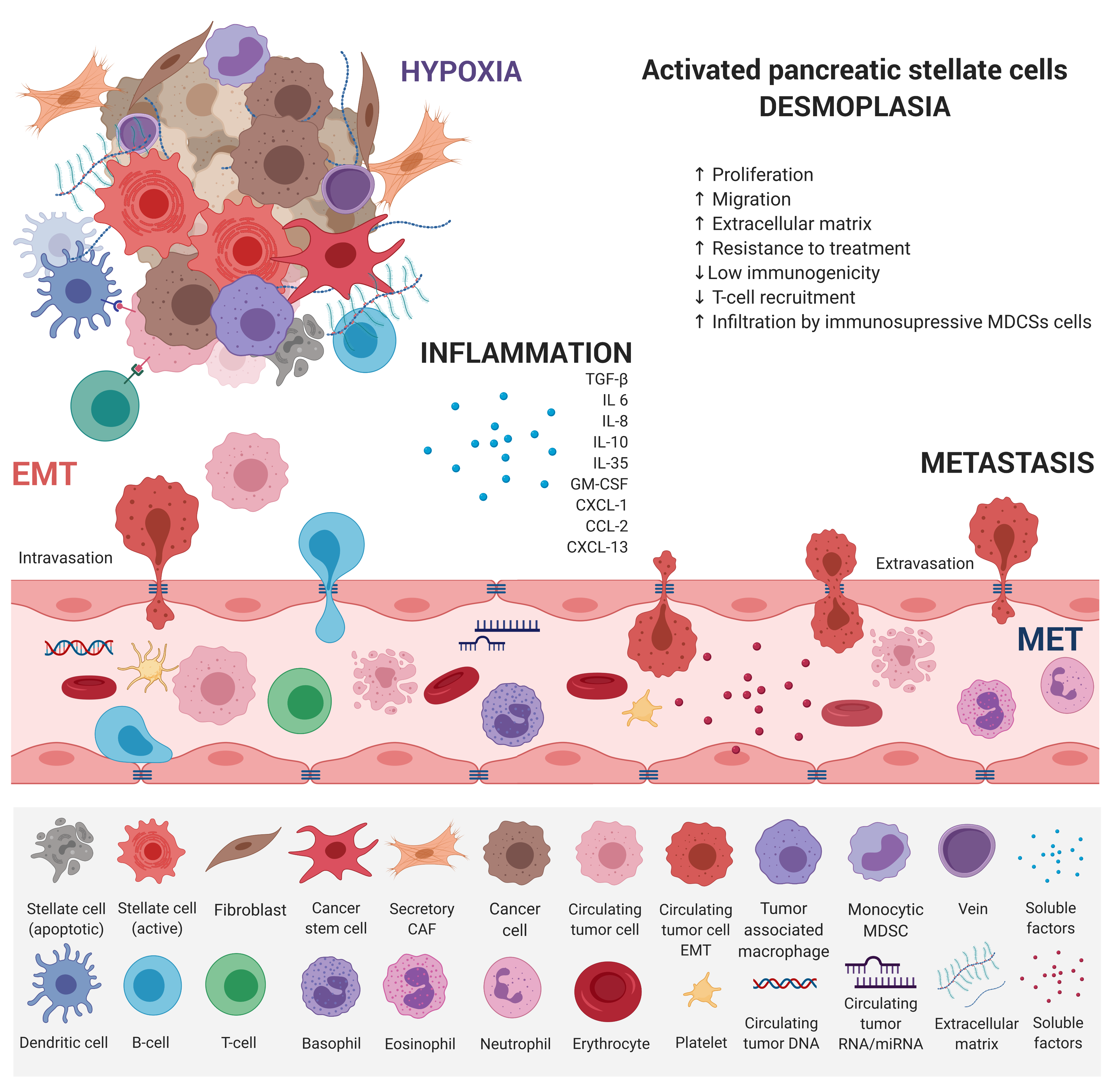
Figure 2.
KRAS mutation is an early oncogenic event that drives the recruitment of B cells early in the development of PDAC, which can exert immunosuppressive effects. Therefore, T cells, both CD4+ and CD8+, are not primed against PDAC antigens and are not excluded from tumors. The paucity of blood vessels leads to high levels of hypoxia in the interior of the tumor. Paracrine interactions between inflammatory cells and hypoxia stimuli induce EMT, which promotes circulating tumor cell (CTC) formation and endows differentiated normal and cancer cells with stem cell properties. They are characterized by a high expression of drug efflux pumps including multi-drug resistance genes, protecting them from chemotherapeutic reagents thus increasing their metastatic potential.
This dedifferentiation process is associated with a loss of functional epithelial cell markers, such as E-cadherin, and increased expression of mesenchymal markers
EMT represents a dedifferentiation process that is associated with a loss of functional epithelial cell markers, such as E-cadherin, and increased expression of mesenchymal markers
[8]. Stem cell-associated marker expression of CD24+, CD44+, and CD133+ cells was found to correlate with EMT in PDAC [9]. TGF-b, TNF-a, as well as Notch, Hedgehog, and Wnt molecular pathways have been shown to induce EMT by the upregulation of Snail, Twist, and Zeb1 transcription factors, which destroy the tight junctions and degrades adhesion molecules [10][11]
. Stem cell-associated marker expression of CD24+, CD44+, and CD133+ cells was found to correlate with EMT in PDAC
[12]
. TGF-b, TNF-a, as well as Notch, Hedgehog, and Wnt molecular pathways have been shown to induce EMT by the upregulation of Snail, Twist, and Zeb1 transcription factors, which destroy the tight junctions and degrades adhesion molecules
. Reversibility of EMT-related changes via epigenetic regulation allows the tumor cell to respond to various external and internal stimuli. Decreased expression of the
CDH1
gene, which encodes the glycoprotein E-cadherin, is considered the hallmark of EMT, characterized by the dedifferentiation of epithelial cells and a loss of intercellular junctions
. Loss of E-cadherin-mediated cell-cell adhesion was associated with higher tumor invasiveness and increased metastatic potential
[1821]. This occurs in 42–60% of PDAC tumors and shows a strong correlation with metastasis to remote organs. However, a mechanism of
. This occurs in 42–60% of PDAC tumors and shows a strong correlation with metastasis to remote organs. However, the mechanism of
CDH1
downregulation in most tumors is unknown. In fact, a decreased expression of the
CDH1
gene is rarely induced by mutation or loss of heterozygosity. According to the published data, hypermethylation of the gene promoter region and interaction with the EMT-inducing transcription factors Slug, Snail, and Twist1 may explain the decreased
CDH1
gene expression
. Moreover, von Burstin et al. demonstrated the downregulation of E-cadherin in metastatic PDAC cells mediated by a repressor complex, containing Snail and HDACs. The Snail protein binds directly to the
CDH1
promoter, resulting in the inhibition of
CDH1
expression, and Snail mRNA overexpression has been shown to correlate with the metastatic potential of cells
. These results were in accordance with previous findings showing an association between EMT and HDAC-mediated transcription control
. Several miRNAs were also identified as modulators of EMT, mainly the members of the miR-200 family and miR-205
, and their participation in PDAC tumors was also confirmed
. The role of epigenetic factors in EMT during malignant progression in PDAC was also confirmed by the epigenetic silencing of FOXA1/2 transcriptions factors, which were determined to be effective antagonists of EMT by promoter hypermethylation
.
The success of PDAC treatment will be determined by the integration of genomic, epigenomic, and transcriptomic data into a biomarker-driven approach. Progress in early disease detection and reliable monitoring, together with a better understanding of EMT-associated epigenetic changes and the possibilities of their modulation will, hopefully soon, help to improve the outcome of patients with this devastating disease.
Acknowledgments: This research was funded by Scientific Grant Agency of the Ministry of Education, Science, Research and Sport of the Slovak Republic and Slovak Academy of Sciences VEGA, project number 2/0052/18 and by European Union´s Horizon 2020 grant agreement No 857381, project VISION (Strategies to strengthen scientific excellence and innovation capacity for early diagnosis of gastrointestinal cancers).
This research was funded by the Scientific Grant Agency of the Ministry of Education, Science, Research and Sport of the Slovak Republic and Slovak Academy of Sciences VEGA, project number 2/0052/18 and by European Union´s Horizon 2020 grant agreement No 857381, project VISION (Strategies to strengthen scientific excellence and innovation capacity for early diagnosis of gastrointestinal cancers).
The article has been published on
References
- Thompson, M.J.; Rubbi, L.; Dawson, D.W.; Donahue, T.R.; Pellegrini, M.; Pancreatic Cancer Patient Survival Correlates with DNA Methylation of Pancreas Development Genes. PLOS ONE Ramakrishnan, S.; Sno (RNA) wing and Pancreatic Cancer Metastasis. Gastroenterology 2015, 7, 10, e0128814, doi:10.1371/journal.pone.0128814.53, 12-14, 10.1053/j.gastro.2017.05.039.
- Liu, Z.; Gao, Y.; Li, X.; Cancer epigenetics and the potential of epigenetic drugs for treating solid tumors. Expert Rev AnticVincent, A.; Herman, J.; Schulick, R.; Hruban, R.H.; Goggins, M.; Pancreatic cancer. Lancer Ther t 2019, 19, 139-149, doi:10.1080/14737140.2019.1552139.1, 378, 607–620, 10.1016/S0140-6736(10)62307-0.
- Henriksen, S.D.; Madsen, P.H.; Krarup, H.; Thorlacius-Ussing, O.; DNA Hypermethylation as a Blood-Based Marker for Pancreatic Cancer: A Literature Review. PSohn, T.A.; Yeo, C.J.; Cameron, J.L.; Koniaris, L.; Kaushal, S.; Abrams, R.A.; Sauter, P.K.; Coleman, J.; Hruban, R.H.; Lillemoe, K.D.; et al. Resected adenocarcinoma of the pancreas—616 patients: Results, outcomes, and prognostic indicators. J. Gastroincreates t. Surg. 2015, 00, 44, 1036-1045, doi:10.1097/mpa.0000000000000487., 567–579, 10.1016/s1091-255x(00)80105-5.
- Syren, P.; Andersson, R.; Bauden, M.; Ansari, D.; Epigenetic alterations as biomarkers in pancreatic ductal adenocarcinoma. Thompson, M.J.; Rubbi, L.; Dawson, D.W.; Donahue, T.R.; Pellegrini, M.; Pancreatic Cancer Patient Survival Correlates with DNA Methylation of Pancreas Development Genes. PLOScand JONE Gastroenterol 2017, 52, 668-673, doi:10.1080/00365521.2017.1301989.5, 10, e0128814, doi:10.1371/journal.pone.0128814.
- Tchio Mantho, C.I.; Harbuzariu, A.; Gonzalez-Perez, R.R.; Histone deacetylases, microRNA and leptin crosstalk in pancreatic cancer. WoLiu, Z.; Gao, Y.; Li, X.; Cancer epigenetics and the potential of epigenetic drugs for treating solid tumors. Experldt J Clin OncolRev Anticancer Ther 2017, 8, 178-189, doi:10.5306/wjco.v8.i3.178.9, 19, 139-149, doi:10.1080/14737140.2019.1552139.
- Krebs, A.M.; Mitschke, J.; Lasierra Losada, M.; Schmalhofer, O.; Boerries, M.; Busch, H.; Boettcher, M.; Mougiakakos, D.; Reichardt, W.; Bronsert, P., et al.; et al. The EMT-activator Zeb1 is a key factor for cell plasticity and promotes metastasis in pancreatic cancer. NHenriksen, S.D.; Madsen, P.H.; Krarup, H.; Thorlacius-Ussing, O.; DNA Hypermethylation as a Blood-Based Marker for Pancreatic Cancer: A Literature Review. Pat Cncrell Biol as 2017, 19, 518-529, doi:10.1038/ncb3513.5, 44, 1036-1045, doi:10.1097/mpa.0000000000000487.
- Neureiter, D.; Jäger, T.; Ocker, M.; Kiesslich, T.; Epigenetics and pancreatic cancer: pathophysiology and novel treatment aspects. WorlSyren, P.; Andersson, R.; Bauden, M.; Ansari, D.; Epigenetic alterations as biomarkers in pancreatic ductal adenocarcinoma. Scand journalJ of gGastroenterology 2014, 7, 520, 7830-7848, doi:10.3748/wjg.v20.i24.7830., 668-673, doi:10.1080/00365521.2017.1301989.
- Zagorac, S.; Garcia-Bermejo, L.; Sainz, B.; The Epigenetic Landscape of Pancreatic Cancer Stem Cells. EpTchio Mantho, C.I.; Harbuzariu, A.; Gonzalez-Perez, R.R.; Histone deacetylases, microRNA and leptin crosstalk in pancreatic cancer. World J Cligen Onomescol 2018, 2, 10.7, 8, 178-189, doi:10.5306/wjco.v8.i3.178.
- Zhang, Y.; Wei, J.; Wang, H.; Xue, X.; An, Y.; Tang, D.; Yuan, Z.; Wang, F.; Wu, J.; Zhang, J.; et al. Epithelial mesenchymal transition correlates with CD24+ CD44+ and CD133+ cells in pancreatic cancer. OncoKrebs, A.M.; Mitschke, J.; Lasierra Losada, M.; Schmalhofer, O.; Boerries, M.; Busch, H.; Boettcher, M.; Mougiakakos, D.; Reichardt, W.; Bronsert, P., et al.; et al. The EMT-activator Zeb1 is a key factor for cell plasticity and promotes metastasis in pancreatic cancer. Nat Cellogy reportsBiol 2012, 27, 1599-1605, doi:10.3892/or.2012.1681.7, 19, 518-529, doi:10.1038/ncb3513.
- Mantovani, A.; Allavena, P.; Sica, A.; Balkwill, F.; Cancer-related inflammation. NNeureiter, D.; Jäger, T.; Ocker, M.; Kiesslich, T.; Epigenetics and pancreatic cancer: pathophysiology and novel treatment aspects. World journal of gasturroe nterology 2008, 454, 436-444, doi:10.1038/nature07205.14, 20, 7830-7848, doi:10.3748/wjg.v20.i24.7830.
- Klymkowsky, M.W.; Savagner, P.; Epithelial-mesenchymal transition: a cancer researcher's conceptual friend and foe. AZagorac, S.; Garcia-Bermejo, L.; Sainz, B.; The Epigenetic Landscape of Pancreatic Cancer Stem Cells. Epigenomes J Pathol 2009, 174, 1588-1593, doi:10.2353/ajpath.2009.080545.18, 2, 10.
- Thiery, J.P.; Acloque, H.; Huang, R.Y.; Nieto, M.A.; Epithelial-mesenchymal transitions in development and disease. CZhang, Y.; Wei, J.; Wang, H.; Xue, X.; An, Y.; Tang, D.; Yuan, Z.; Wang, F.; Wu, J.; Zhang, J.; et al. Epithelial mesenchymal transition correlates with CD24+ CD44+ and CD133+ cells in pancreatic cancer. Oncology rell ports 2009, 139, 871-890, doi:10.1016/j.cell.2009.11.007.12, 27, 1599-1605, doi:10.3892/or.2012.1681.
- Voulgari, A.; Pintzas, A.; Epithelial–mesenchymal transition in cancer metastasis: Mechanisms, markers and strategies to overcome drug resistance in the clinic. BiochimicMantovani, A.; Allavena, P.; Sica, A.; Balkwill, F.; Cancer-related inflammation. Na et Biophysica Acta (BBA) - Rureviews on Cancer 2009, 1796, 75-90, doi:https://doi.org/10.1016/j.bbcan.2009.03.002.8, 454, 436-444, doi:10.1038/nature07205.
- Vetter, G.; Saumet, A.; Moes, M.; Vallar, L.; Le Béchec, A.; Laurini, C.; Sabbah, M.; Arar, K.; Theillet, C.; Lecellier, C.H., et al.; et al. miR-661 expression in SNAI1-induced epithelial to mesenchymal transition contributes to breast cancer cell invasion by targeting Nectin-1 and StarD10 messengers. OncKlymkowsky, M.W.; Savagner, P.; Epithelial-mesenchymal transition: a cancer researcher's conceptual friend and foe. Am J Pathogene l 2010, 29, 4436-4448, doi:10.1038/onc.2010.181.9, 174, 1588-1593, doi:10.2353/ajpath.2009.080545.
- Kotsafti, A.; Farinati, F.; Cardin, R.; Cillo, U.; Nitti, D.; Bortolami, M.; Autophagy and apoptosis-related genes in chronic liver disease and hepatocellular carcinoma. BMThiery, J.P.; Acloque, H.; Huang, R.Y.; Nieto, M.A.; Epithelial-mesenchymal transitions in development and disease. C gastroenterollogy 2012, 09, 12, 118, doi:10.1186/1471-230X-12-118.39, 871-890, doi:10.1016/j.cell.2009.11.007.
- Liu, J.; Hu, G.; Chen, D.; Gong, A.; Soori, G.; Dobleman, T.; Chen, X.-M.; Suppression of SCARA5 by Snail1 is essential for EMT-associated cell migration of A549 cells. OnVoulgari, A.; Pintzas, A.; Epithelial–mesenchymal transition in cancer metastasis: Mechanisms, markers and strategies to overcome drug resistance in the clinic. Biochimica et Biogenesis physica Acta (BBA) - Reviews on Cancer 2013, 2, e73-e73, doi:10.1038/oncsis.2013.37.09, 1796, 75-90, doi:https://doi.org/10.1016/j.bbcan.2009.03.002.
- Yang, M.-H.; Wu, M.-Z.; Chiou, S.-H.; Chen, P.-M.; Chang, S.-Y.; Liu, C.-J.; Teng, S.-C.; Wu, K.-J.; Direct regulation of TWIST by HIF-1α promotes metastasis. Nature Vetter, G.; Saumet, A.; Moes, M.; Vallar, L.; Le Béchec, A.; Laurini, C.; Sabbah, M.; Arar, K.; Theillet, C.; Lecellier, C.H., et al.; et al. miR-661 expression in SNAI1-induced epithelial to mesenchymal transition contributes to breast cancer cell invasion by targeting Nectin-1 and StarD10 messengers. Oncell biology gene 20108, 10, 295-305, doi:10.1038/ncb1691., 29, 4436-4448, doi:10.1038/onc.2010.181.
- Christofori, G.; Semb, H.; The role of the cell-adhesion molecule E-cadherin as a tumour-suppressor gene. TrendsKotsafti, A.; Farinati, F.; Cardin, R.; Cillo, U.; Nitti, D.; Bortolami, M.; Autophagy and apoptosis-related genes in chronic liver disease and hepatocellular carcinoma. BMC in biochemicgal sciencesstroenterology 201999, 2, 124, 73-76, doi:10.1016/s0968-0004(98)01343-7., 118, doi:10.1186/1471-230X-12-118.
- Onder, T.T.; Gupta, P.B.; Mani, S.A.; Yang, J.; Lander, E.S.; Weinberg, R.A; Loss of E-cadherin promotes metastasis via multiple downstream transcriptional pathways. CaLiu, J.; Hu, G.; Chen, D.; Gong, A.; Soori, G.; Dobleman, T.; Chen, X.-M.; Suppression of SCARA5 by Snail1 is essential for EMT-associated cell migration of A549 cells. Oncoger research nesis 2008, 68, 3645-3654, doi:10.1158/0008-5472.CAN-07-2938.13, 2, e73-e73, doi:10.1038/oncsis.2013.37.
- Peinado, H.; Olmeda, D.; Cano, A.; Snail, Zeb and bHLH factors in tumour progression: an alliance against the epithelial phenotype? . Yang, M.-H.; Wu, M.-Z.; Chiou, S.-H.; Chen, P.-M.; Chang, S.-Y.; Liu, C.-J.; Teng, S.-C.; Wu, K.-J.; Direct regulation of TWIST by HIF-1α promotes metastasis. Nature rceviewsll biology cancer 2007, 7, 415-428, doi:10.1038/nrc2131.8, 10, 295-305, doi:10.1038/ncb1691.
- von Burstin, J.; Eser, S.; Paul, M.C.; Seidler, B.; Brandl, M.; Messer, M.; von Werder, A.; Schmidt, A.; Mages, J.; Pagel, P., et al.; et al. E-cadherin regulates metastasis of pancreatic cancer in vivo and is suppressed by a SNAIL/HDAC1/HDAC2 repressor complex. GChristofori, G.; Semb, H.; The role of the cell-adhesion molecule E-cadherin as a tumour-suppressor gene. Trends in biochemical stroenterology 200ciences 1999, 137, 361-71, 371.e1-5, doi:10.1053/j.gastro.2009.04.004., 24, 73-76, doi:10.1016/s0968-0004(98)01343-7.
- Glozak, M.A.; Seto, E.; Histone deacetylases and cancer. OOnder, T.T.; Gupta, P.B.; Mani, S.A.; Yang, J.; Lander, E.S.; Weinberg, R.A; Loss of E-cadherin promotes metastasis via multiple downstream transcriptional pathways. Cancoger rense arch 2007, 28, 6, 5420-5432, doi:10.1038/sj.onc.1210610.8, 3645-3654, doi:10.1158/0008-5472.CAN-07-2938.
- Gregory, P.A.; Bert, A.G.; Paterson, E.L.; Barry, S.C.; Tsykin, A.; Farshid, G.; Vadas, M.A.; Khew-Goodall, Y.; Goodall, G.J.; The miR-200 family and miR-205 regulate epithelial to mesenchymal transition by targeting ZEB1 and SIP1. Peinado, H.; Olmeda, D.; Cano, A.; Snail, Zeb and bHLH factors in tumour progression: an alliance against the epithelial phenotype? . Nature Cell Biolreviews cancer 2008, 10, 593-601, doi:10.1038/ncb1722.7, 7, 415-428, doi:10.1038/nrc2131.
- Paik, W.H.; Song, B.J.; Kim, H.W.; Kim, H.R.; Hwang, J.H.; MicroRNA-200c as a Prognostic Biomarker for Pancreatic Cancer. Korean J von Burstin, J.; Eser, S.; Paul, M.C.; Seidler, B.; Brandl, M.; Messer, M.; von Werder, A.; Schmidt, A.; Mages, J.; Pagel, P., et al.; et al. E-cadherin regulates metastasis of pancreatic cancer in vivo and is suppressed by a SNAIL/HDAC1/HDAC2 repressor complex. Gastroenterol ogy 2015, 66, 215-220, doi:10.4166/kjg.2015.66.4.215.09, 137, 361-71, 371.e1-5, doi:10.1053/j.gastro.2009.04.004.
- Song, Y.; Washington, M.K.; Crawford, H.C.; Loss of FOXA1/2 is essential for the epithelial-to-mesenchymal transition in pancreatic cancer. CaGlozak, M.A.; Seto, E.; Histone deacetylases and cancer. Oncoger Rnes 2010, 70, 2115-2125, doi:10.1158/0008-5472.can-09-2979.7, 26, 5420-5432, doi:10.1038/sj.onc.1210610.
- Gregory, P.A.; Bert, A.G.; Paterson, E.L.; Barry, S.C.; Tsykin, A.; Farshid, G.; Vadas, M.A.; Khew-Goodall, Y.; Goodall, G.J.; The miR-200 family and miR-205 regulate epithelial to mesenchymal transition by targeting ZEB1 and SIP1. Nat Cell Biol 2008, 10, 593-601, doi:10.1038/ncb1722.
- Paik, W.H.; Song, B.J.; Kim, H.W.; Kim, H.R.; Hwang, J.H.; MicroRNA-200c as a Prognostic Biomarker for Pancreatic Cancer. Korean J Gastroenterol 2015, 66, 215-220, doi:10.4166/kjg.2015.66.4.215.
- Song, Y.; Washington, M.K.; Crawford, H.C.; Loss of FOXA1/2 is essential for the epithelial-to-mesenchymal transition in pancreatic cancer. Cancer Res 2010, 70, 2115-2125, doi:10.1158/0008-5472.can-09-2979.