1. Synthesis of Curcumin Nanomaterials
An array of methodologies has been developed to synthesize nanocurcumin. Fessi method, nanoprecipitation, thin-film hydration, microemulsion, emulsion polymerization, ultra-sonication, wet milling, spray drying, antisolvent precipitation, ionic gelation, solvent evaporation, coacervation process, single emulsion, ionic gelation, and solid dispersion are some of the most popular techniques
[1][2][3][4][59,60,61,62]. According to in-depth literature evidence, curcumin nanostructures demonstrate better stability and solubility. The tendency of polymeric materials to cross-link in the existence of counterions is a basis for the ionic gelation procedure. This method has appeared as one of the most effective methods for producing biocompatible, non-toxic, and environmentally friendly natural polymers (alginate and chitosan)
[4][5][6][62,63,64]. Therefore, many experiments have been conducted to examine the ability or utilization of natural-based polymeric nanostructures (alginate/chitosan) for curcumin’s oral delivery
[7][8][9][10][65,66,67,68]. Das et al. designed the curcumin-based nanoformulation of tripolymeric composites (chitosan, alginate, and pluronic) as well as their transmission into cancerous cells utilizing an ionic gelation strategy
[10][11][68,69]. Curcumin-bound chitosan nanostructures were developed by Akhtar et al., who showed the effectiveness of employing this strategy to increase antimalarial function in mice while also improving metabolic durability and biocompatibility or bioavailability
[11][69]. Antisolvent precipitation has become another commonly cost-effective and viable method for making curcumin nanostructures, and its effectiveness is determined by stirring speed, temperature, and time
[12][70]. It also improves the curcumin nanostructures’ stability and solubility. It can be used in the industrial processing of pharmaceutical nanomaterials because it is an easy-to-use method
[13][14][71,72].
2. Nano-Based Formulation Strategies of Curcumin
Previous research found that the hydrophobic nature of nanostructures influenced the durability and bioavailability of nanomedicines. As a result, it is a salient target in terms of the manipulation of drug carriers
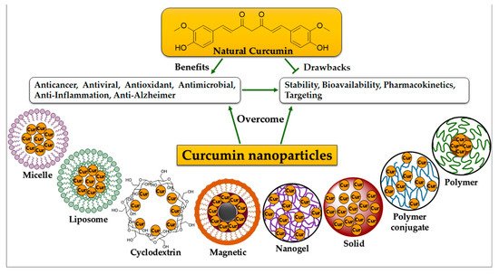
[15][16][73,74]. Many curcumin nanoformulations have been produced over the last few years. Most of them aim to improve curcumin solubility and bioavailability while also protecting it from hydrolysis inactivation. Such formulations are intended for long-term maintenance and circulation throughout the body, whereas others have focused on intracellular release and cellular transportation processes. Multiple curcumin-based nanoformulations have provided a significant effect on medicinal implementations, which are shown to be effective in the detection of many human disorders. They are defined and addressed in the following (
Figure 1).
Figure 1. Nano-based formulations of curcumin. Many curcumin-based nanoformulations have a significant impact on pharmaceutical applications, which are effective in the treatment of a wide range of human disorders due to their anti-cancer, antioxidant, antimicrobial, and antiinflammation, and even anti-Alzheimer properties. Most nanoformulations are capable of overcoming curcumin’s weak hydrophobicity, as well as its poor stability and poor cellular bioavailability. Such nanoformulations are utilized for long-term preservation and circulation throughout the body.
2.1. Micelle Structures
A micelle is a spherical vesicle made up of surfactant molecules with an amphiphilic property that assembles spontaneously in the water
[17][18][75,76]. Commonly, it is chosen to transmit loaded drugs that are not highly soluble in water, such as curcumin. Curcumin-encapsulated polymeric micelles were synthesized using a solid dispersion technique in a one-step process. The efficacy of generated micelles was assayed on breast cancer
[19][77]. They were more effective than unformulated curcumin at inhibiting the development of breast tumors and uncontrolled spontaneous pulmonary metastatic spread. The solid dispersion of micelles comprising curcumin-poly(ethylene glycol) methyl ether improved curcumin’s anti-tumor and anti-angiogenesis effects. Curcumin micelles can be advantageous during the treatment of pulmonary carcinoma
[20][78]. Chang et al. investigated the intracellular localization, cell absorption, and cytotoxic effects of different dimensions of micelles embodied with curcumin in carcinoma cells from the human colon in vitro. The findings showed that smaller micelles loaded with curcumin possess a higher capacity for inducing cytotoxic effects in carcinoma cells of the human colon than bigger micelles. Accordingly, when employing nanoparticles as drug delivery nanosystems, micelle size, drug loading, and release kinetics and cellular uptake are all crucial elements to consider
[21][79]. Curcumin loaded on the ultra-hydrophilic zwitterionic polymeric material poly(sulfobetaine methacrylate) (PSBMA), which conjugated to zein and formed polymeric micelles, had significantly improved durability, cellular absorption, cytotoxicity toward cancerous cells, and pharmacokinetic properties when compared to traditional methods of using curcumin
[22] (Figure 2) [80]. Compared to natural curcumin, encapsulated curcumin in monomethoxy poly(ethylene glycol)-poly(3-caprolactone) micelles inhibits CT26 colon carcinoma cell proliferation in vivo
[23][81].
2.2. Liposome Structures
Liposomes are globular vesicles with multiple or single phospholipid bilayers covering aqueous systems that strongly mimic the architecture of the cellular membranes. They have suitable delivery mechanisms for bioactive compounds both in vivo and in vitro
[24][25][82,83]. Increased biodegradability and biocompatibility, low toxic effects, high durability, improved dissolution rate, controlled release/delivery, ability to target individual cells, ease of production, and versatility are only a few of the benefits of liposomes
[26][27][28][84,85,86]. As a result, researchers have paid attention to liposomes as a potent drug-carrier mechanism. The liposome’s size varies from 0.025 to 2.5 μm. The amount of medication capsulation within a liposome is determined by the number and size of bilayers and the vesicle diameter, which are significant considerations in determining the liposome’s circulation time
[24][82]. Many experiments have demonstrated that curcumin is solubilized inside the phospholipidic bilayer of the liposome, allowing curcumin to be dispersed throughout the aqueous phase, thereby increasing curcumin’s effectiveness
[29][30][87,88]. Liposomal formulations have extended plasma circulation to reach the spleen
, and other organs
, and tissues to provide a major therapeutic effect and limit non-specific toxicity
[31][32][89,90]. Extensive research has revealed that liposome-based curcumin was the best platform for treating various cancer diseases. Liposome-based curcumin prevented the development of the MCF-7 breast cancer cell line and the KHOS OS cell line
, and had a significant antitumor behavior in both in vivo and in vitro conditions
[33][91]. Tian investigated the anticancer efficacy and biochemical pathways caused by liposomal curcumins in PC-3 human prostate cancer cells
[34][92]. In contrast with free curcumin, the rate of survival of liposomes loaded with curcumin administered to PC-3 cells was poor and time-dependent. Tefas et al. manufactured liposomes co-encapsulating curcumin and doxorubicin (Dox), which inhibited C-26 murine colon cancer cell proliferation and exhibited a better cytotoxic impact than Dox-loaded liposomes
[35][93]. Correspondingly, resveratrol and curcumin liposomes had a high encapsulation performance, a higher polydispersity index (PI), and
a narrow size distribution
[36][94]. Liposomal nanocarriers for curcumin delivery and photodynamic treatment mediated by blue light-emitting diode were recently combined to achieve ideal bioactivity and anticancer function. Overall, the findings suggested that liposomes could be a safer transporter for curcumin (
Figure 1)
[37][95]. Chen et al. generated liposome nanostructures loaded with curcumin and tested them in B16BL6 melanoma cells for antitumor behavior. Liposome nanostructures significantly slowed the growth of B16BL6 melanoma cells. This was primarily attributed to the improved drug distribution facilitated by the intracellular lipid fusion of cellular membranes and particulates. Additionally, it blocked the PI3K/AKT pathway, which is involved in skin cancer
[38][96].
2.3. Cyclodextrin Structures
3.2.3. Cyclodextrin Structures
As soluble transporter structures as well as multi-component systems, cyclodextrins (CDs), such as α-, β-, and γ-cyclodextrins, serve to bind drugs non-covalently. They are frequently used to improve pharmaceutical stability and solubility and to administer active medications to cancerous cells. Cyclodextrins are macrocycle-forming oligosaccharides made up of eight (γ-), seven (β-), or six (α-) D-glucopyranose components interconnected by a 1,4-glycosidic bond. Because of their low cost, ease of production, and versatility, γ-CD, β-CD, and their derivatives are commonly employed to deliver pharmaceutical active agents. Many researchers have recently shown the importance of cyclodextrin in the medication delivery systems of curcumin
[39][40][97,98]. Yallapu et al. designed a β-CD-controlled curcumin medication transmission mechanism and demonstrated that β-CD–curcumin improved the dissemination of curcumin within prostate cancer cells and strengthened its therapeutic outcomes when compared to curcumin alone
[41][17]. Using cell cycle suspension and the pro-apoptotic behaviors of lung cancer cells, Zhang et al. discovered that β-CD–curcumin (CD15) composition showed higher cytotoxic effects than pure curcumin
[42][99]. Furthermore, CD15 may be a platform for improving curcumin distribution and its therapeutic effectiveness for lung cancer. Nanostructures can be synthesized from sulfobutyl-ether-β-cyclodextrin hyaluronic acid and chitosan and employed for the treatment of colorectal and intestinal epithelial cancer cells, either alone or in combination with curcumin. Curcumin nanostructure demonstrates excellent stabilization and encapsulation properties. It also reduces tumor cell proliferation and the cytotoxicity effects of curcumin in human intestinal epithelial cells
[43][44][45][100,101,102]. Furthermore, in retinitis pigmentosa, a complex of cyclodextrins with curcumin, having the property of water-solubility, increased the dissolution rate and generated continuous release of the drug. The findings aided in the development of eye drops made from naturally available phytochemicals (
Figure 1)
[46][103].
2.4. Conjugate Structures
Conjugates are complexes created by linking two or several substances through a covalent bond. Researchers have increased the bioavailability and oral bioaccumulation of curcumin by conjugating curcumin into hydrophilic polymers and small molecules. According to Manju and Sreenivasan, the conjugation of hyaluronic acid with curcumin reduces the effectiveness of gold nanoparticles while increasing the stability and water solubility of the substance
[47][104]. Singh et al. found that esterifying four phenolic hydroxyls increased the bioavailability of curcumin conjugates glycine and piperic acid, and triggered apoptotic cell death in MDA-MB-231 and MCF-7 cell lines by a mitochondrion-mediated mechanism
[48][105]. Muangnoi et al. made a conjugate of glutaric acid with curcumin in the form of a curcumin–glutaric acid prodrug and evaluated its function on mice. The dissolution rate and anti-nociceptive activity of the prepared prodrug were found to be higher than curcumin alone. A conjugate of gold nanoparticles with polyvinylpyrrolidone and curcumin has recently been shown to inhibit amyloid Aβ (1–6) agglomeration, with increased curcumin bioavailability and loading capacity (80%), as well as greater medication release. This formulation has the potential to be advantageous in the treatment of Alzheimer’s disease (
Figure 13)
[49][106].
2.5. Nano- and Nanosphere Structures
Nanomaterials, which have a dimension of 1–100 nm and have special biological, chemical, and physical properties, can be used for drug delivery
[50][51][107,108]. Medications encapsulated within nanostructures have improved pharmacokinetics, dissolution rate,
and specific targeting delivery and controlled/guided release
[52][53][54][109,110,111]. Other substances, including solid lipids and polymeric, gold, albumin, and magnetic nanostructures, have also been used to improve the therapeutic benefits of curcumin thus far because they are biodegradable and non-toxic while also possessing a high binding potential and being non-toxic (
Figure 1)
[52][55][56][109,112,113].
Sun et al. discovered that solid lipid nanostructures based on curcumin showed increased cell absorption, inhibition of malignant cell proliferation, and improved chemical durability and medicine dissolution rate
[57][114]. Lipid curcumin nanomaterials were assessed for antitumor behavior in adenocarcinoma breast cancer cells. In contrast to pure curcumin, lipid curcumin nanomaterials demonstrated potent bioavailability and drug-releasing assistance. Furthermore, these nanocurcumins effectively increased apoptotic cell death of breast adenocarcinoma cells (
Figure 1)
[58][59][115,116].
Polymeric-based nanostructures provide the benefit of being lightweight and highly biocompatible, allowing them to circulate throughout the bloodstream over an extended time. Polyvinyl alcohol (PVA),
N-isopropylacrylamide (NIPAAM), hydrophobically modified starch, polyethylene glycol monoacrylate [NIPAAM (VP/PEG A)], silk fibroin, poly(lactic-co-glycolic acid) (PLGA), chitosan, and
N-vinyl-2-pyrrolidone are only a few of the synthetic and natural polymeric materials that have been used to manufacture curcumin nanomaterials. In CAL27 cancer cells, which show resistance to cisplatin, Chang et al. investigated the molecular pathways triggered with PLGA-modified nanoparticles loaded by curcumin
[60][61][62][63][117,118,119,120]. The curcumin-loaded-PLGA nanostructures seemed to regulate reactive oxygen species (ROS) formation as well as the function of multiple drug resistance protein 1 (MDR1) of CAL27 cisplatin-resistant cancerous cells by triggering the endogenous apoptotic route. Compared to pure curcumin, these PLGA-modified curcumin-loaded nanoparticles are more efficient in treating CAL27 cisplatin-resistant cancerous cells, having higher in vitro bioactivity and better in vivo bioavailability. Another study found that polymer-based nanostructures loaded with curcumin designed with cationic-based Eudragit R E100 copolymer had a high binding affinity and cellular absorption, resulting in increased cytotoxic effects. This nanomaterial formula inhibited tumorigenesis and suppressed colon-26 cell growth 19 times more effectively than curcumin alone (
Figure 1)
[62][64][65][66][119,121,122,123].
In a xenografted tumor animal study, Kim et al. discovered that human serum albumin nanostructures loaded with curcumin showed better in vivo anticancer efficacy than unformulated curcumin, with no toxic effects
[67][124]. Furthermore, the results of this study indicated that this formulation could be used as a curcumin-based drug delivery nanosystem for cancer therapy. Thadakapally et al. have demonstrated that nanostructures based on PEG-albumin-curcumin have strong anticancer efficacy for breast cancer lines with a steady long-term circulation threshold and improved dissolution rate
[68][125].
Gold nanomaterials have attracted a lot of attention because of their unique catalytic and optical properties, as well as the fact that they are biocompatible and non-toxic
[69][70][126,127]. In another study, a potential nanocurcumin substance based on gold nanostructures was synthesized by Nambiar et al. in a cell culture medium without or in combination with fetal bovine serum, and its antitumor activity was reported in human prostate cancer cells
[71][128]. The effects of gold nanomaterials containing curcumin on renal cancerous cells were investigated in vitro. These nanocurcumins were effective antitumor candidates, causing apoptotic cell death in the A498 cell line of renal carcinoma
[72][129]. Curcumin-containing gold nanomaterials, obtained by a green-based synthesis, were also assayed on breast cancer (MCF-7) and colon cancer (HCT-116) cell lines. Compared to free curcumin, these nanomaterials showed potent apoptotic and antiproliferative activity against cancer cells
[73][130].
In cancer cells, iron oxide nanomaterial cores coated by curcumin-containing pluronic polymer (F68) and CD demonstrated an increased dissolution rate
[74][75][76][131,132,133]. This composition inhibited the mitochondrial membrane’s capacity and generated more ROS than curcumin. It further displayed a significant antitumor effect, as well as magnetic targeting and resonance imaging capabilities. In lymphocyte cells, sustained release of iron oxide nanostructures comprising curcumin and coated by thiolated starch revealed substantial compatibility of the system. Because of its improved medication encapsulation, durability, and maximum loading capacity, it also induced cytotoxic effects in cancer cells
[77][134]. In another study, Fe
3O
4-magnetic nanostructures loaded with curcumin displayed considerable solubility and were appropriate for drug release throughout tumoral tissues
[78][135]. Furthermore, this formulation is applied for tumor tissue imaging techniques. Magnetic nanostructures covered by PEGylated curcumin have newly been established as biocompatible anticancer drug delivery systems (
Figure 1)
[79][80][136,137].
Other formulations for enhancing curcumin’s biological activities include yeast cells, nanodisks, nanogels, and complexes of metals
[81][82][138,139]. Curcumin was determined to be hydrogen-bonded towards the cellular wall when loaded into the cell membrane of
Saccharomyces cerevisiae. In another study, Paramera et al. investigated the durability of loaded curcumin in yeast cells, discovering that yeast cells protected the curcumin from external stimuli, such as heat, humidity, and light
[83][84][140,141]. Curcumin nanodisk compounds are an efficient adjuvant for treating mantle cell lymphoma and other cancers
[85][142]. The association of a curcumin nanodisk with glioblastoma multiforme cells, promoted by ApoE primes, resulted in improved curcumin absorption as well as
an enhanced biological function
[86][143]. Compared to curcumin alone, the curcumin–nanogel hybrid could destroy cancer cells. Dandekar et al. combined polyvinyl pyrrolidone and hydroxypropyl methylcellulose to create hydrogel nanostructures loaded with curcumin and evaluated their antimalarial behavior in mice
[87][144]. It was discovered that curcumin-loaded hydrogel nanostructures function much better than unformulated curcumin. As compared to pure curcumin, curcumin stacked into nanogels demonstrated better cellular absorption and improved cytotoxicity on MCF7 and huh7 cell lines
[88][145]. As self-assembled capsules of casein nanogels and carboxymethyl cellulose, curcumin is being delivered and manufactured with casein and folic acid using a layer-by-layer procedure to combat skin cancer. The findings revealed improved cytotoxicity, cellular uptake, and apoptotic cell death in melanoma cancer cells (MEL-39)
[89][146]. Palladium (II) complexes produced with curcumin showed a significant anticancer impact on HeLa, MCF-7, and A549 tumoral cells (
Figure 1)
[90][147].
3. Anti-Cancer Activity of Curcumin
Due to the strong therapeutic results of curcumin, as many as 68 clinical studies (as of 3 May 2012) have investigated curcumin focusing on cancer. Curcumin has a relatively minimal toxicity potential in both humans and animals
[91][28]. Like gemcitabine and cisplatin as chemotherapeutic medications, it inhibits the development of malignant cells with IC
50 ranging from 5 to 30 μM
[92][93][94][148,149,150]. A clinical trial involving 15 people with colorectal cancer found that the cancer was unresponsive to curcumin at 3.6 g/day over four months
[95][151]. According to this re
seapor
cht, tumor biomarkers and tumorigenesis did not alter. Generally, the experiments found that while curcumin has anti-tumor properties at a rate of 5–30 μM for 1–2 days, owing to curcumin’s better metabolic functions and its poor bioavailability, reaching these levels at the tumor location in human populations has not been possible
[96][152]. Therefore, curcumin delivery must be developed to address these important matters.
Curcumin nanoformulations have been produced and adapted by different generic and pharmaceutical companies to improve solubility and dissolution rates. To mitigate this problem, cyclodextrin, adjuvant, and some proprietary innovations were originally employed
[97][98][99][100][153,154,155,156]. Piperine, for example, is strongly advised due to its inhibition effect on intestinal and hepatic glucuronidation. Its inclusion resulted in bioavailability increases of 2000% and 154% in humans and rats, respectively
[101][102][157,158]. There have been several efforts to enhance the bioavailability of curcumin with the aim of increasing its effectiveness, but these approaches may not have the potential to deliver curcumin into tumors. Therefore, curcumin encapsulation within nanostructures is necessary for cancer treatment with potential target-specific moieties
[99][103][104][155,159,160]. Curcumin has been shown in several experiments to serve as an inhibiting factor, preventing cancer development in its early stages. It also acts as a suppressive factor, preventing the proliferation of malignant cells during the progression of carcinogenesis. Curcumin’s antitumor pathways are extensive but diverse, involving various phases of the apoptosis processes and cellular growth. Because curcumin has a plethora of targets and functionalities on cellular growth regulatory pathways, it holds a lot of promise as a chemotherapeutic agent against human solid tumors
[105][106][107][161,162,163]. Furthermore, curcumin’s activity on a variety of transcription factors, oncogenes, and signaling proteins affects metastatic spread, tumorigenesis, and cancer development at various levels of carcinogenesis, beginning with early repercussions that cause DNA mutation
[108][109][164,165]. Curcumin slows tumor development by blocking specific signaling pathways, including inhibiting transcription factors involved in tumorigenesis viz. signal transducers, activating protein-1 (AP-1), and transcription (STAT) protein activators
[110][166]. It causes apoptosis by prohibiting misfolded
N-CoR protein breakdown and disruption of the ubiquitin–proteasome pathway. Protein kinases are another target of curcumin. Curcumin inhibited the mitogen-activated protein kinase function and epidermal growth
-factor receptor activity in lung and pancreatic adenocarcinoma cells
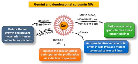
[111][167]. Delivery of curcumin and other phytocompounds to the tumor niche is a significant difficulty. Zibaei et al. investigated the tumoricidal potential of Gemini surfactant nanostructures loaded with curcumin in 3D spheroid HT-29 cells.
In vitro, the findings revealed that Gemini curcumin has the ability to inhibit cell growth and metastatic spread in 3D spheroid HT-29 cells
[103][159]. In another study, the antiproliferative effects of Gemini curcumin and free curcumin were investigated against ovarian cancer (OVCAR-3 cells). The results showed that compared with pure curcumin, Gemini surfactant nanoparticles increase cellular uptake and effectively suppress the proliferation of OVCAR-3 cells via induction of apoptosis
[112][168].
Additionally, Sobhkhizi et al. revealed that dendrosomal nanocurcumin could be used as an anti-tumor medicine in p53-mutant tumor malignancies
[113][169]. Curcumin’s capacity to induce apoptosis, disrupt cell cycle activity and inhibit proliferation of cancer cells makes it a possible therapy for human breast (especially MCF-7, MDA-MB-231, MDA-MB-468, and SkBr-3 cells), lung, colorectal, prostate, carcinoma, melanoma liver, myeloma, and pancreatic cancers
[114][115][116] (Figure 2) [170,171,172]. Curcumin has been shown to inhibit cancer cell metastasis development. It prevents cancer cells from destroying healthy tissue by inhibiting the action of matrix metalloproteinases, which control the mechanism. Curcumin inhibits the expression of genes participating during tumor development, apoptosis, and proliferation, such as c-myc, cyclin D1, Bcl-xL, and Bcl-2. NF-κB suppression is essential in proliferation and carcinogenesis. Curcumin inhibits the function of NF-κB, which may enhance the expression of genes involved in invasion (for example, matrix metalloproteinases), proliferation (for example, c-myc and cyclin D1), and antiapoptosis
[117][173].
Figure 2. Anti-cancer activity of Gemini and dendrosomal nanocurcumin against various cancer cell lines was examined [155]. Curcumin’s ability to provoke apoptotic cell death, disturb cell cycle activity, and suppress proliferative behavior in cancer cells makes it a promising therapeutic target for human breast, colorectal, lung, carcinoma, prostate, melanoma, myeloma, liver, and pancreatic cancers.
Basniwal et al. investigated the antitumor characteristics of curcumin nanostructures in cancer cell lines from the skin (A431), liver (HepG2), and lungs (A549)
[118][174]. In aqueous environments, curcumin nanomaterials have been shown to have a much better impact on cancer cells than natural curcumin. Another study found that PLGA–curcumin nanomaterials increased apoptosis, lysosomal function, suppression of nuclear β-catenin action, and androgen receptor in prostate cancer cells as a consequence of a development blockade
[119][175]. One of the more common genomic subgroups of breast cancer with a metastatic form is triple-negative breast cancer. Exogenous p53 and dendrosomal nanocurcumin have been shown to have antitumor activity on triple-negative breast cancer cells, especially when combined
[120][176]. NF-κB and HIF-1 are both needed for the control of cancer cell development. In the hypoxic microenvironment, PLGA nanostructures packed with curcumin increased the expression of NF-κB and HIF-1 subunits (nuclear p65 (Rel A) and HIF-1α) throughout the lung and breast cancerous cells
[121][122][177,178].
In cancer therapy, providing appropriate concentrations of therapeutic drugs around the tumor region is essential to eradicate the malignant cells while causing the least
amount of harm to normal cells
[123][124][179,180].
TIn our opinion, the development of curcumin nanoformulations displaying better anticancer properties is a critical step forward. Different types of nanocarriers have been employed to obtain better characteristics for potential application in cancer therapy
[125][181]. In the case of cancer therapies, the type of preparation determines the durability, effectiveness, and selectivity of a formulation. Researchers believe that because of their biocompatibility, liposomal, poly(caprolactone) (PCL) or poly(lactide-co-glycolide) (PLGA) nanomaterials, and self-assembly curcumin formulations should be given the greatest priority in cancer therapeutic strategies
[126][127][182,183].
The majority of tabulated formulations (Table 1) report the passive targeting mechanism of curcumin nanostructures rather than the active targeting mechanism. The main aspect of curcumin nanomaterials is that they are passively targeted, and it is this trait that encourages their accumulation in tumors. Several significant characteristics, including particle diameter, zeta potential, and the solubility or dispersion of nanostructures, may influence the effectiveness of passive targeting techniques. All that is observed in nanoformulations of ideal size is the enhanced permeability and retention (EPR) effect, which in turn causes a rise in the concentrations of accumulation in tumors. Additionally, a hydrophilic coating using poly(ethylene glycol) inhibits the interaction between proteins and cells, resulting in a reduction in the opsonization response.
Table 1.
In vivo and in vitro anticancer potential and mechanism of action of some kinds of curcumin nanoformulations.
| Curcumin Nanoforms |
In Vitro Cytotoxic Activity |
Molecular Mechanism |
In Vivo Results |
Ref. |
| Poly(lactide-co-glycolide); PLGA |
Cytotoxicity against HCT116, DU145, MDA-MB-231, SEG-1, Jurkat, and KBM-5 cells with IC50 < 5 μM. |
NF-κB-induced inactivation of and decrease in cyclin D1, MMP-9, and VEGF production. |
The half-life of curcumin nanoparticles was 1.75 times longer than curcumin. |
[128][184] |
| Poly(lactide-co-glycolide); PLGA |
Equal cytotoxicity of nanocurcumin and curcumin toward SKBr3, HeLa, and A549 cells. |
Increase in Annexin V staining, cleaved PARP expression.
Decrease in NF-κB activation. |
Not available. |
[129][185] |
| Poly(lactide-co-glycolide); PLGA |
Cytotoxicity against PC-3, LNCaP, and DU145 cells;
curcumin-loaded PLGA nanostructures: IC50 = 20–22.5 μM;
free curcumin: IC50 = 32–34 μM. |
Inhibition of NF-κB function. |
Not available. |
[130][186] |
| β-cyclodextrin self-assembly of curcumin |
In C4-2 and DU145 cells, the curcumin self-assembly concentration was 16.8 μM and 17.6 μM, respectively, which is slightly less than the free curcumin concentration. |
Increase in cleaved PARP expression. |
Increased curcumin levels in serum concentrations by up to twofold (Unpublished data with Subhash Chauhan Lab) |
[41][17] |
| MPEG-PCL micelle |
Cytotoxicity against C-26 colon cancer cells;
Cur-MPEG-PCL micelles: IC50 = 5.78 mg·mL−1.
Free curcumin: IC50 = 3.95 mg·mL−1. |
Not available. |
Increase in curcumin concentrations in rat plasma (>2 times) and suppression of subcutaneous C-26 colon cancer development in a xenograft mice model. |
[131][187] |
| Poly(butyl cyanoacrylate) nanomateriales |
Cytotoxicity against Bel7402, HepG2, and Huh7 cells (IC50 ≈ 15 μg/mL). |
Suppression of VEGF and downregulation of COX-2 expression. |
A 2.2-fold reduction in HepG2 tumor volume in a xenograft mice model. |
[132][188] |
| Dendrosomal curcumin |
Cytotoxicity against WEHI-164 cells; IC50 = 16.8 & 7.5 μM after 24 & 48 h.
Cytotoxicity against A431 cells: (IC50 = 19.2 and 14.3 μM after 24 & 48 h. |
Increase in cleaved PARP expression and further Annexin V staining (apoptosis). |
Reduction in tumor development. |
[133][189] |
| Self-microemulsifying medication delivery device enhanced with folic acid. |
Effective cytotoxicity of folate curcumin-nanoemulsion, curcumin-emulsion, and free curcumin against HeLa cells at concentrations of 18.27, 36.69, and 30.4 μM, respectively.
Effective cytotoxicity of folate curcumin-nanoemulsion, curcumin-emulsion, and free curcumin against HT-29 cells at concentrations of 20.57, 38.59, and 25.62 μM, respectively. |
Not available. |
Increase in folate curcumin-nanoemulsion adsorbsion from 58.41% to 73.38% in 6 h (in situ colon-perfused rats) |
[134][190] |
| Thermo-sensitive nanocarrier |
Showing particular toxic effects on cancer cell lines (KB, MCF-7, and PC-3 cells) while being nontoxic to the L929 cell line. |
Increase in apoptosis due to Annexin-A and PI binding. |
Not available. |
[135][191] |
| NanoCurc™ |
Little or inhibited growth of JHH-GBM14, D283Med, DAOY, and glioblastoma neurosphere lines HSR-GBM1. |
G(2)/M arrest and apoptosis induction via the inhibition of STAT3 and Hedgehog signaling pathways. |
~0.5% localization of the injected drug within the brain. |
[136][137][192,193] |
| Amphiphilic mPEG-palmitic acid polymer |
Cytotoxicity against HeLa cells;
nanocurcumin; IC50 = 15.58 μM,
curcumin; IC50 = 14.32 μM. |
Increasing the anticancer activity in vitro by enzyme-catalyzed release. |
Not available. |
[138][194] |