Reactive Species-Activatable AIEgens for Biomedical Applications: Comparison
Please note this is a comparison between Version 1 by Ji Qi and Version 2 by Vivi Li.

Precision medicine requires highly sensitive and specific diagnostic strategies with high spatiotemporal resolution. Accurate detection and monitoring of endogenously generated biomarkers at the very early disease stage is of extensive importance for precise diagnosis and treatment. Aggregation-induced emission luminogens (AIEgens) have emerged as a new type of excellent optical agents, which show great promise for numerous biomedical applications. AIn this review, we highlight the recent advances of AIE-based probes for detecting reactive species (including reactive oxygen species (ROS), reactive nitrogen species (RNS), reactive sulfur species (RSS), and reactive carbonyl species (RCS)) and related biomedical applications are introduced. The molecular design strategies for increasing the sensitivity, tuning the response wavelength, and realizing afterglow imaging are summarized, and theranostic applications in reactive species-related major diseases such as cancer, inflammation, and vascular diseases are reviewed.

  • aggregation-induced emission
  • reactive oxygen nitrogen species
  • activatable probe
  • theranostics
  • fluorescence
  • photoacoustic
  • afterglow
  • bioimaging

1. Introduction

Precision medicine requires highly sensitive and specific diagnostic methods with high accuracy at the very early disease stage [1][2][3][1,2,3]. Some traditional imaging modalities such as ultrasound, computed tomography (CT), and magnetic resonance imaging (MRI) have been widely used in clinic [4][5][6][4,5,6]. However, most of them suffer from low sensitivity, and it is usually difficult to recognize tiny pathological changes when the lesion is small [7][8][7,8]. Optical imaging techniques such as fluorescence and photoacoustic imaging have significant advantages such as high sensitivity, real-time monitoring, noninvasive imaging, and portable instruments, which are very promising for disease diagnosis and therapy [9][10][11][12][13][14][9,10,11,12,13,14]. Fluorescence has been used for in vitro examination of diseased samples and in vivo image-guided tumor surgery clinically. However, due to interference from the strong light–tissue interaction (e.g., absorption, scattering, and reflection) and autofluorescence, the sensitivity of fluorescence is significantly reduced [15][16][15,16]. Therefore, the development of new imaging agents that could improve the therapeutic performance (e.g., recognition of disease-related markers) is highly desirable.
Numerous materials have been used for optical imaging, for example, carbon nanomaterials, metal nanostructures, rare earth-doped nanoparticles (NPs), and organic materials [17][18][19][20][21][17,18,19,20,21]. Among them, organic compounds possess unique intrinsic merits including excellent reproducibility, specific chemical structures, and good biocompatibility [22][23][24][25][26][22,23,24,25,26]. Currently, small-molecule dyes, i.e., indocyanine green (ICG) and methylene blue (MB) have been approved by the Food and Drug Administration (FDA) for clinical use, highlighting the great clinical translation potential of organic optical materials [27][28][29][27,28,29]. Nevertheless, most conventional organic dyes are planar structures, which face the obstacle of aggregation-caused quenching (ACQ) effect in aggregate state due to strong intermolecular interactions (e.g., π-π stacking) [30][31][30,31]. The ACQ problem seriously hinders the applications of these hydrophobic molecules in a hydrophilic living environment. In 2001, Tang’s group first coined the the concept of aggregation-induced emission (AIE), representing a new type of optical materials that were weak or non-luminescent in dilute solution, but became highly emissive in aggregate form [32][33][34][35][36][37][32,33,34,35,36,37]. For AIE luminogens (AIEgens), the excited-state energy is consumed by the intensive intramolecular motion through non-radiative decay in solution, while the molecular motion is restricted in aggregate form, thus, the non-radiative pathway is closed and the radiative process is open [38][39][40][41][42][38,39,40,41,42]. As a result, restriction of intramolecular motion (RIM) is considered to be the working principle of the AIE phenomenon, and a library of AIEgens with various properties have been developed [43][44][45][46][43,44,45,46]. AIEgens have been used in many areas such as optoelectronic devices, chemo/biosensing, and biological imaging [47][48][49][50][47,48,49,50]. In the biomedical field, AIEgens have shown excellent performance in organelle imaging, in vivo high-resolution imaging, disease theranostics, and activatable detection [51][52][53][54][55][51,52,53,54,55].
Excessive expression of various reactive species can lead to oxidative stress, which is known to cause DNA, protein, cell, and tissue damage, and affect signaling pathways [56][57][58][56,57,58]. These processes are closely associated with many diseases including inflammation, cancers, diabetes, and neurodegeneration diseases [59][60][61][62][59,60,61,62]. Thus, accurate detection and monitoring of these endogenously generated biomarkers is extensively important for precise disease diagnostics and therapeutics at an early stage [63][64][65][63,64,65]. According to their nature, reactive species can be divided into reactive oxygen species (ROS) including hydrogen peroxide (H2O2), hypochlorite/hypochlorous acid (HOCl/ClO), hydroxyl radical (OH), superoxide anion radical (O2•−), singlet oxygen (1O2), and peroxy radical (ROO); reactive nitrogen species (RNS) including nitric oxide (NO), peroxynitrite (ONOO), S-nitrosothiol (RSNO), and S-nitrosoglutathione (GSNO); reactive sulfur species (RSS) including hydrogen sulfide (H2S), thiyl radical (RS), thiol (RSH), S-nitrosothiol, sulfenic acid, and sulfite; reactive carbonyl species (RCS) including carbon monoxide (CO), formaldehyde (FA), glyoxal (GO), acrolein, and glucosone [66][67][68][69][70][71][72][73][74][75][66,67,68,69,70,71,72,73,74,75]. Reactive species have gained great interest from both fundamental biological scientists and clinical doctors, and more and more new phenomena about their functions have been discovered [76][77][78][76,77,78]. Numerous molecular probes for detecting ROS, RNS, RSS, and RCS have been exploited, focusing on understanding the physiological/pathological effects and disease theranostics [79][80][81][82][83][84][79,80,81,82,83,84]. Recently, the development of reactive species-responsive AIEgens has attracted considerable attention, which are advantageous for applications in the biomedical field [85][86][87][88][85,86,87,88]. Thanks to the salient merits of good stability, large Stokes shift, facile structure modification, and excellent sensitivity, AIEgens have emerged as a new type of potent probes for detecting various reactive species. Although there are many review papers that have focused on AIEgens [89][90][91][92][93][94][89,90,91,92,93,94], to the best of ouresearchers' knowledge, comprehensive summaries of reactive species-responsive AIEgens are very rare. In this entry, researchersreview, we highlight the recent advances of AIEgen-based reactive species-activatable systems. The recent development of AIEgens for sensing reactive species such as ROS, RNS, RSS, and RCS are discussed. The molecular design strategies for increasing sensitivity, tuning the response wavelength, increasing the afterglow imaging efficiency, as well as different biomedical applications are reviewed. The challenges and outlooks for the reactive species-activatable AIE systems for biomedical applications are also discussed. 

2. Detection of Reactive Oxygen Nitrogen Species

When designing a specific chemical/biological probe, a usually requisite is to synthesize molecules with specific recognition groups or moieties.The boronate subunit is a popularly used building block for H2O2 sensors, as the boronate cage is nonfluorescent and the conversion of arylboronates to phenols results in turn-on emission [95][96][97][95,96,97]. The deprotonated H2O2 is a potent nucleophile, which can attack the boron center to generate a labile borate species that hydrolyses to the corresponding phenol [98]. For O2•− detection, the diphenyl phosphinyl group can be introduced into an organic compound, in which the fluorescence is strongly quenched at first, and obvious turn-on fluorescent signal is realized in the presence of O2•− [99][100][99,100]. The oxidative properties of ClO can be utilized to destroy C=C or C=N bonds rapidly, therefor, the conjugation of fluorescence quencher through C=C or C=N bonds has turned out to be an efficient strategy to construct ClO probes [101][102][101,102]. Some arylboronate groups, diphenylphosphinate groups, and nitrophenyloxoacetamide moieties have been employed as the response substitutes for ONOO detection [103][104][105][103,104,105]. The tunability of molecular structure will alter the photophysical properties and biomedical applications as well. H2O2 is an overexpressed molecule in many serious diseases, and thus, it is regarded as a pivotal biomarker for some biological processes and disease diagnoses [106][107][108][106,107,108]. A variety of H2O2-activatable probes have been exploited based on AIEgens, which exhibit excellent performance for both in vitro and in vivo applications [109][110][111][112][109,110,111,112]. Xia and Lou et al. developed a H2O2-responsive AIEgen for peroxidase-mediated selective imaging and inhibition of inflammatory cells [113].The probe consisted of a TPE core and two tyrosine (Tyr) moieties, which could undergo enzyme-catalyzed dityrosine formation in the presence of peroxidase and H2O2. By conjugating two hydrophilic Tyr groups, the hydrophobic TPE molecule became hydrophilic TT, which showed weak fluorescence in aqueous solution due to the excited-state energy consumption via intense molecular motion. As a result, the H2O2-responsive and myeloperoxidase (MPO)-mediated TT self-assembly enabled turn-on fluorescence, which could be used for selectively imaging and inhibiting inflammatory cells containing overexpressed H2O2 and MPO. The AIE process could be activated through dityrosine linkage-induced hydrophobic aggregates formation, which helped to distinguish between inflammatory and normal cells. Additionally, the in situ formation of TT aggregates could inhibit RAW264.7 cell growth through inducing mitochondria damage and cell apoptosis. Wang and Li et al. reported a ROS-responsive theranostic nanoplatform for accurate diagnosis and therapy of inflammation diseases [114]. A two-photon AIEgen (TP) was conjugated with the widely used anti-inflammatory glucocorticoid, prednisolone (Pred) with the ROS-sensitive linkage to afford the compound TPP. Then, the TPP was encapsulated with an amphiphilic block copolymer PMPC−PMEMA (PMM) to give polymeric micelles (TPP@PMM). Noteworthy, the PMEMA part served as the hydrophobic block in the NPs formation, which could be oxidized in response to ROS to yield the hydrophilic sulphone product. The ROS-triggered hydrophobic-to-hydrophilic conversion was able to realize ROS-mediated drug delivery at an inflammatory site. This shell-core dual ROS-responsive nanoplatform was used in three different inflammatory murine models including acute lung injury, atherosclerosis, and arthritis. The deep-penetration two-photon fluorescence diagnosis and efficient serial ROS sensitive anti-inflammation could be used for both acute and chronic inflammation theranostics. Two-photon imaging with the AIEgen helped to provide unambiguous delineation of inflammatory tissue with minimum autofluorescence interference. Moreover, TPP@PMM also possessed excellent anti-inflammatory effect that reduced the inflammatory response and decreased inflammatory cytokines expression.

3. Detection of Gasotransmitters

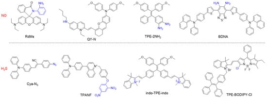
Small gaseous molecules including NO, CO, and H2S, function as important signal transmitters in living systems as they are associated with many biological functions and major diseases [115][116][117][118][148,149,150,151]. NO is a neutral diatomic free radical that is produced from L-arginine by NO synthase (NOSs) isoforms such as neuronal NOS (nNOS), inducible NOS (iNOS), and endothelial NOS (eNOS) [119][120][152,153]. CO is the second gasotransmitter that is generated as a byproduct of haem cleavage by two distinct haem oxygenases [121][154]. H2S is predominantly formed from Cys or its derivatives by the enzymes cystathionine β-synthase and cystathionine γ-lyase [122][155]. All these gasotransmitters play vital roles in vasorelaxation and inflammatory responses, thus, numerous molecular probes have been developed for precise monitoring of related diseases [123][124][125][156,157,158]. For example, the o-diamino aromatic moiety is a recognition group for NO, and the cyclization reaction of o-diamine with NO produces a triazole moiety, which alters the electronic property and conjugation nature [126][127][128][159,160,161]. For H2S detection, the popularly used approaches include reduction of azides into amines and nucleophilic addition of H2S to the electrophilic group [129][130][162,163]. Some representative AIEgens for sensing gasotransmitters are listed Figure 13, which show great potential for applications in biological imaging and disease diagnosis.
Figure 13.
 Chemical structures of different types of gasotransmitter-responsive molecular probes.
Wu’s group developed a NO-activatable AIEgen for precisely diagnosing herbal medicine-induced liver injury with NIR-II fluorescence and PA imaging [131][164]. They designed and synthesized a D-π-A-type probe (QY-N) consisting of an electron-rich bismethoxyphenyl-amine-containing dihydroxanthene group and an electron-deficient quinolinium moiety. The linking of electron-donating butylamine to the quinolinium group weakened the electron-accepting capability, and thus, quenched the fluorescence, and butylamine also served as a NO-responsive group based on the N-nitrosation reaction of aromatic secondary amine. In the presence of NO, the electron-donating butylamine was transformed into an electron-withdrawing butyl-N-nitroso group, which resulted in a bathochromic shift of absorption in the range of 700–850 nm for PA imaging, and boosted NIR-II fluorescence at 910–1110 nm. The AIE probe was able to detect and assess the severity of herbal medicine-induced liver injury in vivo in a high-contrast manner for significantly enhanced NIR-II fluorescence and PA signals via reacting with the overexpressed NO at a disease site. In addition, the probe was also capable of monitoring the rehabilitation of liver injury during the treatment process.
Video Production Service