PTEN Dual Lipid- and Protein-Phosphatase Function in Tumor: Comparison
Please note this is a comparison between Version 1 by Yanlin Yu and Version 3 by Lindsay Dong.

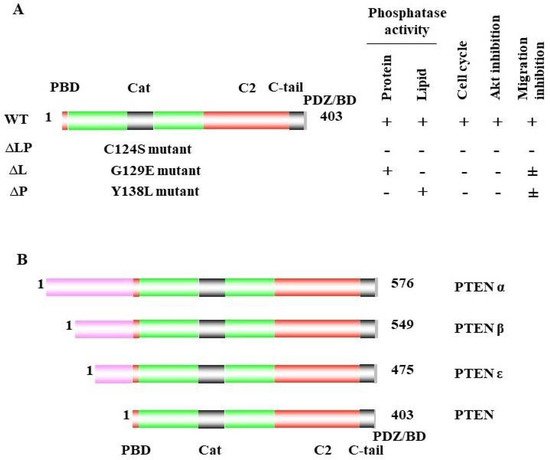
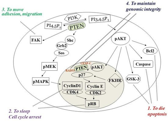
Phosphatase and tensin homolog deleted on chromosome ten (PTEN) is a multifunctional tumor suppressor with protein- and lipid-phosphatase activities. The inactivation of PTEN is commonly found in all human cancers and is correlated with tumor progression. PTEN-lipid-phosphatase activity has been well documented to dephosphorylate phosphatidylinositol-3, 4, 5-phosphate (PIP3), which hinders cell growth and survival by dampening the PI3K and AKT signaling activity. PTEN-protein-phosphatase activity dephosphorylates the different proteins and acts in various cell functions. 

  • PTEN
  • PTEN lipid phosphatase
  • PTEN protein phosphatase
  • mutation
  • PTEN protein substrate
  • tumorigenesis

1. PTEN Dual Lipid and Protein Phosphatase

In 1984, scientists discovered that the loss of part or all of chromosome 10 was associated with brain, bladder, and prostate cancer [1][2][3][1,2,3]. Conversely, the reintroduction of wild-type chromosome 10 into glioblastoma-cell lines reduced the ability of the tumor formation in nude mice [4], which suggested an important tumor-suppressor role for chromosome 10. Later, a chromosome loss-of-heterozygosity (LOH) analysis identified region 10q23 as the most common region of loss on chromosome 10 in prostate cancer [5], which suggested that this region contains a critical tumor-suppressor gene. Then, in 1997, several laboratories identified a putative tumor-suppressor gene at 10q23 that encodes a 403 amino acid protein with a protein-tyrosine-phosphatase domain and homology to chicken tensin and bovine auxilin, which was then named phosphatase and tensin homolog deleted on chromosome ten (PTEN) for phosphatase and tensin homolog deleted on chromosome 10 [6][7][8][9][6,7,8,9]. The PTEN protein contains five major domains: the N-terminal PIP2-binding domain (PBD), the catalytic domain of the phosphatase, the C2 domain, the C-tail domain, and the PDZ-binding domain (PDZ/BD); the C-terminal PDZ/BD domain can inter- act with other proteins [10][11][12][10,11,12] (Figure 1A). The PTEN phosphatase domain contains a signature CX5R motif that forms its catalytic pocket, which is also known as the P- loop. The cysteine at the base of this pocket allows the phosphatase to react with substrates. Moreover, later studies confirmed that PTEN not only specifically dephosphorylates protein substrates at tyrosine-, serine-, and threonine sites, but al- so dephosphorylates lipid phosphatidylinositol 3,4,5-trisphosphate [PtdIns(3,4,5)P3] to form phosphatidylinositol 4,5-bisphosphate [PtdIns(4,5)P2]. PTEN also contains a TI-loop that contributes to its lipid-phosphatase activity by determining the size of the catalytic pocket [10][13][10,13].
Figure 1. Schematic of the PTEN protein. (A) PTEN contains five functional domains: two key domains that are required for its tumor-suppressor function: the phosphatase (catalytic) domain (amino acids 14–189), with an active site included within the residues 123 and 130 (black), and the C2 (lipid-membrane-binding) domain (amino acids 190–350) (red); two binding domains that are the N-terminal PIP2-binding domain PBD (amino acids 1–14) and C-terminal PDZ-binding domain (grey; amino acids 401–403), which binds proteins containing PDZ domains; the carboxy-terminal region (amino acids 351–400), which contains PEST sequences and contributes to PTEN stability and activity, and is less well defined in the tumor-suppressor functions of PTEN. Wild-type PTEN with both lipid- and protein-phosphatase activity inhibits the cell cycle, AKT activity, and cell migration. The mutation at C124S (∆LP) inactivates both PTEN lipid and protein phosphatase, which provokes the loss of the inhibition of cell-cycle arrest and AKT and cell migration. The G129E (∆L) mutant loses only its lipid-phosphatase activity and can still inhibit cell migration. The mutation of Y138L is deficient in its protein phosphatase, which may lose the capacity to inhibit cell migration. (B) Three PTEN alternative translational isoforms, PTENα, PTENβ, and PTENε, which are produced from the same mRNA as canonical PTEN and are generated due to non-AUG translational initiation. Each has a longer N-terminal extension than the canonical PTEN protein.
PTEN protein is mainly found in the cytoplasm. However, the study of the crystal structure of PTEN suggested that its C2 domain helps it bind to phospholip-id membranes [10]. SUMO1 modification at the K266 and K254 sites in the C2 domain promotes the cooperative binding of PTEN to the plasma membrane via electrostatic interactions, downregulating the PI3K/AKT pathway [14][15]. PTEN localized in the cy- toplasmic membrane performs its lipid-phosphatase activity by converting PIP3 to PIP2. PTEN protein can also shuttle to the nucleus to maintain genomic stability. By physically associating with CENP-C (centromere proteins), which are an integral component of the kinetochore, PTEN regulates RAD51 expression, which reduces the incidence of spontaneous double-stranded breaks (DSBs) and plays a role in DNA repair [15][14]. Nuclear PTEN also exhibits its tumor-suppressive effect through G1-phase cell-cycle arrest by preventing cyclin D1 localization and decreasing the level of cyclin D1 [16], together with the effects of PTEN on p27Kip1, to suppress the cell cycle [17][18][17,18] (Figure 2).
Figure 2. PTEN has four major cellular functions. By dephosphorylating PIP3 and negatively regulating the activation of AKT, PTEN can both prevent the activation of Bcl2 and GSK-3 and promote the activation of caspase, thereby inducing cell apoptosis (1), and against both the AKT and MAPK signaling pathways to promote cell-cycle arrest (2). The protein phosphatase dephosphorylates the FAK proteins to reduce cell adhesion, movement, and migration (3). It can also shuttle into the nucleus to maintain genomic integrity (4).
PTEN functions as a haploinsufficient tumor suppressor, where the protein produced after the deletion of one allele on chromosome 10 is insufficient for proper function [19][22] (Figure 3). The complete loss of PTEN will trigger senescence and often leads to early death in genetically engineered mice (GEM) [20][23]. Mammary epithelial cells from PTEN hypermorphic (Ptenhy/+) mice with 80% of the regular PTEN expression showed enhanced proliferation and increased resistance to apoptosis after ultraviolet irradiation [21][24].
Figure 3. PTEN functions as a haploinsufficient tumor suppressor. PTEN loss correlates with the incidence of tumorigenesis and increased activity of AKT in a dose-dependent manner (the more loss of PTEN (left panel), the higher tumor incidence (middle panel) and AKT activity (right panel)). PTEN deficiency (0% of PTEN) leads to embryonic lethality and hyperactivated AKT, while the hypomorphic allelic loss of PTEN (Hypo 30%) revealed more increased cancer phenotypes than the heterozygous loss of a PTEN allele (Het 50%) and the hypermorphic allelic loss of PTEN (Hyper 80%, with small 20% reductions in PTEN doses) (the tumor incidence from high to low: Hypo 30% > Het 50% > Hyper 80%). In contrast, elevated PTEN (>100%) protects against tumorigenesis and promotes longevity in GEM models [20][21][22][23,24,25].

2. PTEN Inactivation in Cancer Progression

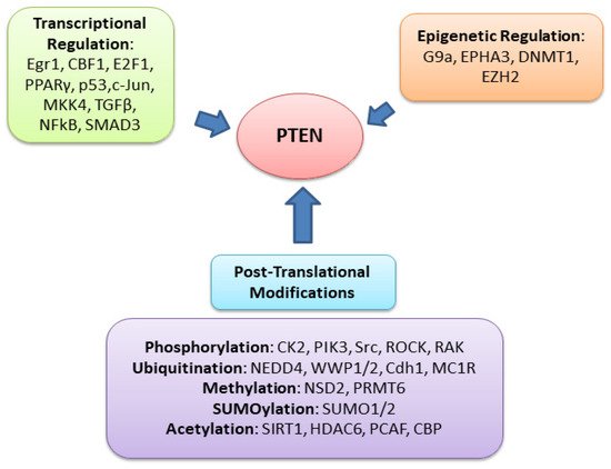
PTEN is one of the most highly mutated genes in human cancers. The dysfunction of PTEN by genetic mutation or epigenetic silencing contributes to most cancer development and progression (Figure 4, Table 1) [7][8][23][7,8,33]. It is often associated with high-grade and metastatic potential drug resistance [24][25][34,35] and poor patient prognosis [26][27][28][36,37,38]. The inactivation of PTEN is caused by various mechanisms, including genetic loss, point mutation, epigenetic regulation, and posttranslational modifications. Most alterations result in the loss of or reduction in PTEN protein.  
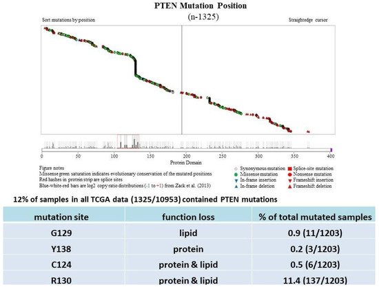
Figure 4. PTEN mutations cause PTEN inactivation and loss of the tumor-suppressive function. Most PTEN mutations in cancer (TCGA dataset) are found in its phosphatase domain, including G129, Y138, C124, and R130, which lose lipid or protein or both phosphatase activities.
Table 1.
PTEN Lesions (Deletion and/or Mutation) in Sporadic Human Malignancies.
Site/Tissue Tumor Type Range Average Comment Reference(s)
Brain Glioblastoma 12–84% 29% (88/303) Mostly LOH [29][30][53,54]
Breast Ductal carcinoma 11–55% 33% (415/1257) Mostly LOH [31][32][33][55,56,57]
Endometrium Endometrioid carcinoma 19–82% 67% (352/529) LOH and mutation [34][58[35],59]
Prostate Adenocarcinoma 12–63% 33% (88/267) Mostly LOH [36][37][60,61]
Ovary Cystadenocarcinoma 9–61% 30% (33/112) LOH and mutation [38][39][62,63]
Skin Melanoma 11–39% 30% (57/190) Mostly LOH [40][41][64,65]
Thyroid Carcinoma (ATC) 10–41% 11% (21/196) Mostly LOH [42][43][44][66,67,68]

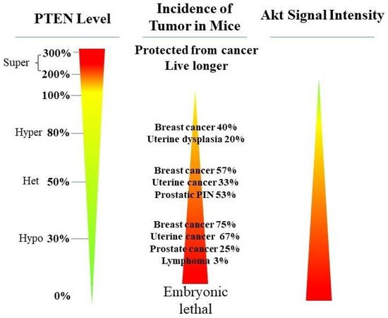
PTEN genetic loss is involved in various tumor types, such as glioblastoma, breast ductal carcinoma, endometrial carcinoma, prostate adenocarcinoma, ovary cystadenocarcinoma, melanoma, pancreatic adenocarcinoma, colorectal cancer, etc. (Table 1). In GEM mice, PTEN deficiency leads to embryonic lethality, and even a small reduction in the PTEN levels enhances the cancer incidence [20][21][45][46][23,24,39,40]. Contrarily, the systemic overexpression of PTEN in GEM models amplifies its tumor-suppressive function and protects against tumorigenesis [47][41], which suggests that the precise level of PTEN expression is a critical factor for the tumor-suppressor function, and that the reduction in PTEN activity is a driving mechanism for tumor progression. The pathologic characteristics that are associated with PTEN deletion in mice are phenocopied in a wide range of human tumors with loss-of-heterozygosity (LOH) mutations. In cancer, the PI3K/PTEN/Akt pathway has been identified as one of the critical molecular axes driving tumorigenesis [48][49][50][51][42,43,44,45]. The loss of PTEN activity has been reported to be responsible for many of the phenotypes of cancers, and it affects the development of 15–70% of human cancers [25][49][50][35,43,44] (Table 1).

Much has been learned about the features of PTEN point mutation through the analysis of PTEN germline mutations found in patients with Cowden disease (CD) [52][46]. Patients with CD, who harbor missense mutations in the PTEN phosphatase domain, are cancer-prone and develop more lesions than patients with PTEN deletion, which causes the complete loss of the PTEN function [53][47].

There are multiple posttranslational modifications (PTMs) for PTEN, such as phosphorylation, methylation, ubiquitination, SUMOylation, and acetylation [11] (Figure 5). The PTEN C2 domain and its C-terminal can be phosphorylated by several kinases, such as protein kinase CK2 [54][80], activated Src kinases [55][81], ROCK1 (RhoA- associated kinase) [56][57][82,83], Rak tyrosine kinase [58][84], GSK3β (glycogen synthase kinase β) [59][85], and ATM (ataxia telangiectasia mutated) [60][86]. The phosphorylation of the PTEN C2 domain and its C-terminal regulates the PTEN function. Specifically, the PTEN phosphorylation of Ser370 and the S/T cluster (Ser380, Thr382, The383, Ser385) (also called the A4 cluster) modulates the PTEN stability and activity [61][62][21,87]. This phosphorylation of the C-terminal tail region interferes with the electrostatic binding of PTEN to the plasma membrane, controlling the membrane translocation of PTEN [63][88]. Deletion in the tail impairs phosphorylation and increases PTEN activity. Serine/threonine-to-alanine substitutions of PTEN block phosphorylation and alter its stability and activity. Interestingly, aspartic acid substitutions at the phosphorylation sites will restore the stability of PTEN [62][87].
Figure 5. The function of PTEN can be regulated by different mechanisms through various genes, including transcriptional regulation, post-transcriptional regulation, epigenetic regulation, and posttranslational modifications. The transcriptional downregulation of PTEN causes protein levels to decrease by epigenetic regulation (promoter methylation) transcriptional factors.
PTEN methylation at its protein level is found in the cytoplasm and the nucleus, regulating the PTEN function. At DSB sites, NSD2 (MMSET/WHSC1) mediates the dimethylation of PTEN at K349 [64][89]. Methylated PTEN allows PTEN to be recruited into DNA-damage sites by the 53BP1 Tudor domain. By studying wild-type PTEN and the PTEN mutants G129E, C124S, and Y138L, it was confirmed that protein-phosphatase activity is required for efficient DSB repair, along with the dimethylation of PTEN at K349.
Ubiquitination is an essential posttranslational modification for PTEN [65][91]. NEDD4-1 was identified as the major E3 ubiquitin ligase of PTEN; NEDD4-1 ubiquitinates PTEN and regulates tumorigenesis. As NEDD4-1 negatively regulates PTEN, the deletion of NEDD4-1 inhibits tumor growth in a PTEN-dependent manner [66][92]. K289 and K13 are major sites for PTEN monoubiquitination. These two lysine sites are critical for PTEN nuclear shuttling and import. The PTEN function can be suppressed by the ubiquitin ligase WWP1 (WW domain-containing ubiquitin E3 ligase via nondegradative ubiquitination [67][93]).
The inactivation of PTEN by mutation and posttranslational modification, or the reduction in the PTEN protein levels by LOH and epigenetic mechanisms, results in the activation of PI3K/AKT signaling by the loss of PTEN lipid phosphatase as a major driver for susceptibility in various human cancers [68][94]. PTEN protein phosphatase has been proposed to dephosphorylate focal adhesion kinase (FAK) [69][48], c-Src [70][95], and PTEN itself [61][21], thereby inhibiting cell adhesion and migration, and it has been implicated in the maintenance of genomic integrity [15][71][14,96].

34. PTEN-Protein-Phosphatase Substrates and PI3K-Independent Function

3.1. PTEN

4.1. PTEN

PTEN has been reported to play a vital role in regulating cell migration, invasion, and metastasis [69][72][73][74][75][48,115,116,117,118]. As indicated above, the naturally derived PTEN point mutant G129E [9][75][76][9,118,119], which loses lipid but maintains protein-phosphatase activity, retains the ability to inhibit cell migration and invasion, as well as metastasis [69][48]. To study the other mechanisms involved in cell migration, Reftopoulou and colleagues found that PTEN protein phosphatase is required for migration. They further demonstrated that PTEN protein phosphatase dephosphorylates its C domain at Thr383, inhibiting the migration independent of its effects on the PI3K pathway [61][21].

3.2. Abi1

4.2. Abi1

Abl-interactor 1 (Abi1) is a core component of the WASP-family verprolin homologous protein (WAVE) regulatory complex (WRC). Abi1 acts as a core scaffold protein to mediate the membrane recruitment and stabilization of WRC subunits [77][78][122,123], and it is regulated by extracellular cues and intracellular signaling pathways. PTEN can bind and dephosphorylate Abi1at Y213 and S216, triggering its degradation through the calpain pathway, thereby promoting epithelial differentiation and polarization [79][99], as well as epithelial–mesenchymal transition and cancer-stem-cell activity [80][124]. PTEN dephosphorylates Abi1 and downregulates the WRC at the cell cortex, thereby reorganizing the actin cytoskeleton to facilitate the formation of the apical actin belt and adherent junctions.

3.3. Β-Catenin

4.3. Β-Catenin

Transforming growth factor β (TGF-β) is a pleiotropic cytokine that plays a role in growth suppression in normal epithelial cells, but it supports metastasis formation in many tumors [81][125].

3.4. Cofilin-1

4.4. Cofilin-1

Cofilin-1 is an essential actin regulator. As an actin-depolymerizing factor (ADF)/cofilins family protein, Cofilin-1 regulates the rapid depolymerization of actin microfilaments that give actin its characteristic dynamic instability and its central role in muscle contraction, cell motility, and transcription regulation [82][83][84][126,127,128]. The activity of cofilin is regulated by a variety of mechanisms, including specific phosphorylation and dephosphorylation [84][128].

3.5. CREB

4.5. CREB

The transcription factor cyclic AMP response element-binding protein (CREB) is activated via phosphorylation at serine133, which mediates gene transcription and promotes cell proliferation and survival. PTEN protein phosphatase is required for the dephosphorylation of CREB in the nucleus. Under PTEN-deficient conditions, CREB phosphorylation is enhanced independently of the PI3K/AKT pathway. The inhibition of the PI3K/AKT pathway does not affect the CREB phosphorylation in PTEN-deficient cells. C124S-mutant PTEN cannot dephosphorylate CREB, while G129E and wild-type PTEN can, which demonstrates that the protein-phosphatase activity of PTEN is essential for dephosphorylating and colocalizing with CREB in the nucleus [85][102], which suggests that PTEN regulates gene expression through this mechanism.

3.6. Drebrin

4.6. Drebrin

Drebrin is a protein that is encoded by the DBN1 gene. It is a crucial regulator of the actin cytoskeleton in neuronal cells for synaptic plasticity, neurogenesis, and neuronal migration, as well as in cancer cells for tumor invasion [86][87][88][132,133,134]. The defect of Drebrin in expression and activation contributes to the pathogenesis.

3.7. Dvl

4.7. Dvl

PTEN is an essential regulator of multicilia formation and cilia disassembly via Dishevelled (DVL2) phosphorylation. DVL is an important component of the WNT signaling pathways that plays a role during convergent extension movements. DVL is a ciliogenesis regulator in Xenopus and human epithelial cells, and it has been identified as a direct substrate for PTEN. Among the DVL proteins, DVL2 and DVL3 have the strongest associations with PTEN. The knockdown of PTEN increases the DVL2 phosphorylation on serine143 during cilia formation. By studying wild-type PTEN and mutants of PTEN (C124S, G129E, Y138L), it was confirmed that the protein-phosphatase activity of PTEN is responsible for directly dephosphorylating DVL2 on serine 143 [89][104], which implicates PTEN in multicilia formation and movement.

3.8. FAK

4.8. FAK

Focal adhesion kinase (FAK) is one of the earliest confirmed substrates for PTEN. PTEN overexpression inhibits cell migration via reducing the phosphorylation of FAK. The effect of PTEN on cell spreading was examined on fibronectin (FN) in multiple cell lines: NIH 3T3 cells, human fibroblast cells, DBTRG-05MG cells (glioblastoma), and U-87MG (glioma) cells. The overexpression of PTEN delayed or inhibited the spreading in these cell lines [69][48]. FAK was first found to be a substrate of the Src proto-oncogene. It is an important regulator of cell adhesion and motility. The tyrosine phosphorylation of FAK is associated with focal contacts that form at ECM integrin junctions, and integrin-binding proteins recruit FAK to the focal contacts. It has been reported that PTEN suppresses cell migration, invasion, and metastasis through the dephosphorylation of FAK at Tyr397 [69][90][48,105]; however, specific FAK dephosphorylation sites have not been observed in other cell types [91][136], and other mechanistic details have yet to be uncovered.

3.9. Glucocorticoid Receptor (GR)

4.9. Glucocorticoid Receptor (GR)

GR is a pleiotropic nuclear receptor and transcriptional regulatory factor that controls the network of glucocorticoid (GC)-responsive genes in a positive or negative manner for regulating numerous physiological and cellular processes [92][137]. In cancer, GR activation also appeared as tumor-suppressing [93][138] and tumor-promoting effects [94][139].

3.10. IRS1

4.10. IRS1

PTEN protein phosphatase can dephosphorylate insulin receptor substrate-1 (IRS1). IRS1 is a mediator of insulin and IGF signaling, which is negatively affected by NEDD4. In NEDD4-deficient cells, IGF signaling becomes defective, while AKT activation is unimpaired. PTEN ablation rescues impaired IGF signaling by dephosphorylating IRS1. Although NEDD4 is required for IGF signaling, the role of NEDD4 in IGF signaling is PTEN-dependent. NEDD4 inhibits the PTEN function to enable IGF signaling. NEDD4 is responsible for the ubiquitination of PTEN, and for suppressing its phosphatase activity. The C124S mutant of PTEN is not able to dephosphorylate IRS1, while the G129E mutant and wild-type PTEN are, which proves that IRS1 is a direct substrate of PTEN’s protein phosphatase [95][106].

3.11. MCM2

4.11. MCM2

The MCM2-7 complex is one of the core components of the replisome [96][97][140,141], and it plays crucial roles in replication origin firing, elongation, termination, and the replication-stress response [98][99][142,143]. MCM2 is a critical component of the MCM2-7 complex, and it is a core replication helicase of the replisome that is regulated by the MCM2 phosphorylation status.

3.12. NKX3.1

4.12. NKX3.1

NKX3.1, which is a prostate-specific homeobox gene, is a gatekeeper suppressor and is commonly deleted in prostate cancer. NKX3.1 inhibits cell proliferation and mediates cell apoptosis and DNA repair.

3.13. PLK1

4.13. PLK1

Polo-like kinase 1 (PLK1) is a mitotic kinase that regulates mitotic entry and exit. PLK1 controls spindle bipolarity and is involved in cytokinesis. PTEN physically associates with PLK1 and dephosphorylates PLK1, maintaining genomic stability during cell division. PTEN loss or deficiency causes failure in cytokinesis through nondisjunction chromosomes and cleavage-furrow regression, which leads to spontaneous polyploidy and resistance to spindle disruption.

3.14. PTK6

4.14. PTK6

PTEN inhibits protein tyrosine kinase 6 (PTK6/BRK/Sik) activity in prostate cancer cells by dephosphorylating PTK6 at tyrosine 342 (PY342). In the absence of PTEN, PTK6 is activated and downstream oncogenic signaling is promoted [100][111].

3.15. Pol II

4.15. Pol II

The RNA polymerase II (Pol II) C-terminal domain (CTD) constantly undergoes cycles of phosphorylation/dephosphorylation during gene transcription. It was demonstrated that PTEN dephosphorylates Pol II CTD with specificity for Ser5, and that the phosphorylation of Pol II CTD Ser5 is inversely related to PTEN expression. When there is an overexpression of PTEN, there will be a decrease in the Pol II CTD phosphorylation; global Pol II CTD phosphorylation increases along with the PTEN loss. Pol II CTD is a significant platform for posttranslational modifications, such as elongation, termination, and co-transcriptional processes. PTEN, as a Pol II CTD phosphatase, raises the possibility that PTEN can regulate global transcription [101][110].

3.16. Rab7

4.16. Rab7

Rab7 is a GTPase for endosome maturation that is involved in epidermal growth factor receptor (EGFR) signaling. PTEN dephosphorylates Rab7 on two residues, S72 and Y183, and it promotes late endosome maturation, which reduces EGFR signaling. The residues are required to associate Rab7 with GDP dissociation inhibitor (GDI)-dependent recruitment to late endosomes and maturation. EGFR is a receptor tyrosine kinase that regulates cell proliferation, growth, and motility. PTEN controls the EGFR-endocytic-trafficking pathway via the dephosphorylation of Rab7 and the localization of Rab7. The loss of PTEN causes EGFR transport from early to late endosomes because PTEN is needed for Rab7-endosomal-membrane targeting. PTEN-mediated Rab7 dephosphorylation allows Rab7 to interact with GDI, GEF, and effector proteins. Rab7 has been identified as a protein substrate for PTEN, which provides a new mechanism for controlling the EGFR signaling in cells via PTEN [102][112].

3.17. Shc

4.17. Shc

Src homology collagen (Shc) is a direct substrate for PTEN protein phosphatase. Shc is an SH2-binding adaptor molecule that activates the Raf and MAPK pathway. PTEN inhibits the MAPK pathway by dephosphorylating Shc in positions Tyr239/240, which reduces cell proliferation and inhibits Shc-mediated tumor metastasis in renal-cell carcinoma (RCC) [103][113].

3.18. SRC

4.18. SRC

SRC is a membrane-anchored tyrosine kinase that is activated following the engagement of many different classes of cellular receptors, and it regulates various biological activities, including cell proliferation, adhesion, migration, and transformation [104][144]. The study reported that SRC modulates the antibody Trastuzumab that targets the human epithermal growth factor receptor-2 (HER-2 or ERBB2) response in breast cancer, and that activating SRC by phosphorylation at Tyr416 was required for regulating the multiple resistance pathways [105][114].
 
Video Production Service