Ivermectin during the COVID-19 Pandemic: Comparison
Please note this is a comparison between Version 2 by Amina Yu and Version 1 by Arnoldo Aquino-Gálvez.

Ivermectin (IVM) is a broad-spectrum antiparasitic agent, developed and funded by Merck & Co. in 1974 to control and eradicate onchocerciasis caused by the parasitic worm Onchocerca volvulus in West Africa, which in the 1980s infected approximately 340,000 people. At the time, Africa did not have the resources necessary to seek treatments for this condition. The avermectins, of which IVM is a member, were discovered by Professor Satoshi Ōmura as fermentation products of the bacterium Streptomyces avermitilis at the Kitasato Institute in Tokyo. For this discovery, he received the 2015 Nobel Prize in Physiology and Medicine, which he shared with William Campbell. IVM is used to treat onchocerciasis, lymphatic filariasis, strongyloidiasis and scabies, and, very recently, has been used to combat lice. The drug’s low cost, high efficacy, safety, and marked tropism for helminths, as well as the fact that it has almost no impact on human biochemistry, have led to the inclusion of IVM in the twentieth list of essential medicines and sixth list of vital medicines in children, a recommendation made by the expert committee of the World Health Organization (WHO) in 2019. The safety profile is attributed to its selective affinity for ion channels.

  • SARS-CoV-2
  • ivermectin
  • antiviral

1. Mechanisms of Action of Ivermectin

Ivermectin is a broad-spectrum drug with numerous effects on parasites, nematodes, arthropods, flavivirus, mycobacteria and mammals through a variety of mechanisms. The mechanism of action of Ivermectin as an anthelmintic agent at various stages in the life cycle works by binding to glutamate-gated chloride ion channels in the nerve cells and invertebrate muscles of microfilaria [42,43][1][2]. The union of Ivermectin (IVM) with the channels causes an increment in the cell membrane permeability for chloride ions, hyperpolarizing the membrane and interrupting the motility, feeding and reproduction, leading to the paralysis and death of the parasite. In addition to the glutamate-gated ion channels, ivermectin is also an agonist of the neurotransmitter gamma-aminobutyric acid (GABA)-activated channels. Since GABA channels in mammals are exclusively found in the central nervous system, and IVM does not readily cross the blood–brain barrier, it has a favorable safety profile in regard to the treatment doses used in humans [6,42,43][1][2][3]. Ivermectin achieves adequate levels of availability when administered orally, and due to its high lipid solubility, IVM is widely distributed, with a volume of distribution of 46.8 L. The metabolism of IVM is hepatic, primarily effected by the CYP3A4, and it is removed through the feces and only 1% through urine [2,42,44][1][4][5].

2. Possible Benefits of Ivermectin in SARS-CoV-2

2.1. Antiviral Activity

One of the most exciting effects of IVM is its possible role as an antiviral against COVID-19 [45][6]. Certain reviews have emphasized the antiviral effect of IVM in vitro and in vivo against RNA and DNA viruses [46][7]. An experiment in which cell cultures were treated with and without IVM (20 μmol/L) over 24 h identified increases in the gene expression of proteins participating in four antiviral pathways that were statistically significant, including the routes of infection of HCMV, HPV, EBV and HIV1. These results support the broad-spectrum antiviral activity of IVM [47][8]. It is essential to highlight that the movement of proteins between the cytoplasm and the nucleus is mediated by the superfamily of proteins called importins, which are essential for cellular processes, such as differentiation and development, and are fundamental in the pathological states of viral diseases and oncogenesis [48][9]. The specific viral proteins enter the nucleus of infected cells to perform essential functions as part of the viral replication cycle [49][10]. An example is the interaction between the HIV-1 integrase protein and the importin α/β1 heterodimer, which is blocked by IVM, thus inhibiting the nuclear import of the integrase protein and, therefore, damaging the viral replication mechanisms [48][9].
The broad-spectrum antiviral activity of IVM is related to the fact that RNA viruses, to transport viral proteins to the nucleus of the host cell, depend on the importin alpha-beta (IMPα/β1) heterodimer during the viral infection process. This importin is blocked by IVM. The transport of viral proteins through IMPα/β1 to the nucleus occurs in order to inhibit the antiviral response that is assembled by a portion of the host cells. This mechanism has been observed in viruses such as Zika, Dengue, HIV-1, yellow fever, Chikungunya and many more [45,48,50][6][9][11]. Regarding the antiviral response of IVM to DNA viruses, it has been shown that, if the viral proteins necessary for viral replication require entry to the nucleus through IMPα/β1, then this can have an antiviral effect, as in the case of the pseudorabies virus and polyomavirus BK [51][12].
In the case of SARS-CoV-2, it is known that there is no transport of viral proteins to the cell nucleus, as in the case of the infection mechanisms of other viruses. This is because the viral replication cycle takes place exclusively in the cytoplasm of infected cells. However, it is also known that, as part of the antiviral response, there is a communication that involves the transport of proteins related to the regulation of the antiviral responses of infected cells [52,53,54][13][14][15] (Figure 21). There is great controversy regarding antiviral activity in the case of SARS-CoV-2; thus, it weis believed that more studies are required to clarify the mechanism by which a molecule can be considered to have an antiviral capacity.
Figure 21. Proposed mechanism of action of ivermectin in COVID-19. IVM blocks the binding complex of the SARS-CoV-2 S protein and the ACE2 receptor, and additionally it blocks the TMPRSS2 protein, inhibiting viral entry into the host cell. IVM could also inhibit the TLR receptors and block NF-κβ, inhibiting the production of the cytokines TNF-α, IL-1 and IL-6 and ROS. TLR also activates IRF3 and IRF7, which initiate the production of type-I and -III IFNs. IFNs activate the JAK/STAT pathway, while IVM can lower the expression of JAK2 and the activity of STAT3. Moreover, in the cytosol, IVM blocks the 3CLpro, the main protease that participates in the viral replication, and blocks the importin complex α/β1 that transports the VP to the nucleus. Furthermore, IVM blocks the overexpression of HIF-1α, induced by the viral protein ORF3a. IVM has also been shown to mitigate the proinflammatory state, where the cytokine storm activates the participation of monocytes, dendritic cells and macrophages, and IVM also promotes the polarization of M2 macrophages over M1. IVM, ivermectin; IC, intracellular; EC, extracellular; VP, viral protein; TLRs, toll-like receptors; NF-κβ, nuclear factor-kappa beta; TNF-α, tumor necrosis factor-alpha; IL-1, interleukin-1, IL-6; interleukin-6; ROS, reactive oxygen species; IRF 3/7, interferon regulatory factors; DC, dendritic cells; M1, M1 macrophage; M2, M2 macrophage; 3CLpro, 3-chymotrypsin-like protease. Created with BioRender.com.
One of the first studies to suggest that IVM might have an effect against SARS-CoV-2 reported that IVM caused SARS-CoV-2 viral RNA to be reduced approximately 5000 fold within 48 h of its administration in infected cell cultures [45][6]. Unfortunately, controversy arose after the publication of this studyerein, after observations that the IVM concentration used in this study herein was 35 times higher than that approved by the FDA (Food and Drug Administration) for parasitic diseases, which raised doubts about the drug’s efficacy at the FDA-approved doses [55][16]. It should be noted that the study questioning the dosage was based on an in silico analysis, meaning that the results obtained in an in vivo model could differ. Furthermore, the virus infects the alveolar epithelial cells [55][16], and in the referred work, the African green monkey kidney cell line, Vero/hSLAM, was used, which does not express the ACE2 receptor, as expressed in the lung tissue.
What is currently known is that one of the mechanisms by which IVM could be effective against SARS-CoV-2 is its interference with the viral entry, since it was shown that IVM interacts with the SARS-CoV-2 spike protein and the ACE2 protein, binding to the spike protein at leucine 91 and the receptor ACE2 at histidine 378 [56,57][17][18]. Therefore, it is likely that high doses are not required in order to treat patients with COVID 19, as suggested by some authors. Similarly, Choudhury et al., through in silico studies, indicated that IVM could inhibit the formation of the spike-ACE2 complex formation, targeting the S2 subunit in the spike protein, as well as having a high binding affinity for TMPRSS2, interfering with viral entry. Though these findings are promising, further experimental studies are required to corroborate them [58][19].
Through a computational analysis, in which 2447 drugs were analyzed to determine their capacity for interfering with the main protease (3CL pro), which is essential for the replication of SARS-CoV-2, IVM, Diosmin, and Selinexor were identified as candidates for use as anti-COVID-19 drugs through this mechanism of action [59][20] .
Another option that has been proposed is to enhance the effect of IVM by combining it with other molecules that could enhance its effects. As seen on the Clinicaltrials.gov platform, there are clinical trials of IVM in combination with different molecules. 
Some preprints describe therapeutic benefits with safe doses of IVM. Thus, more attention should be paid and research devoted to IVM as a possible antiviral agent against SARS-CoV-2. A clinical trial reported that, after five days of IVM treatment, there was an earlier virological clearance (9.7 days) in the IVM-treated group than in the placebo group (12.7 days) [60][21]. One study reported that IVM treatment did not affect the viral load of SARS-CoV-2 in the respiratory tracts of infected hamsters and attributed its beneficial effect to its anti-inflammatory effect, as described in the work [61][22].

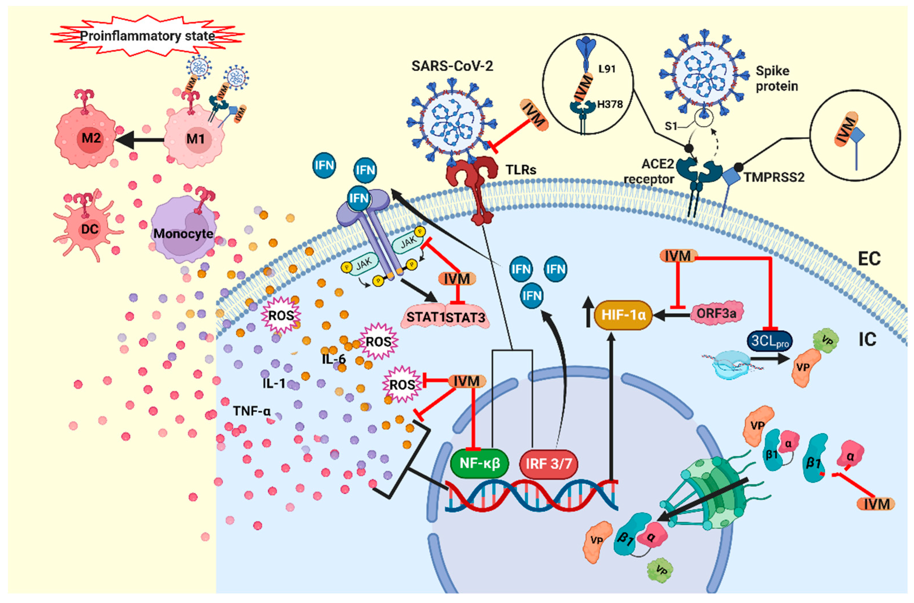
2.2. Immunomodulatory Effects

An interesting study showed that the standard dose of IVM (400 µg/kg) presented with an immunomodulatory activity through the cholinergic anti-inflammatory pathway, preventing clinical deterioration, reducing the olfactory deficit, and limiting the inflammation of the upper and lower respiratory tract in infected golden hamsters. IN the case of SARS-CoV-2, it was also observed that the IL-6/IL-10 ratio in the lung decreased dramatically [61][22]. Macrophage polarization towards the M2 subpopulation was observed [61,68][22][23]. On the other hand, it was observed that the anti-inflammatory effect is influenced by sex, since the treatment led to a better response in women [61][22]. It has been suggested that this positive allosteric effect of IVM is caused by the activation of neuronal α7 nicotinic acetylcholine receptors (α-7 nAChR) [69][24] expressed in the subpopulation of the M2 macrophages [70,71][25][26]. Another study performed using a rat spinal cord injury model found that treatment with a combination of IVM and carbon nanotubes led to a decrease in the pro-inflammatory cytokines and oxidative stress modulated by the M1/M2 macrophage subpopulations [72][27]. Over the past few years, there have been several reports on the anti-inflammatory effects of IVM [73,74][28][29], with reports indicating that, to achieve this effect in humans, 36 mg should be administered in a single dose with a standard weight of 70 kg [75][30].
In in vitro and in vivo models, IVM has been observed to inhibit immune cell recruitment and to suppress mucus hypersecretion and cytokine liberation based on bronchoalveolar lavage in a mouse model of allergic asthma [76][31]. Similarly, in a model sensitized by lipopolysaccharides (LPS), IVM inhibits the production of TNF-alpha, IL-1 and IL-6 [77][32]. This effect is probably due to the suppression of pro-inflammatory factors, such as NF-κB and the MAP pathway kinase [74][29]. Similarly, in an in vitro study, IVM was demonstrated to cause a significant reduction in TNF-α production, induced by TLR agonists, suggesting that IVM could block TLR activity [58,78][19][33]. In the pathogenesis of SARS-CoV-2, STAT1 activity is inhibited by the viral proteins NSP1 and ORF6, favoring the activation of STAT3 and enhancing the production of IL-6 [36,79][34][35]. IVM decreases the expression of JAK2 [80][36] and the activity of STAT3 [36,80[34][36][37],81], leading to a reduction in IL-6 production and inflammation. IVM has also been shown to modulate the immune activity in mast cells and macrophages [3][38] and limit the production of nitric oxide and prostaglandin E2 [82][39]. Furthermore, in animals infected with SARS-CoV-2, IVM treatment improves clinical outcomes and is associated with a reduction in the inflammatory state, though without impacting the viral load in the upper and lower respiratory tract [61][22]. Additionally, the effect of IVM is being explored in the context of its participation in the pathogenesis of SARS-CoV-2 [59,83][20][40] (Figure 21).

2.3. Antitumoral Mechanisms

The effect of IVM as an anti-tumor agent has been explored, and the concentrations necessary for achieving these effects in vivo are within the clinically approved dosages for the treatment of parasitosis [84][41]. Some of the anti-tumor mechanisms attributed to IVM are the inhibition of the Akt/mTOR and WNT-TCF pathways [85[42][43],86], inhibition of MDR proteins, PAK1 helicase, DDX23 and the SIN3 domain [87,88][44][45], the activation of the P2X4/P2X7 [89[46][47],90], an increment in the chloride channel activity [91][48], the downregulation of Nanog/Sox2/Oct4 genes [92][49], and an antimitotic activity (through the damage of tubulin dynamics) [93][50]. In the breast cancer cell lines MDA-MB-231, MDA-MB-468 and MCF-7, and the ovarian cancer cell line SKOV-3, IVM was demonstrated to have a more significant anti-tumor effect (the induction of the cell cycle arrest at the G0-G1 phase and reductions in the cell viability and tumor size) and a synergistic effect combined with docetaxel, cyclophosphamide and tamoxifen [86][43]. In glioma cells, it was observed that it stimulated the activity of caspase-3 and -9, enhancing the expression of p53 and Bax, thus causing apoptosis and blocking the cell cycle in the G0/G1 phase [94][51]. On the other hand, IVM increased TFE3-dependent autophagy via ROS signaling pathways in melanoma cells, inducing apoptosis [95][52]. In porcine trophectoderm and uterine luminal epithelial cells, IVM has also been shown to cause apoptosis through the loss of calcium ion overload, the mitochondrial membrane potential, and the generation of reactive oxygen species [96][53]. Furthermore, hypoxia, through hypoxia-inducible factors (HIF), plays an essential role in drug resistance [97[54][55],98], since, through HIF, cancer cells can resist the decrease in the oxygen concentration and even proliferate. In particular, HIF-1α is translocated to the nucleus by IMPα/β1, and IVM has been shown to block this mechanism [99][56], making it a viable target for cancer treatments [100][57]. Furthermore, Tian et al. found that the SARS-CoV-2 protein ORF3a elevates the production of HIF-1α, promoting an inflammatory state, and IVM could potentially mitigate the inflammatory response through the inhibition of HIF-1α [101][58].

3. Ivermectin in COVID-19 Comorbidities

3.1. Nosocomial Pneumonia

Bacterial coinfections are common in respiratory viral infections [122[59][60],123], and patients with COVID-19 are no exception [124][61]. Of the various studies that report on this situation, only a few representative studies are mentioned here. One study reported that, of 340 COVID-19 patients, 12% had secondary bacterial infections, and of these, 25.59% belonged to the species Klebsiella, 20.93% to methicillin-sensitive Staphylococcus aureus, 16.28% to Escherichia coli, 13.95% to methicillin-resistant Staphylococcus aureus, 11.63% to Enterobacter, 2.32% to Streptococcus pneumonia and 9.30% to Pseudomonas aeruginosa. Of the Enterobacteriaceae isolates, 74% were resistant to cotrimoxazole, 67% to piperacillin, 47.5% to ceftazidime and 42% to cefepime [123][60].
It should be noted that atypical bacteria (Mycoplasma pneumoniae, Chlamydia pneumoniae, and Legionella pneumophila) may be masked by the presentation of COVID-19, as they have overlapping clinical and imaging features, and the timely identification of this co-infection could be vital in critically ill patients [125][62]. Severely and critically ill patients are especially susceptible to co-infections. In one study, serum fungal antigens were observed more frequently in the critical group than in the severe group, and the positive frequency rate of serum fungal antigens increased with a prolonged stay in the intensive care unit (ICU) [126][63]. These findings were replicated in seven ICUs in England, where an increase in the proportion of pathogens was correlated with the length of stay in the ICU, with the identification of mainly Gram-negative bacteria, particularly Klebsiella pneumonia and Escherichia coli. Patients with co-infections/co-colonization were more likely to die in the ICU than those without co-infections [127][64].
Many factors can influence the development of co-infections in terms of the frequency and type of pathogens, including the level of development of the country or region—such is the case of COVID-associated Mucormycosis in India [128][65]. Some studies even agree that antibiotic therapies targeting respiratory pathogens should be considered in severe cases [129][66]. However, there developed a growing concern during the pandemic that the widespread use of empirical antibiotics could contribute to the rise of multidrug-resistant microorganisms, and antimicrobial administration programs are required to minimize and reduce this threat [130][67]. Although, in theory, antibiotics do not directly affect SARS-CoV-2, viral respiratory infections often result in bacterial pneumonia. Some patients may die from bacterial coinfection rather than the viral infection itself; therefore, bacterial coinfections are considered critical risk factors for COVID-19 severity and mortality [131][68]. Inversely, a predictor of rapid recovery from COVID-19 is the absence of bacterial coinfections [132][69].
Some antibiotics are obtained from fermentation carried out by Gram-positive bacteria of the genus Streptomyces, as in the case of Streptomyces griseus, from which the well-known streptomycin is derived. Therefore, it is not surprising that the fermentation products of Streptomyces avermitilis have antibiotic properties. Strategies have been proposed that aim to search for new antimicrobials in order to combat multidrug resistance, and the repurposing of IVM as an antibiotic has potential. Among the avermectins group, IVM stands out for its antibacterial effects. In clinical isolates of multidrug-resistant Mycobacterium tuberculosis, IVM has shown bactericidal effects [133][70]. Additionally, avermectins such as doramectin, IVM, moxidectin and selamectin inhibit the growth of strains of Mycobacterium Bovis BCG, Mycobacterium tuberculosis from H37Rv, CDC 1551, Erdman, and Mycobacterium smegmatis at concentrations ranging from 1 to 8 μg/mL [133][70]. It has also been observed to have an antibacterial effect against Staphylococcus aureus at concentrations of 6.25 and 12.5 μg/mL [134][71]. Macrolide antibiotics have a distinctive macrocyclic lactone ring, and their mechanism of action works through the inhibition of bacterial protein synthesis. However, they also have modulatory effects on the host defense responses and inflammatory responses [135][72]. An example of this is the activation of the P2X4 receptors by IVM in macrophages, increasing the destruction of bacteria and protecting against sepsis [136][73], which is most likely the most prominent antibacterial effect of IVM.

3.2. Wound Healing

Many COVID-19 patients show symptoms of acute lung injury that can eventually lead to pulmonary fibrosis [137][74]. The treatment of inflammation with corticosteroids reduces inflammation and the likelihood of developing fibrosis [138][75]. Regarding the effect of IVM on wound healing, a study reported that IVM cream, at low dosages (0.03–0.1%), induced wound healing, with minimal scarring, and decreased the macroscopic indices of wounds, such as exudation, the edge of oedema, hyperemia and granulation tissue deposits [139][76]. Other works report a decrease in skin inflammation under certain conditions [140,141,142,143][77][78][79][80], which can help to avoid scar formation. It would be interesting to explore this mechanism of IVM directed against post-COVID-19 pulmonary fibrosis.

References

  1. IVERMECTIN. Micromedex® (Electronic Version); IBM Watson Health: Greenwood Village, CO, USA; Available online: https://www.micromedexsolutions.com/ (accessed on 14 August 2022).
  2. González, P.; González, F.A.; Ueno, K. Ivermectin in human medicine, an overview of the current status of its clinical applications. Curr. Pharm. Biotechnol. 2012, 13, 1103–1109.
  3. Kircik, L.H.; Del Rosso, J.Q.; Layton, A.M.; Schauber, J. Over 25 Years of Clinical Experience with Ivermectin: An Overview of Safety for an Increasing Number of Indications. J. Drugs Dermatol. 2016, 15, 325–332.
  4. Canga, A.G.; Prieto, A.M.S.; Diez Liébana, M.J.; Martínez, N.F.; Sierra Vega, M.; García Vieitez, J.J. The pharmacokinetics and interac-tions of ivermectin in humans—A mini-review. AAPS J. 2008, 10, 42–46.
  5. Ashour, D.S. Ivermectin: From theory to clinical application. Int. J. Antimicrob. Agents 2019, 54, 134–142.
  6. Caly, L.; Druce, J.D.; Catton, M.G.; Jans, D.A.; Wagstaff, K.M. The FDA-approved drug ivermectin inhibits the replication of SARS-CoV-2 in vitro. Antivir. Res. 2020, 178, 3–6.
  7. Heidary, F.; Gharebaghi, R. Ivermectin: A systematic review from antiviral effects to COVID-19 complementary regimen. J. Antibiot. 2020, 73, 593–602.
  8. Li, N.; Zhao, L.; Zhan, X. Quantitative proteomics reveals a broad-spectrum antiviral property of ivermectin, benefiting for COVID-19 treatment. J. Cell Physiol. 2021, 236, 2959–2975.
  9. Wagstaff, K.; Sivakumaran, H.; Heaton, S.; Harrich, D.; Jans, D. Ivermectin is a specific inhibitor of importin α/β-mediated nuclear import able to inhibit replication of HIV-1 and dengue virus. Biochem. J. 2012, 443, 851–856.
  10. Wagstaff, K.M.; Rawlinson, S.M.; Hearps, A.C.; Jans, D.A. An AlphaScreen®-based assay for high-throughput screening for spe-cific inhibitors of nuclear import. J. Biomol. Screen. 2011, 16, 192–200.
  11. Yang, S.N.Y.; Atkinson, S.C.; Wang, C.; Lee, A.; Bogoyevitch, M.A.; Borg, N.A.; Jans, D.A. The broad spectrum antiviral ivermectin targets the host nuclear transport importin α/β1 heterodimer. Antivir. Res. 2020, 177, 104760.
  12. Lv, C.; Liu, W.; Wang, B.; Dang, R.; Qiu, L.; Ren, J.; Yan, C.; Yang, Z.; Wang, X. Ivermectin inhibits DNA polymerase UL42 of pseudorabies virus entrance into the nucleus and proliferation of the virus in vitro and vivo. Antivir. Res. 2018, 159, 55–62.
  13. Wolff, G.; Limpens, R.W.; Zevenhoven-Dobbe, J.C.; Laugks, U.; Zheng, S.; de Jong, A.W.; Koning, R.I.; Agard, D.A.; Grünewald, K.; Koster, A.J.; et al. A molecular pore spans the double membrane of the coronavirus replication organelle. Science 2020, 369, 1395–1398.
  14. Snijder, E.J.; Limpens, R.W.A.L.; de Wilde, A.H.; de Jong, A.W.M.; Zevenhoven-Dobbe, J.C.; Maier, H.J.; Faas, F.F.G.A.; Koster, A.J.; Bárcena, M. A unifying structural and functional model of the coronavirus replication organelle: Tracking down RNA synthesis. PLoS Biol. 2020, 18, e3000715.
  15. Fehr, A.R.; Perlman, S. Coronaviruses: An overview of their replication and pathogenesis. Methods Mol. Biol. 2015, 1282, 1–23.
  16. Schmith, V.D.; Zhou, J.; Lohmer, L.R. The Approved Dose of Ivermectin Alone is not the Ideal Dose for the Treatment of COVID-19. Clin. Pharmacol. Ther. 2020, 108, 762–765.
  17. Lehrer, S.; Rheinstein, P.H. Ivermectin docks to the SARS-CoV-2 spike receptor-binding domain attached to ACE2. In Vivo 2020, 34, 3023–3026.
  18. Elalfy, H.; Besheer, T.; El-Mesery, A.; El-Gilany, A.H.; Soliman, M.A.A.; Alhawarey, A.; Alegezy, M.; Elhadidy, T.; Hewidy, A.A.; Zaghloul, H.; et al. Effect of a combination of nitazoxanide, ribavirin, and ivermectin plus zinc supplement (MANS.NRIZ study) on the clearance of mild COVID-19. J. Med. Virol. 2021, 93, 3176–3183.
  19. Choudhury, A.; Das, N.C.; Patra, R.; Bhattacharya, M.; Ghosh, P.; Patra, B.C.; Mukherjee, S. Exploring the bind-ing efficacy of ivermectin against the key proteins of SARS-CoV-2 pathogenesis: An in silico approach. Future Virol. 2021, 16, 277–291.
  20. Yuce, M.; Cicek, E.; Inan, T.; Dag, A.B.; Kurkcuoglu, O.; Sungur, F.A. Repurposing of FDA-approved drugs against active site and potential allosteric drug-binding sites of COVID-19 main protease. Proteins Struct. Funct. Bioinform. 2021, 89, 1425–1441.
  21. Ahmed, S.; Karim, M.M.; Ross, A.G.; Hossain, M.S.; Clemens, J.D.; Sumiya, M.K.; Phru, C.S.; Rahman, M.; Zaman, K.; Somani, J.; et al. A five-day course of ivermectin for the treatment of COVID-19 may reduce the duration of illness. Int. J. Infect. Dis. 2021, 103, 214–216.
  22. de Melo, G.D.; Lazarini, F.; Larrous, F.; Feige, L.; Kornobis, E.; Levallois, S.; Marchio, A.; Kergoat, L.; Hardy, D.; Cokelaer, T.; et al. Attenuation of clinical and immunological outcomes during SARS-CoV-2 infection by ivermectin. EMBO Mol. Med. 2021, 13, e14122.
  23. Laura CMiller, Y.S. Macrophage Polarization in Virus-Host Interactions. J. Clin. Cell Immunol. 2015, 6, 311.
  24. Miyazaki-Krause, R.; Buisson, B.; Bertrand, S.; Corringer, P.-J.; Galzi, J.-L.; Changeux, J.-P.; Bertrand, D. Ivermectin: A Positive Allosteric Effector of the α7 Neuronal Nicotinic Acetylcholine Receptor. Mol. Pharmacol. 1998, 53, 283–294.
  25. Wang, H.; Yu, M.; Ochani, M.; Amella, C.A.; Tanovic, M.; Susarla, S.; Li, J.H.; Wang, H.; Yang, H.; Ulloa, L.; et al. Nicotinic acetylcholine receptor α7 subunit is an essen-tial regulator of inflammation. Nature 2003, 421, 384–388.
  26. Galvis, G.; Lips, K.S.; Kummer, W. Expression of Nicotinic Acetylcholine Receptors on Murine Alveolar Macrophages. J. Mol. Neurosci. 2006, 30, 107–108.
  27. Rahbar, A.; Shakyba, S.; Ghaderi, M.; Kazemi, K.; Fagheh, A.F.; Farsinejad, P.; Khosravi, A.; Louyeh, P.A.; Mirzaeyian, E.; Chamanara, M.; et al. Ivermectin-functionalized multiwall carbon nanotube enhanced the locomotor activity and neuropathic pain by modulating M1/M2 macrophage and decrease oxidative stress in rat model of spinal cord injury. Heliyon 2021, 7, e07311.
  28. Ebbelaar, C.C.F.; Venema, A.W.; Van Dijk, M.R. Topical Ivermectin in the Treatment of Papulopustular Rosacea: A Systematic Review of Evidence and Clinical Guideline Recommendations. Dermatol. Ther. 2018, 8, 379–387.
  29. Ci, X.; Li, H.; Yu, Q.; Zhang, X.; Yu, L.; Chen, N.; Song, Y.; Deng, X. Avermectin exerts anti-inflammatory effect by downregulating the nuclear transcription factor kappa-B and mitogen-activated protein kinase activation pathway. Fundam. Clin. Pharmacol. 2009, 23, 449–455.
  30. Di Nicolantonio, J.J.; Barroso, J.; Mccarty, M. Ivermectin may be a clinically useful anti-inflammatory agent for late-stage COVID-19. Open Heart 2020, 7, e001350.
  31. Zhang, X.; Li, J.; Chen, C.; Ci, X.; Yu, Q.; Zhang, X.; Deng, X. Protective effect of abamectin on acute lung injury induced by lipopoly-saccharide in mice. Fundam. Clin. Pharmacol. 2011, 25, 700–707.
  32. Zhang, X.; Song, Y.; Ci, X.; An, N.; Ju, Y.; Li, H.; Wang, X.; Han, C.; Cui, J.; Deng, X. Ivermectin inhibits LPS-induced production of inflammatory cytokines and improves LPS-induced survival in mice. Agents Actions 2008, 57, 524–529.
  33. Rynkiewicz, E.C.; Clerc, M.; Babayan, S.; Pedersen, A.B. Variation in Local and Systemic Pro-Inflammatory Immune Markers of Wild Wood Mice after Anthelmintic Treatment. Integr. Comp. Biol. 2019, 59, 1190–1202.
  34. Low, Z.Y.; Yip, A.J.W.; Lal, S.K. Repositioning Ivermectin for COVID-19 treatment: Molecular mechanisms of action against SARS-CoV-2 replication. Biochim. Biophys. Acta Mol. Basis Dis. 2022, 1868, 166294.
  35. Matsuyama, T.; Kubli, S.P.; Yoshinaga, S.K.; Pfeffer, K.; Mak, T.W. An aberrant STAT pathway is central to COVID-19. Cell Death Differ. 2020, 27, 3209–3225.
  36. Feng, Y.; Wang, J.; Cai, B.; Bai, X.; Zhu, Y. Ivermectin accelerates autophagic death of glioma cells by inhibiting glycolysis through blocking GLUT4 mediated JAK/STAT signaling pathway activation. Environ. Toxicol. 2021, 37, 754–764.
  37. Lu, H.; Zhou, L.; Zuo, H.; Le, W.; Hu, J.; Zhang, T.; Li, M.; Yuan, Y. Ivermectin synergizes sorafenib in hepatocellular carcinoma via targeting multiple oncogenic pathways. Pharmacol. Res. Perspect. 2022, 10, e00954.
  38. Batiha, G.E.-S.; Alqahtani, A.; Ilesanmi, O.; Saati, A.; El-Mleeh, A.; Hetta, H.; Beshbishy, A.M. Avermectin Derivatives, Pharmacokinetics, Therapeutic and Toxic Dosages, Mechanism of Action, and Their Biological Effects. Pharmaceuticals 2020, 13, 196.
  39. Zhang, X.; Song, Y.; Xiong, H.; Ci, X.; Li, H.; Yu, L.; Zhang, L.; Deng, X. Inhibitory effects of ivermectin on nitric oxide and prostaglandin E2 pro-duction in LPS-stimulated RAW 264.7 macrophages. Int. Immunopharmacol. 2009, 9, 354–359.
  40. Siemieniuk, R.A.C.; Bartoszko, J.J.; Ge, L.; Zeraatkar, D.; Izcovich, A.; Kum, E.; Pardo-Hernandez, H.; Rochwerg, B.; Lamontagne, F.; Han, M.A.; et al. Drug treatments for COVID-19: Living systematic review and network meta-analysis. BMJ 2020, 370, m2980.
  41. Juarez, M.; Schcolnik-Cabrera, A.; Dueñas-Gonzalez, A. The multitargeted drug ivermectin: From an antiparasitic agent to a repositioned cancer drug. Am. J. Cancer Res. 2018, 8, 317–331.
  42. Liu, Y.; Fang, S.; Sun, Q.; Liu, B. Anthelmintic drug ivermectin inhibits angiogenesis, growth and survival of glioblastoma through inducing mitochondrial dysfunction and oxidative stress. Biochem. Biophys. Res. Commun. 2016, 480, 415–421.
  43. Juarez, M.; Schcolnik-Cabrera, A.; Dominguez-Gomez, G.; Chavez-Blanco, A.; Diaz-Chavez, J.; Duenas-Gonzalez, A. Antitumor effects of ivermectin at clinically feasible concentrations support its clinical development as a repositioned cancer drug. Cancer Chemother. Pharmacol. 2020, 85, 1153–1163.
  44. Didier, A.; Loor, F. The abamectin derivative ivermectin is a potent P-glycoprotein inhibitor. Anti-Cancer Drugs 1996, 7, 745–751.
  45. Yin, J.; Park, G.; Lee, J.E.; Choi, E.Y.; Park, J.Y.; Kim, T.-H.; Park, N.; Jin, X.; Jung, J.-E.; Shin, D.; et al. DEAD-box RNA helicase DDX23 modulates glioma malignancy via elevating miR-21 biogenesis. Brain 2015, 138, 2553–2570.
  46. DraDraganov, D.; Gopalakrishna-Pillai, S.; Chen, Y.R.; Zuckerman, N.; Moeller, S.; Wang, C.; Ann, D.; Lee, P.P. Modulation of P2X4/P2X7/Pannexin-1 sensitivity to extracellular ATP via Ivermectin induces a non-apoptotic and inflammatory form of cancer cell death. Sci. Rep. 2015, 5, 16222.
  47. Nörenberg, W.; Sobottka, H.; Hempel, C.; Plötz, T.; Fischer, W.; Schmalzing, G.; Schaefer, M. Positive allosteric modulation by ivermectin of human but not murine P2X7 receptors. J. Cereb. Blood Flow Metab. 2012, 167, 48–66.
  48. Sharmeen, S.; Skrtic, M.; Sukhai, M.A.; Hurren, R.; Gronda, M.; Wang, X.; Fonseca, S.B.; Sun, H.; Wood, T.E.; Ward, R.; et al. The antiparasitic agent ivermectin induces chlo-ride-dependent membrane hyperpolarization and cell death in leukemia cells. Blood 2010, 116, 3593–3603.
  49. Dominguez-Gomez, G.; Chavez-Blanco, A.; Medina-Franco, J.L.; Saldivar-Gonzalez, F.; Flores-Torrontegui, Y.; Juarez, M.; Díaz-Chávez, J.; Gonzalez-Fierro, A.; Dueñas-González, A. Ivermectin as an inhibitor of cancer stem-like cells. Mol. Med. Rep. 2017, 17, 3397–3403.
  50. Ashraf, S.; Prichard, R. Ivermectin exhibits potent anti-mitotic activity. Vet. Parasitol. 2016, 226, 1–4.
  51. Song, D.; Liang, H.; Qu, B.; Li, Y.; Liu, J.; Zhang, Y.; Li, L.; Hu, L.; Zhang, X.; Gao, A. Ivermectin inhibits the growth of glioma cells by inducing cell cycle arrest and apoptosis in vitro and in vivo. J. Cell. Biochem. 2018, 120, 622–633.
  52. Deng, F.; Xu, Q.; Long, J.; Xie, H. Suppressing ROS-TFE3-dependent autophagy enhances ivermectin-induced apoptosis in hu-man melanoma cells. J. Cell Biochem. 2019, 120, 1702–1715.
  53. Lee, J.-Y.; Lim, W.; Ham, J.; Kim, J.; You, S.; Song, G. Ivermectin induces apoptosis of porcine trophectoderm and uterine luminal epithelial cells through loss of mitochondrial membrane potential, mitochondrial calcium ion overload, and reactive oxygen species generation. Pestic. Biochem. Physiol. 2019, 159, 144–153.
  54. Kilic, M.; Kasperczyk, H.; Fulda, S.; Debatin, K.-M. Role of hypoxia inducible factor-1 alpha in modulation of apoptosis resistance. Oncogene 2006, 26, 2027–2038.
  55. Rankin, E.B.; Giaccia, A.J. The role of hypoxia-inducible factors in tumorigenesis. Cell Death Differ. 2008, 15, 678–685.
  56. Kosyna, F.K.; Nagel, M.; Kluxen, L.; Kraushaar, K.; Depping, R. The importin α/β-specific inhibitor Ivermectin affects HIF-dependent hypoxia response pathways. Biol. Chem. 2015, 396, 1357–1367.
  57. Vishnoi, K.; Viswakarma, N.; Rana, A.; Rana, B. Transcription Factors in Cancer Development and Therapy. Cancers 2020, 12, 2296.
  58. Tian, M.; Liu, W.; Li, X.; Zhao, P.; Shereen, M.A.; Zhu, C.; Huang, S.; Liu, S.; Yu, X.; Yue, M.; et al. HIF-1α promotes SARS-CoV-2 infection and aggravates inflammatory responses to COVID-19. Signal Transduct Target Ther. 2021, 6, 308.
  59. Gil, E.; Martyn, E.; Rokadiya, S.; Jain, S.; Chin, T.L. Bacterial Coinfection in COVID-19. Clin. Infect. Dis. 2021, 73, e843–e845.
  60. Mahmoudi, H. Bacterial co-infections and antibiotic resistance in patients with COVID-19. GMS Hyg. Infect. Control 2020, 15, 35.
  61. Rawson, T.M.; Moore, L.S.; Zhu, N.; Ranganathan, N.; Skolimowska, K.; Gilchrist, M.; Satta, G.; Cooke, G.; Holmes, A. Bacterial and fungal co-infection in individuals with coronavirus: A rapid review to support COVID-19 antimicrobial prescribing Timothy. Clin Infect. Dis. 2020, 71, 2459–2468.
  62. Chaudhry, R.; Sreenath, K.; Batra, P.; Vinayaraj, E.V.; Rathor, N.; Saikiran, K.; Aravindan, A.; Singh, V.; Brijwal, M.; Soneja, M.; et al. Atypical bacterial co-infections among patients with COVID-19: A study from India. J. Med. Virol. 2022, 94, 303–309.
  63. Yang, S.; Hua, M.; Liu, X.; Du, C.; Pu, L.; Xiang, P.; Wang, L.; Liu, J. Bacterial and fungal co-infections among COVID-19 patients in intensive care unit. Microbes Infect. 2021, 23, 104806.
  64. Baskaran, V.; Lawrence, H.; Lansbury, L.E.; Webb, K.; Safavi, S.; Zainuddin, N.I.; Huq, T.; Eggleston, C.; Ellis, J.; Thakker, C.; et al. Co-infection in critically ill patients with COVID-19: An observational cohort study from England. J. Med. Microbiol. 2021, 70, 001350.
  65. Gambhir, R.S.; Aggarwal, A.; Bhardwaj, A.; Kaur, A.; Sohi, R.K.; Mehta, S. COVID-19 and mucormycosis (Black Fungus): An epidemic within the pandemic. Rocz. Państwowego Zakładu Hig. 2021, 72, 239–244.
  66. Elabbadi, A.; Turpin, M.; Gerotziafas, G.T.; Teulier, M.; Voiriot, G.; Fartoukh, M. Bacterial coinfection in critically ill COVID-19 pa-tients with severe pneumonia. Infection 2021, 49, 559–562.
  67. Chong, W.H.; Saha, B.K.; Ramani, A.; Chopra, A. State-of-the-art review of secondary pulmonary infections in patients with COVID-19 pneumonia. Infection 2021, 49, 591–605.
  68. Mirzaei, R.; Goodarzi, P.; Asadi, M.; Soltani, A.; Aljanabi, H.A.A.; Jeda, A.S.; Dashtbin, S.; Jalalifar, S.; Mohammadzadeh, R.; Teimoori, A.; et al. Bacterial co-infections with SARS-CoV-2. IUBMB Life 2020, 72, 2097–2111.
  69. Ny, P.; Kelsom, C.; Chron, A.; Lou, M.; Nieberg, P.; Shriner, K.; Huse, H.; Wong-Beringer, A. Factors associated with prompt recovery among hospitalised patients with coronavirus disease 2019. Int. J. Clin. Pract. 2021, 75, e14818.
  70. Lim, L.E.; Vilchèze, C.; Ng, C.; Jacobs, W.R.; Ramón-García, S.; Thompson, C.J. Anthelmintic avermectins kill mycobacterium tu-berculosis, including multidrug-resistant clinical strains. Antimicrob. Agents Chemother. 2013, 57, 1040–1046.
  71. Ashraf, S.; Chaudhry, U.; Raza, A.; Ghosh, D.; Zhao, X. In vitro activity of ivermectin against Staphylococcus aureus clinical isolates. Antimicrob. Resist. Infect. Control 2018, 7, 7–12.
  72. Čulić, O.; Eraković, V.; Parnham, M.J. Anti-inflammatory effects of macrolide antibiotics. Eur. J. Pharmacol. 2001, 429, 209–229.
  73. Csóka, B.; Németh, Z.H.; Szabó, I.; Davies, D.L.; Varga, Z.V.; Pálóczi, J.; Falzoni, S.; Di Virgilio, F.; Muramatsu, R.; Yamashita, T.; et al. Macrophage P2X4 receptors augment bacterial killing and protect against sepsis. JCI Insight 2018, 3, e99431.
  74. Yim, J.; Lim, H.H.; Kwon, Y. COVID-19 and pulmonary fibrosis: Therapeutics in clinical trials, repurposing, and potential development. Arch. Pharmacal Res. 2021, 44, 499–513.
  75. Travis, W.D.; Costabel, U.; Hansell, D.M.; King, T.E.; Lynch, D.A.; Nicholson, A.G.; Ryerson, C.J.; Ryu, J.H.; Selman, M.; Wells, A.U.; et al. An official American Thoracic Socie-ty/European Respiratory Society statement: Update of the international multidisciplinary classification of the idiopathic interstitial pneumonias. Am. J. Respir. Crit. Care Med. 2013, 188, 733–748.
  76. Sia, D.K.; Mensah, K.B.; Opoku-Agyemang, T.; Folitse, R.D.; Darko, D.O. Mechanisms of ivermectin-induced wound healing. BMC Vet.-Res. 2020, 16, 397.
  77. Feaster, B.; Cline, A.; Feldman, S.R.; Taylor, S. Clinical effectiveness of novel rosacea therapies. Curr. Opin. Pharmacol. 2019, 46, 14–18.
  78. Charnowski, S.; Wollenberg, A.; Reinholz, M. Perioral Dermatitis Successfully Treated with Topical Ivermectin. Ann. Dermatol. 2019, 31, S27–S28.
  79. Ventre, E.; Rozières, A.; Lenief, V.; Albert, F.; Rossio, P.; Laoubi, L.; Dombrowicz, D.; Staels, B.; Ulmann, L.; Julia, V.; et al. Topical ivermectin improves allergic skin inflammation. Allergy 2017, 72, 1212–1221.
  80. Barańska-Rybak, W.; Kowalska-Olędzka, E. New indications for topical ivermectin 1% cream: A case series study. Adv. Dermatol. Allergol. 2019, 36, 58–62.
More