Sex Dimorphism in Body Fat Distribution: Comparison
Please note this is a comparison between Version 1 by Yu Wang and Version 2 by Rita Xu.

Body fat distribution is a well-established predictor of adverse medical outcomes, independent of overall adiposity. Studying body fat distribution sheds insights into the causes of obesity and provides valuable information about the development of various comorbidities. Compared to total adiposity, body fat distribution is more closely associated with risks of cardiovascular diseases.

  • obesity
  • adipose tissue
  • sexual dimorphism
  • body fat distribution

1. Introduction

Of the world’s population, one-third are currently overweight or obese (https://www.worldometers.info/obesity/, accessed on 1 July 2022). The rapid increase in obesity is threatening public health globally, including in China [1], where the prevalence has risen from approximately 0 to 16.4% (1982–2019) over the past ~40 years [2]. The pandemic of obesity has greatly burdened individuals, society, and the healthcare system. Obesity is responsible for approximately five million premature deaths each year and represents an independent risk factor for cardiovascular disease, the leading cause of global mortality and a major contributor to the disability [3][4][3,4]. The global prevalence of obesity in women is higher than in men. In 2020, the overall global obesity rate for women was estimated at 25% (vs. 17% for men), of which 54 million (vs. 22 million for men) are severe (Class III) BMI ≥ 40 kg/m2. By 2030, this number of women could be as high as 30% (vs. 20% for men) and 77 million (vs. 34 million) being severely obese [5]. Under obese conditions, the dysfunctional adipose tissue contributes to various pathologies in the cardiovascular system in a sex-dependent manner [6][7][8][6,7,8]. There is increasing interest in the pathophysiological differences between males and females in the incidence and consequence of obesity [4][9][4,9].

2. Adipose Tissue: Classification, Distribution, and Function

Adipose tissues, also known as body fats, are energy-processing endocrine organs that are classically classified by their functions or anatomical distributions. Functionally, while energy-storing white adipose tissues (WAT) are distributed in almost every part of the human body, thermogenic-controlling brown adipose tissues (BAT) are mainly located in the interscapular and mediastinal regions with rich nerves and blood vessels [10][11][12][10,11,12]. Under the condition of an increasing energy intake, excessive triglycerides deposited in WAT lead to obesity. In contrast, BAT oxidises glucose and lipids through uncoupled mitochondrial respiration to generate heat, thus dissipating energy via adaptive heat production [13][14][13,14]. Anatomically, adipose tissues are classified as subcutaneous adipose tissues (SAT) making up over 80% of total fat in the body [15], and visceral adipose tissues (VAT) surrounding the different thoracic and abdominal organs. WAT surrounding the heart comprises the epicardial (ECAT) and pericardial adipose tissue (PCAT) [16]. The abdominal VAT, including the omental, mesenteric, and retroperitoneal fat depots, are highly metabolically active. Most blood vessels are surrounded by perivascular adipose tissues (PVAT). Depending on the anatomical positions, the cellular compositions and the properties of PVAT are different. For example, PVAT associated with the thoracic aorta resembles BAT, whereas those surrounding the abdominal aorta exhibit similarities with WAT [17]. Adipose tissue is not only an energy source, but also the largest endocrine organ in the body [18][19][18,19]. The protein factors secreted from adipose tissue are collectively referred to as adipokines. Emerging evidence suggests that adipose tissue has more colours. Beige adipocytes are a distinct type of WAT sharing similarities with the classic cells in BAT. Brown adipocytes and myocytes, which are derived from a Myf5-expressing cell lineage, exhibit similar developmental origins [20][21][20,21]. The beige adipocytes originate from different and heterogeneous populations of cell lineages and are characteristic of both white and brown fat cells [22]. Pink adipose tissues (PAT) are sex specific. During pregnancy, the female SAT of the mammary gland begins to transform into a reservoir as PAT, which gradually replaces the WAT during the lactation period. PAT turns into WAT again when breastfeeding ends [23][24][23,24]. The whole process is referred to as alveolarogenesis, which involves the development of the lobule-alveolar gland structure to produce milk [23][24][23,24]. PAT secretes leptin and adiponectin that act to prevent neonatal obesity [25][26][25,26]. The yellow adipocytes are related to those of the marrow adipose tissues (MAT) in bone. MAT accounts for more than 10% of the total fat mass in healthy lean people [27][28][27,28]. Similar to WAT, MAT also acts as a large endocrine organ that secretes leptin and adiponectin [28][29][28,29], which increase or decrease under pathological conditions such as osteoporosis [30][31][30,31], diabetes [32], and obesity [33]. Depending on the anatomical locations, adipose tissue depots show different metabolic and endocrine properties. The propensity to generate new adipocytes in different adipose depots varies, thus their expansions are intrinsically different leading to a diversified cellular composition, function, and cardiometabolic consequences [34]. Adipose-derived factors, including adipokines, are key mediators of the alterations in body fat composition with age [35][36][35,36]. Different fat depots produce a distinct profile of mediators, which is affected by age and pathophysiological conditions. For example, the VAT expresses a greater amount of inflammatory adipokines [17][37][38][17,37,38]. Even in the same individual, the ob (obese) mRNA level in the adipose tissue varies from region to region [39]. As a result, the production of leptin as well as other inflammatory cytokines such as angiotensinogen, interleukin 6 (IL-6), and plasmin activator inhibitor 1 from SAT and VAT are different [40]. Leptin produced in the SAT is closely related to the circulating concentration [41][42][41,42]. Overall, the heterogeneity in the distribution and functions of adipose tissues exerts different effects on body fat distribution (Figure 1).
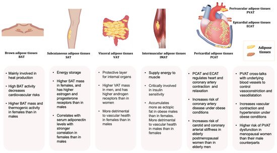
Figure 1. Typical adipose tissues, main functions, and some sex-related differences.

3. Sex Dimorphism in Body Fat Distribution

Sex is defined as the biological feature of males and females determined by genetics, regardless of social or environmental influences. The XX (female) or XY (male) chromosomes make individuals’ distinct sex from each other genetically and physiologically. Sex dimorphism, which refers to the characteristic differences between males and females in a species, helps clinicians and researchers classify, treat, and offer the prognosis of diseases differently. Over the last decade, sex-specific medicine has been drawing great attention as one of the first and foremost advancements of personalised medicine. To date, sex dimorphism has been intensively investigated in obesity, cancers, neurodegenerative disorders, and cardiovascular, bone, and infectious diseases, as well as pain management. The sex disparities in different pathologies, with the development of sex-omics technology, particularly sheds light on personalised management in chronic and severe diseases [43]. Adiposity refers to the distribution of body fat while obesity is a more measurable parameter, emphasising the stratification metrics related to the BMI (ratio of weight to the square of height) and waist circumference [44]. Sexual dimorphism of body fat distribution is subtle in the early stages of life, more distinct in adolescence, and strongly present throughout adult life, but attenuated later in life [45][46][47][48][49][45,46,47,48,49]. Of those with the same BMI and similar age, women have a significantly higher amount of adipose tissue deposition, especially the lower extremity fat, than men [50]. By contrast, men often develop central obesity with an increased fat deposition around the abdomen [51]. With advancing age, fat mass increases and peaks around the age of 60–79 years, later in women than men [52]. Age-associated changes in body composition are manifested not only by an increase in VAT, a decrease in SAT, and an accumulation of ectopic fat, but also by a significant reduction in the lean mass [53][54][55][56][53,54,55,56]. With age, the muscle loses its mass, strength, and physical functionality, leading to a high-risk geriatric syndrome known as sarcopenic obesity (SO), which contributes to various medical complications [57]. SO shows the sex variation and is more prevalent in elderly women [58]. However, the epidemiological findings are heterogeneous due largely to the lack of consensus on a standard definition of SO [59]. The prevalence of SO ranges from 4.4% to 84.0% in men and 3.6% to 94.0% in women when assessed with dual-energy X-ray absorptiometry [60]. In Europe and the US, the prevalence of SO is greater in men than women when using the appendicular lean muscle mass divided by squared height (ALM/h2) to define SO [61][62][63][64][65][61,62,63,64,65]. An opposite conclusion is drawn by a study in Korea using ALM/weight (%) as the criterion [66]. A cross-sectional study from China shows men were more likely than women to have sarcopenia and SO, as assessed by the Asian Working Group for Sarcopenia (AWGS) [67]. Women with SO may have higher glucose, while men with SO are more likely to develop osteoporosis and dyslipidaemia [68]. Body fat distribution is modulated by sex hormones and their receptors [69][70][71][69,70,71]. For example, in women, augmented VAT changes the body shape and composition towards a more android type after menopause [69]. The phenomenon is due at least partly to the withdrawal of estrogen levels, which regulate the sexually dimorphic expression of genes involved in adipose tissue development, distribution, and function [72][73][74][72,73,74]. Sex-specific hormonal factors play an important role in the development of SO. In women, the decrease in estrogen levels after menopause leads to an increase in adiposity and a change in the fat distribution pattern, with a shift from subcutaneous to visceral deposits and muscle tissue [75]. In older men, the development of SO is more strongly associated with a decrease in the total testosterone levels, which causes a reduction in both muscle mass and strength [76]. The expression of sex hormone receptors also affects the distribution pattern of adipose depots. Sex hormone-related receptors are differentially expressed in SAT and VAT [77]. The expression levels of estrogen and progesterone receptors are high in SAT, whilst VAT show an increased amount of androgen receptors [78]. Estrogen acts as an antagonist to decrease the expression of androgen receptors [79]. Low total testosterone can also lead to visceral obesity [80]. The decrease in total and bioavailable testosterone is a more direct predictor of VAT accumulation and cardiovascular risk than the decrease in estradiol levels [81]. The sex hormones interact with transcription factors to regulate gene activity in a sex-dependent manner [69][82][69,82]. However, animal studies do not support the correlations between circulating sex hormones and obesity-related genes [83]. Intermuscular adipose tissue (IMAT) has been recognised as an independent fat depot in assessing insulin sensitivity, lipid and lipoprotein metabolism, and predicting cardiovascular risk [84][85][86][84,85,86]. Men with overweight and obesity have significantly higher neck IMAT accumulation as an ectopic fat [87]. The ratio between subcutaneous and intramuscular adipose tissue (SAT/(SAT + IMAT) is significantly associated with serum adiponectin levels in both men and women, but more strongly in the latter, while the correlations with SAT or IMAT alone are not significant for both sexes [88]. The different distribution of adipose tissue affects body shape, but not necessarily the overall BMI in women and men. Under thermoneutral conditions, women exhibit more BAT mass and greater thermogenic responses than men [89][90][91][89,90,91]. However, the sex differences in BAT diminish with age or in cold conditions [92][93][94][95][96][97][92,93,94,95,96,97]. Compared with men, PET-CT can identify more UCP1-immunopositive regions, represented as BAT, and higher 18F-fluorodeoxyglucose (18F-FDG) uptake activity in the area extending from the neck to the chest of women [98]. Men display a decreased response to cold exposure due to the lower mitochondrial function [99]. In addition, ageing in men induces a faster functional decline of BAT activity than in women [100]. Fat deposition of the tongue is higher in men than women and associated with decreased upper airway patency [101]. Compared to women, there is a significantly higher amount of PCAT in the men’s [102]. On the contrary, the ECAT volume is significantly increased in middle-aged and older Japanese women [103].