CTCF and Its Partners in 3D Genome: Comparison
Please note this is a comparison between Version 2 by Amina Yu and Version 1 by sun xiaoyue.

The 3D genome organization and its dynamic modulate genome function, playing a pivotal role in cell differentiation and development. CCCTCF-binding factor (CTCF) and cohesin, acting as the core architectural components involved in chromatin looping and genome folding, can also recruit other protein or RNA partners to fine-tune genome structure during development. Moreover, systematic screening for partners of CTCF has been performed through high-throughput approaches. In particular, several novel protein and RNA partners, such as BHLHE40, WIZ, MAZ, Aire, MyoD, YY1, ZNF143, and Jpx, have been identified, and these partners are mostly implicated in transcriptional regulation and chromatin remodeling, offering a unique opportunity for dissecting their roles in higher-order chromatin organization by collaborating with CTCF and cohesin.

  • CTCF
  • 3D genome
  • protein partners
  • RNA partners
  • post-translational modifications

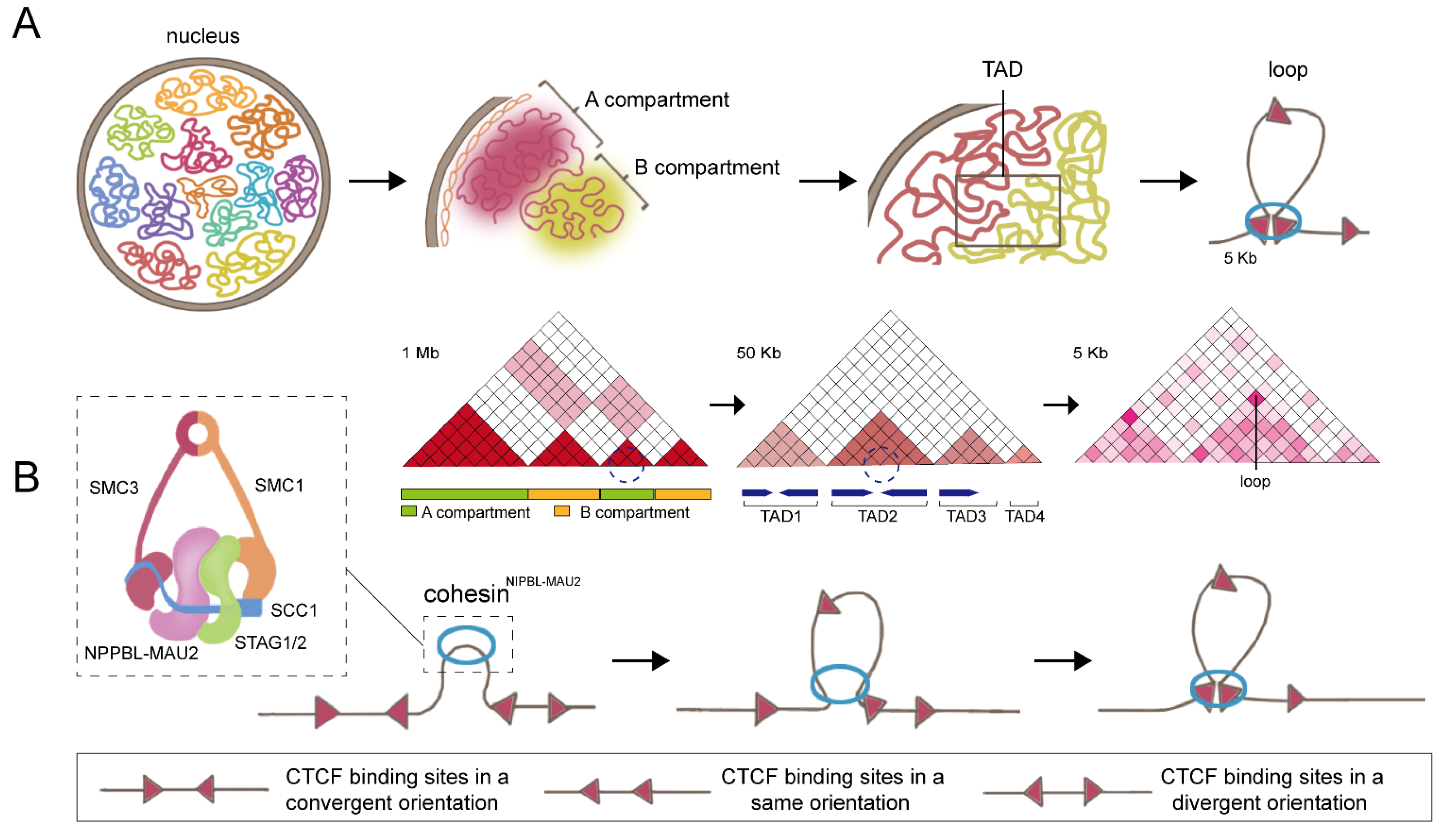
1. Cohesin: The Key Partner of CCCTC-Binding Factor (CTCF)

Regarding the formation of the basic three-dimensional structural loops, the “loop extrusion model” has been proposed by several groups (Figure 1B) [67,68,69][1][2][3]. According to the hypothesis, cohesin extrudes chromatin loops bidirectionally, and these chromatin loops will initially be small and increase over time until they are blocked by CTCF boundaries. Moreover, depending on the loop extrusion model, several modified models, including the walking model, pumping model, and scrunching model, have been proposed for the explanation of the loop extrusion process in eukaryotic mitosis cells, which improves our understanding of chromatin loop formation [70][4].
Figure 1. Three-dimensional genome organization and loop extrusion model. (A) The topological structures of the 3D genome are organized at four levels, comprising chromosome territories, A/B compartments, TAD, and chromatin loops. (B) DNA loop extrusion model. Cohesin binds to DNA and begins to extrude symmetrically until it encounters a convergent-oriented CTCF to form a chromatin loop.
Cohesin, a circular protein complex, was originally identified in eukaryotic mitosis for its function in sister chromatids cohesion [71][5]. Moreover, cohesin also plays crucial roles in regulation of DNA loops formation and 3D genome dynamics. The cohesin complex typically contains four subunits: the ring-forming subunits SMC1, SMC3, and SCC1, and one HEAT repeat proteins associated with kleisins (HAWK) protein (STAG1 or STAG2) [72][6]. Cohesin binds genomic sequences in a cis manner and extrudes DNA bidirectionally to form chromatin loops until encountering the boundaries that are preferentially bound by CTCF [73][7]. Degradation of cohesin resulted in the elimination of nearly all loop domains, showing that cohesin is the key factor responsible for loop formation [74][8]. Indeed, several pieces of evidence have indicated that CTCF works together with cohesin in modulating chromatin structure. In particular, almost 90% of the cohesin ChIP-Seq peaks co-localized with CTCF binding sites [10,11][9][10] and ChIP-seq as well as Hi-C experiments found that cohesin and CTCF are enriched at the TAD boundary region, which support their coordinating function in TAD and loop establishment [12,13][11][12]. The orientations of CTCF motifs are divided into three categories: convergent orientations, same orientations, and divergent orientations. Most loop- or TAD-bound CTCF motif pairs appear in mutually convergent orientations and are critical for loop formation (Figure 1B). A single reversal of the orientation of the CTCF motif is sufficient to make the loop disappear and alter the DNA folding [75][13]. To visualize the dynamics of CTCF- and cohesin-mediated cycling, Hu et al. selected a relatively simple 505-kb TAD in mouse embryonic stem cells containing only one gene, Fbn2, as a model for study. Interestingly, direct observation of dynamic Fbn2 TAD chromatin loops via super-resolution live-cell imaging revealed that cohesin extrusion loops within TADs fail to bridge two CTCF boundaries approximately 92% of the time, suggesting that a single CTCF boundary can also create functional interactions [76][14].
Recently, several other factors, such as WAPL and NIPBL-MAU2, have been identified to participate in mediating loop formation or expansion. WAPL promotes the release of cohesin from DNA template, which restrains loop formation. Depletion of WAPL, in turn, increases the residence time of cohesin on chromatin, where cohesin bypasses CTCF binding sites and generates larger loops [14,15][15][16]. On the contrary, NIPBL forms a heterodimer with MAU2, and this complex works by loading cohesin onto DNA, which is required for loop extrusion. After removing NIPBL-MAU2, cohesin is barely detectable on chromatin in cells, showing that NIPBL-MAU2 functions as a key partner of cohesin. Moreover, NIPBL-MAU2 can stimulate the ATPase activity of cohesin, which is required for cohesin loading onto chromosomes [16][17].

2. Protein Partners of CTCF

2.1. Systemic Discovery of CTCF Partners

Recently, several studies have performed systematic investigation of CTCF’s partners. Hu et al. took advantage of 1306 ChIP-seq data for 431 human protein factors and characterized the genome-wide DNA-binding patterns of these factors, including transcription factors, histone variants, and histone-modifying enzymes, as well as CTCF, in 23 cell lines [52][18]. All factors with ChIP-seq data were screened by co-binding or co-localization analysis with human hyperconserved CTCF binding sites using computational methods. In addition to previously reported co-factors such as cohesin subunits (RAD21, SMC3), histone demethylase KDM5B, and transcription factors YY1 and ZNF143, a number of novel co-binding factors overlapping with CTCF-binding sites were identified. For example, RCOR1 and TEAD4 showed up to 40% overlapping with CTCF binding sites. These novel factors may be important candidate partners that are essential for the establishment of CTCF-mediated chromatin looping.

2.2. Transcriptional Regulatory Protein Partners Related to Embryonic Stem Cell Development

CTCF has been reported to interact with RNA polymerase II both in vivo and in vitro [17][19], which is involved in the regulation of alternative splicing and transcription initiation processes [18][20]. In addition, a great number of recent studies indicated that CTCF also interacts with various transcription factors to regulate transcription and the three-dimensional genome structure, which is essential for embryonic stem (ES) cell self-renewal and differentiation. The pluripotency factor OCT4, acting as a key regulator at the top of the X chromosome inactivation (XCI) hierarchy, can interact with CTCF to regulate XCI by triggering X chromosome pairing and counting, which is indispensable to ES cell differentiation [19][21]. Moreover, CTCF, in addition to acting as an upstream transcription regulator of WD repeat domain 5 (Wdr5), can also physically bind to Wdr5, which plays important roles in maintaining ES cell pluripotency and somatic reprogramming [20][22].

2.3. Transcriptional Regulatory Protein Partners Regulating Immune Cell Development

T and B cells comprise the main forces of adaptive immunity of the immune system, and T cells play a major role in the body’s anti-infection, anti-tumor, and autoimmune diseases. Naïve T cells can differentiate into Th1, Th2, and Th17 cells expressing IFN-γ, IL-4, and IL-17, respectively. Kim et al. showed that Th17 lineage differentiation is restrained by the Th2 locus, and the regulation is attributed to the interaction between Oct-1 and CTCF, which mediates the interchromosomal contacts between the locus control region (LCR) of the Th2 cytokine locus on chromosome 11 and the IL-17 locus on chromosome 1 [25][23]. The high mobility group (HMG) transcription factor TCF-1 functions at the early thymic progenitor (ETP) stage by regulating Gata3 and Bcl11b expression and remains highly expressed until maturation, which is essential for early T-cell development [26][24]. Wang et al. demonstrated that TCF-1 and CTCF co-occupy recombined TAD boundaries during T-cell development, weakening the insulation between adjacent neighbors and enhancing the interaction between regulatory elements and target genes located on previously insulating domains. The promotion of chromatin interactions mediated by TCF-1 is associated with the deposition of the active enhancer marker H3K27ac and the recruitment of NIPBL [27][25].

2.4. Transcriptional Regulatory Protein Partners Associated with Muscle Cell Development

MyoD and Myf5, acting as fundamental helix-loop-helix transcription factors, are required for myogenic initiation during early embryogenesis and functionally complement each other [34][26]. MyoD inactivation in mice had no apparent effect on muscle development [35][27] but resulted in severely deficient muscle regeneration [36][28] and reduced differentiation potential [37][29]. Wang et al. have shown that MyoD cooperated with CTCF, thus driving the formation of different types of chromatin loops in muscle cells. Notably, four types of chromatin loops, namely, MyoD-MyoD (noCTCF), MyoD-MyoD (CTCF), MyoD-CTCF, and CTCF-CTCF, have been resolved. The MyoD-binding loop was significantly shorter than the CTCF-CTCF loop, with the MyoD-MyoD (noCTCF) loop being the shortest, and MyoD inactivation significantly reduced loop strength in all four types of chromatin loops. Taken together, MyoD functionally contributes to the formation of MyoD-bound and CTCF-bound chromatin loops. Of note, MyoD regulates chromatin loops independently of H3K27ac levels, suggesting that MyoD should be a key organizer in establishing the unique 3D genomic architecture of muscle cells [38][30].

2.5. Transcriptional Regulatory Protein Partners Involved in Multiple Developmental Processes

The zinc finger transcription factor Yin Yang 1 (YY1) is essential for both early embryogenesis and adult tissue development [39][31], and biallelic loss of function variants in YY1 cause embryonic lethality in mice [40][32]. In embryonic stem cells, YY1 activates transcription by targeting promoters and super-enhancers through the BAF complex [41][33]. YY1 is also a master regulator to coordinate multidimensional epigenetic crosstalk associated with expanded pluripotency, and depletion of YY1 disrupts specific enhancer–promoter interactions in expanded pluripotent stem cells (EPSC) [42][34]. Further, YY1 directly interacts with CTCF and they work together to regulate X chromosome binary switch [43][35]. Moreover, YY1 deletion in EPSCs reduces DNA methylation, promotes CTCF binding to hypomethylated DNA regions, and promotes gene expression. In B cells, YY1 mediated long-range DNA contacts [44][36] and is necessary for the formation of specific 3D interactions [45][37]. Furthermore, YY1 is also a key regulator of neuron differentiation of neural progenitor cells (NPC) to myelinated oligodendrocytes [46][38]. YY1 can function as a structural protein, linking NPC-specific genes and enhancers, and is implicated in regulation of 3D interactions. More importantly, as a CTCF’s partner, the interactions mediated by YY1 between regulatory elements are often in CTCF-anchored constituent loops [47][39].
The Y-box DNA/RNA binding factor (YB-1) is a multifunctional protein involved in transcription, replication, and RNA processing, and several studies have identified it as a CTCF partner.

2.6. Transcriptional Regulatory Protein Partners Showing Potential Roles in 3D Genome Organization and Transcriptional Regulation

Xiao et al. have shown that Myc-associated zinc finger protein (MAZ) is a physically interacting partner of CTCF, and it can function as a genome architecture protein, one that participates in genome organization. Remarkably, MAZ shares core properties with CTCF, including insulation activity and interaction with cohesin subunit Rad21, supporting the fact that MAZ and CTCF have complementary roles in organizing genome structure [51][40]. In addition, ChIP-seq and co-IP experiments showed that most of the basic helix-loop-helix family member e40 (BHLHE40) binding sites are also occupied by CTCF, and BHLHE40 can physically interact with CTCF, indicating that BHLHE40 should be a CTCF partner [52][18]. Furthermore, loss of BHLHE40 results in a reduced number of CTCF binding sites, decreased CTCF loop strength, and disruption of CTCF-mediated long-range chromatin interactions, demonstrating that BHLHE40 acts as a partner to regulate CTCF-mediated chromatin interactome.
In addition, several other protein partners of CTCF have been identified; nevertheless, the detailed mechanisms underlying the roles of CTCF/partner complex in regulation of 3D genome structure still require further information.

2.7. Chromatin Remodeling Associated Protein Partners

The nucleosome is the basic unit of chromatin, consisting of histone octamers (two copies of H2A, H2B, H3, and H4) and 146 base pairs (bp) of DNA. In addition to these classic histone variants, many other variants have been found, such as H2A.X, H2A.Z, H3.5, and H4.G [78][41]. Moreover, a great number of proteins, which have the capacities of modifying the chromatin architecture, have been identified, and these proteins are defined as chromatin-remodeling-associated proteins [79,80][42][43]. Until now, studies have identified several CTCF’s partners that are related to chromatin remodeling. H2A.Z is a histone variant that shows specific properties in regulating higher-order chromatin structures [81][44]. In vitro studies have shown that the structure of nucleosomes is not static but undergoes spontaneous structural transitions, including DNA respiration (spontaneous opening of DNA ends on nucleosomes) and open states (opening of interfaces between histone subcomplexes), and this dynamic change of nucleosomes is called nucleosome unwrapping. By employing micrococcal nuclease (MNase) digestion of crosslinked chromatin, chromatin immunoprecipitation, and the paired-end sequencing (MNase-X-ChIP-seq) approach, Wen et al. investigated the genome-wide unwrapping state of H2A.Z nucleosomes in mouse embryonic stem cells. Interestingly, compared with canonical nucleosomes, H2A.Z is enriched with nucleosome unwrapping, indicating that H2A.Z is essential to nucleosome unfolding, and H2A.Z may affect CTCF binding regulation and gene expression by modulating the unwrapping states of nucleosomes [56][45].

2.8. Interplay between Nuclear Receptor and CTCF

The nuclear receptor superfamily is a family of ligand-activated transcription factors that regulate cell growth and differentiation by establishing links between signaling molecules and transcriptional responses [82][46]. Warwick et al. demonstrated that activation of the vitamin D receptor (VDR) and its high-affinity ligand 1,25(OH)2D3 induced cellular 3D chromatin changes. Interestingly, their results indicated that more than 3000 CTCF interactions were altered, and VDR binding sites and vitamin D target genes are preferentially located at loop anchors, implying the potential interaction between CTCF and VDR [63][47]. Furthermore, upon estrogen stimulation in breast cancer cells, CTCF binds to enhancer regions and prevents the formation of estrogen receptor (ER)-mediated chromatin loops to regulate ER target transcription [64][48]. During chromatin remodeling, switching between active A and inactive B compartments in endocrine-resistant breast cancer is associated with reduced ER binding and aberrant ER-mediated enhancer–promoter interactions [65][49]. Therefore, VDR and ER may be partners of CTCF, and their interactions may play important roles in regulating the organization of the 3D genome.

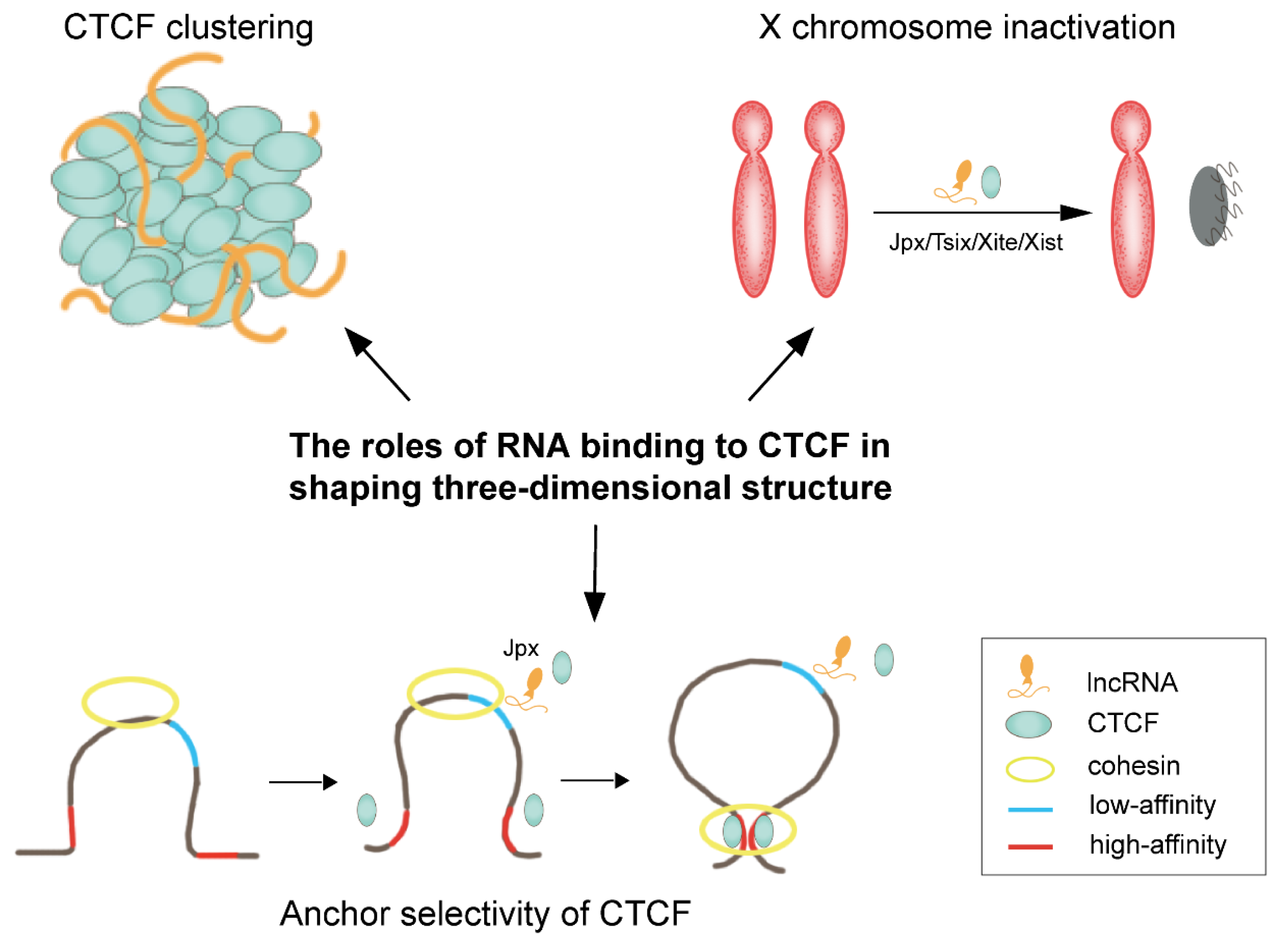
3. RNA Partners of CTCF

In addition to binding to DNA, several recent studies confirmed that CTCF also has the ability to bind to RNA. Moreover, CTCF targets thousands of transcripts throughout the genome and has a higher binding affinity for RNA than DNA [83][50]. ZF1 and ZF10 domains, but not the major DNA binding domains (ZF3-7 domains), are responsible for the RNA-binding property of CTCF [84][51]. Hansen et al. reports that CTCF’s RNA-binding region (RBR) plays crucial roles in CTCF clustering in vivo and is associated with chromatin loop formation. More importantly, RBR suppression in mESCs causes disruption in about half of the chromatin loops, called RBRi-dependent loops [85][52].
Currently, high-throughput-sequencing-based methods, such as UV-crosslinking immunoprecipitation and deep sequencing (CLIP-seq), have been developed to identify CTCF-bound transcripts systemically [83][50]. Additionally, Kuang et al. discovered a novel RNA-binding motif (AGAUNGGA) of CTCF and identified 4925 candidate CTCF-binding lncRNAs by a deep learning model DeepLncCTCF, extending our understandings of CTCF in 3D genome organization [86][53]. Furthermore, to measure higher-order RNA and DNA contacts within 3D structures, the RNA and DNA split-pool via label-extended recognition interaction (RD-SPRITE) method was developed. Depending on this method, hundreds of noncoding RNAs (ncRNAs) were found to form regions of high concentration within the nucleus, and these higher-order RNA-chromatin structures are related to regulation of long-range DNA contacts, heterochromatin assembly, and gene expression [87][54].
Until now, several CTCF’s RNA partners have been identified, and their regulatory roles in chromatin structure provide novel insights into 3D genome organization. X-chromosome inactivation (XCI) is a classical epigenetic reprogramming process that is essential for mammalian development [88][55]. Studies indicated that CTCF is implicated in the XCI process, which is largely driven by Tsix, Xite, and Xist RNAs, and CTCF directly interacts with these RNAs in the X inactivation center during XCI, thereby mediating long-range chromosomal interactions [83,89,90][50][56][57]. Jpx RNA, another CTCF partner, also plays roles in regulating the initiation of X chromosome inactivation (XCI) by expelling CTCF from the Xist promoter [89][56]. Notably, a recent study demonstrated that Jpx/CTCF complex modulates the chromatin structure on a genome-wide manner, not limited to XCI. Importantly, Jpx can act as a CTCF release factor and determine the anchoring selectivity of CTCF. Specifically, Jpx selectively binds to low-affinity CTCF motifs and expels CTCF through competitive inhibition (Figure 3). Thus, knockdown of Jpx RNA results in substantial changes in chromosomal loops, most likely due to the ectopic CTCF binding [90][57].
Figure 3.
RNA/CTCF complexes play important roles in organizing chromatin structure.
In addition, interactions between CTCF and multiple ncRNAs, such as the steroid receptor RNA activator (SRA), Wrap53, HOTTIP, and GATA6-AS1, have been identified, playing potential roles in shaping 3D genome structure. For example, Yao et al. revealed that CTCF/SRA/p68 (DEAD-box RNA helicase) complex can stabilize the interaction between CTCF and cohesin [91][58]. CTCF regulates p53 expression by physically interacting with Wrap53 RNA, which is the natural antisense transcript of p53. Deletion of CTCF not only resulted in a decrease in p53 mRNA levels, but also in Wrap53 levels [92][59]. The HOXA transcript at the distal tip (HOTTIP) was previously identified as a lncRNA located at the 5′ end of the HOXA locus [93][60].

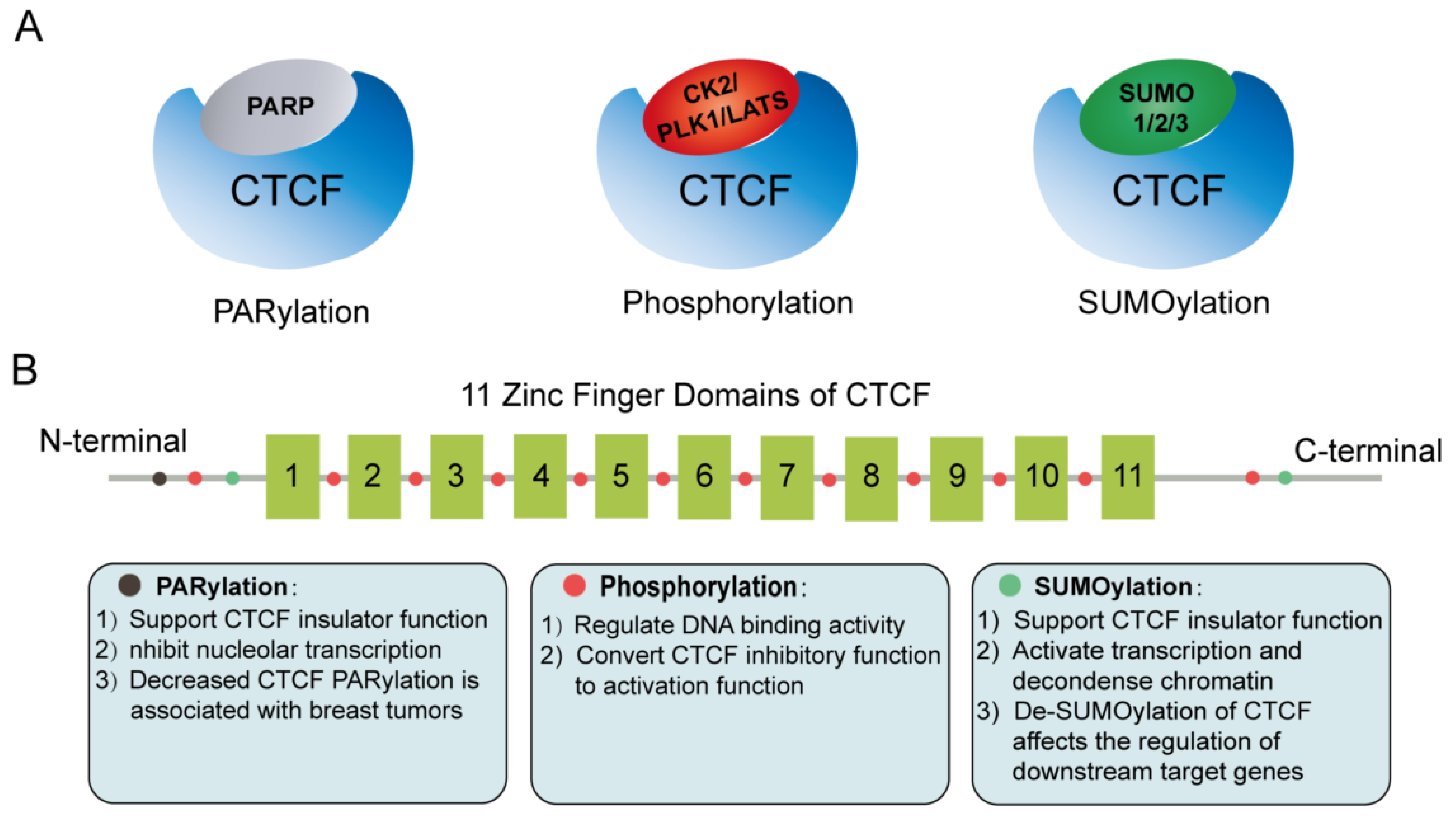
4. Post-Translational Modifications of CTCF

Protein post-translational modifications (PTMs), functioning as a key mechanism for increasing proteome diversity, play critical roles in nearly all biological processes. Indeed, many studies indicate that PTM enzymes, such as PARP, SUMO, CK2, PLK1, and LATS, are also CTCF’s protein partners. In fact, CTCF is regulated by serving as a substrate for these PTM enzymes. PTMs of CTCF are associated with dynamic regulation of the stability and function of CTCF in response to an external or internal stimulus (Figure 4A,B).
Figure 4. Post-translational modification (PTM) enzymes are CTCF’s partners. (A) CTCF is regulated by three types of PTM, namely, PARylation, SUMOylation, and phosphorylation. (B) PTM sites in CTCF protein, and their roles in modulating CTCF insulator function.
Poly(ADP-ribosyl) ation (PARylation) is mediated by poly(ADP-ribose) polymerase (PARP) [97][61], and an increasing number of studies suggest that PARylation is involved in CTCF function regulation. The N-terminal domain of CTCF is a preferred target for PARylation in vitro [98][62], and CTCF/PARP1 complex can function through a dynamic reversible PARylation modification model to regulate CTCF function [99][63], and is associated with the contacts between clock-controlled genes and lamina-associated chromatin [100][64]. Intriguingly, the PARylated CTCF isoform (180 kDa) preferentially located at nucleolus and PARylation of CTCF represses nucleolar transcription [101][65]. Furthermore, CTCF PARylation affects the binding of CTCF to chromatin, and the decrease in CTCF PARylation has been reported to be linked to breast tumorigenesis and cell proliferation [102,103][66][67].
CTCF protein can be modified by small ubiquitin-like proteins, including SUMO 1, 2, and 3. Currently, two major SUMOylation sites of CTCF, located in the COOH and NH2 terminal domains, separately, have been resolved. SUMOylation of CTCF commonly contributes to its repressive functions, such as repression of the cMYC P2 promoter activity [104][68]. Specially, a 107-amino-acid domain was identified in the N-terminal region of CTCF that activates transcription and depolymerizes chromatin, and complete sumoylation of this domain abolishes the transcriptional activity of CTCF and prevents chromatin opening [105][69]. Furthermore, stress-induced hypoxic desumoylation of lysines 74 and 689 in CTCF proteins regulated both the activity of CTCF and its downstream target genes [106][70].
Moreover, it is widely accepted that phosphorylation is a functional determinant of transcription factors and appears to be one of the most studied forms of PTM. The phosphorylation profile of CTCF is dynamic during development and cell differentiation [107][71]. CK2-mediated phosphorylation at several functional phosphorylation sites within CTCF C-terminal region can convert CTCF inhibitory function to activating function [108][72]. In addition, amino acid residues (Thr289, Thr317, Thr346, Thr374, Ser402, Ser461, and Thr518) of the CTCF linker domain are phosphorylated during mitosis to regulate its DNA-binding activity [109][73]. In addition, another Polo-like kinase 1 (PLK1)-mediated phosphorylation of CTCF at serine 224 (Ser224-P) is enriched in the G2/M phase of the cell cycle, especially at pericentric regions. Of note, the CTCF phospho-depletion mutant S224E resulted in dysregulation of hundreds of target genes, including p53 and p21 [110][74]. CTCF was found to be a substrate for LATS kinase, and cellular-stress-induced activity of LATS directly phosphorylates CTCF’s zinc finger (ZF) linker and selectively dissociates CTCF from a small fraction of its genomic binding sites, impairing its DNA-binding activity [111][75]. Therefore, external signals may modulate 3D genome structure through the phosphorylation of CTCF’s ZF linker.

References

  1. Nasmyth, K. Disseminating the genome: Joining, resolving, and separating sister chromatids during mitosis and meiosis. Annu. Rev. Genet. 2001, 35, 673–745.
  2. Alipour, E.; Marko, J.F. Self-organization of domain structures by DNA-loop-extruding enzymes. Nucleic Acids Res. 2012, 40, 11202–11212.
  3. Sanborn, A.L.; Rao, S.S.; Huang, S.C.; Durand, N.C.; Huntley, M.H.; Jewett, A.I.; Bochkov, I.D.; Chinnappan, D.; Cutkosky, A.; Li, J.; et al. Chromatin extrusion explains key features of loop and domain formation in wild-type and engineered genomes. Proc. Natl. Acad. Sci. USA 2015, 112, E6456–E6465.
  4. Davidson, I.F.; Peters, J.M. Genome folding through loop extrusion by SMC complexes. Nat. Rev. Mol. Cell Biol. 2021, 22, 445–464.
  5. Yatskevich, S.; Rhodes, J.; Nasmyth, K. Organization of Chromosomal DNA by SMC Complexes. Annu. Rev. Genet. 2019, 53, 445–482.
  6. Sumara, I.; Vorlaufer, E.; Gieffers, C.; Peters, B.H.; Peters, J.M. Characterization of vertebrate cohesin complexes and their regulation in prophase. J. Cell Biol. 2000, 151, 749–762.
  7. Wendt, K.S.; Yoshida, K.; Itoh, T.; Bando, M.; Koch, B.; Schirghuber, E.; Tsutsumi, S.; Nagae, G.; Ishihara, K.; Mishiro, T.; et al. Cohesin mediates transcriptional insulation by CCCTC-binding factor. Nature 2008, 451, 796–801.
  8. Rao, S.S.P.; Huang, S.C.; Glenn St Hilaire, B.; Engreitz, J.M.; Perez, E.M.; Kieffer-Kwon, K.R.; Sanborn, A.L.; Johnstone, S.E.; Bascom, G.D.; Bochkov, I.D.; et al. Cohesin Loss Eliminates All Loop Domains. Cell 2017, 171, 305–320.e24.
  9. Rubio, E.D.; Reiss, D.J.; Welcsh, P.L.; Disteche, C.M.; Filippova, G.N.; Baliga, N.S.; Aebersold, R.; Ranish, J.A.; Krumm, A. CTCF physically links cohesin to chromatin. Proc. Natl. Acad. Sci. USA 2008, 105, 8309–8314.
  10. Hansen, A.S.; Pustova, I.; Cattoglio, C.; Tjian, R.; Darzacq, X. CTCF and cohesin regulate chromatin loop stability with distinct dynamics. Elife 2017, 6, e25776.
  11. Pugacheva, E.M.; Kubo, N.; Loukinov, D.; Tajmul, M.; Kang, S.; Kovalchuk, A.L.; Strunnikov, A.V.; Zentner, G.E.; Ren, B.; Lobanenkov, V.V. CTCF mediates chromatin looping via N-terminal domain-dependent cohesin retention. Proc. Natl. Acad. Sci. USA 2020, 117, 2020–2031.
  12. Fudenberg, G.; Imakaev, M.; Lu, C.; Goloborodko, A.; Abdennur, N.; Mirny, L.A. Formation of Chromosomal Domains by Loop Extrusion. Cell Rep. 2016, 15, 2038–2049.
  13. Guo, Y.; Xu, Q.; Canzio, D.; Shou, J.; Li, J.; Gorkin, D.U.; Jung, I.; Wu, H.; Zhai, Y.; Tang, Y.; et al. CRISPR Inversion of CTCF Sites Alters Genome Topology and Enhancer/Promoter Function. Cell 2015, 162, 900–910.
  14. Gabriele, M.; Brandão, H.B.; Grosse-Holz, S.; Jha, A.; Dailey, G.M.; Cattoglio, C.; Hsieh, T.S.; Mirny, L.; Zechner, C.; Hansen, A.S. Dynamics of CTCF- and cohesin-mediated chromatin looping revealed by live-cell imaging. Science 2022, 376, 496–501.
  15. Kueng, S.; Hegemann, B.; Peters, B.H.; Lipp, J.J.; Schleiffer, A.; Mechtler, K.; Peters, J.M. Wapl controls the dynamic association of cohesin with chromatin. Cell 2006, 127, 955–967.
  16. Haarhuis, J.H.I.; van der Weide, R.H.; Blomen, V.A.; Yáñez-Cuna, J.O.; Amendola, M.; van Ruiten, M.S.; Krijger, P.H.L.; Teunissen, H.; Medema, R.H.; van Steensel, B.; et al. The Cohesin Release Factor WAPL Restricts Chromatin Loop Extension. Cell 2017, 169, 693–707.e14.
  17. Rhodes, J.; Mazza, D.; Nasmyth, K.; Uphoff, S. Scc2/Nipbl hops between chromosomal cohesin rings after loading. Elife 2017, 6, e30000.
  18. Hu, G.; Dong, X.; Gong, S.; Song, Y.; Hutchins, A.P.; Yao, H. Systematic screening of CTCF binding partners identifies that BHLHE40 regulates CTCF genome-wide distribution and long-range chromatin interactions. Nucleic Acids Res. 2020, 48, 9606–9620.
  19. Chernukhin, I.; Shamsuddin, S.; Kang, S.Y.; Bergstrom, R.; Kwon, Y.W.; Yu, W.; Whitehead, J.; Mukhopadhyay, R.; Docquier, F.; Farrar, D.; et al. CTCF interacts with and recruits the largest subunit of RNA polymerase II to CTCF target sites genome-wide. Mol. Cell. Biol. 2007, 27, 1631–1648.
  20. Shukla, S.; Kavak, E.; Gregory, M.; Imashimizu, M.; Shutinoski, B.; Kashlev, M.; Oberdoerffer, P.; Sandberg, R.; Oberdoerffer, S. CTCF-promoted RNA polymerase II pausing links DNA methylation to splicing. Nature 2011, 479, 74–79.
  21. Donohoe, M.E.; Silva, S.S.; Pinter, S.F.; Xu, N.; Lee, J.T. The pluripotency factor Oct4 interacts with Ctcf and also controls X-chromosome pairing and counting. Nature 2009, 460, 128–132.
  22. Wang, F.; Han, J.; Wang, L.; Jing, Y.; Zhu, Z.; Hui, D.; Wang, Z.; Wang, Y.; Dong, Y.; Tan, T. CCCTC-Binding Factor Transcriptionally Targets Wdr5 to Mediate Somatic Cell Reprogramming. Stem Cells Dev. 2017, 26, 743–750.
  23. Kim, L.K.; Esplugues, E.; Zorca, C.E.; Parisi, F.; Kluger, Y.; Kim, T.H.; Galjart, N.J.; Flavell, R.A. Oct-1 regulates IL-17 expression by directing interchromosomal associations in conjunction with CTCF in T cells. Mol. Cell 2014, 54, 56–66.
  24. Li, L.; Leid, M.; Rothenberg, E.V. An early T cell lineage commitment checkpoint dependent on the transcription factor Bcl11b. Science 2010, 329, 89–93.
  25. Wang, W.; Chandra, A.; Goldman, N.; Yoon, S.; Ferrari, E.K.; Nguyen, S.C.; Joyce, E.F.; Vahedi, G. TCF-1 promotes chromatin interactions across topologically associating domains in T cell progenitors. Nat. Immunol. 2022, 23, 1052–1062.
  26. Pikarsky, E.; Porat, R.M.; Stein, I.; Abramovitch, R.; Amit, S.; Kasem, S.; Gutkovich-Pyest, E.; Urieli-Shoval, S.; Galun, E.; Ben-Neriah, Y. NF-kappaB functions as a tumour promoter in inflammation-associated cancer. Nature 2004, 431, 461–466.
  27. Rudnicki, M.A.; Braun, T.; Hinuma, S.; Jaenisch, R. Inactivation of MyoD in mice leads to up-regulation of the myogenic HLH gene Myf-5 and results in apparently normal muscle development. Cell 1992, 71, 383–390.
  28. Megeney, L.A.; Kablar, B.; Garrett, K.; Anderson, J.E.; Rudnicki, M.A. MyoD is required for myogenic stem cell function in adult skeletal muscle. Genes Dev. 1996, 10, 1173–1183.
  29. Sabourin, L.A.; Girgis-Gabardo, A.; Seale, P.; Asakura, A.; Rudnicki, M.A. Reduced differentiation potential of primary MyoD-/- myogenic cells derived from adult skeletal muscle. J. Cell Biol. 1999, 144, 631–643.
  30. Wang, R.; Chen, F.; Chen, Q.; Wan, X.; Shi, M.; Chen, A.K.; Ma, Z.; Li, G.; Wang, M.; Ying, Y.; et al. MyoD is a 3D genome structure organizer for muscle cell identity. Nat. Commun. 2022, 13, 205.
  31. Vella, P.; Barozzi, I.; Cuomo, A.; Bonaldi, T.; Pasini, D. Yin Yang 1 extends the Myc-related transcription factors network in embryonic stem cells. Nucleic Acids Res. 2012, 40, 3403–3418.
  32. Donohoe, M.E.; Zhang, X.; McGinnis, L.; Biggers, J.; Li, E.; Shi, Y. Targeted disruption of mouse Yin Yang 1 transcription factor results in peri-implantation lethality. Mol. Cell. Biol. 1999, 19, 7237–7244.
  33. Wang, J.; Wu, X.; Wei, C.; Huang, X.; Ma, Q.; Huang, X.; Faiola, F.; Guallar, D.; Fidalgo, M.; Huang, T.; et al. YY1 Positively Regulates Transcription by Targeting Promoters and Super-Enhancers through the BAF Complex in Embryonic Stem Cells. Stem Cell Rep. 2018, 10, 1324–1339.
  34. Dong, X.; Guo, R.; Ji, T.; Zhang, J.; Xu, J.; Li, Y.; Sheng, Y.; Wang, Y.; Fang, K.; Wen, Y.; et al. YY1 safeguard multidimensional epigenetic landscape associated with extended pluripotency. Nucleic Acids Res. 2022, gkac230.
  35. Donohoe, M.E.; Zhang, L.F.; Xu, N.; Shi, Y.; Lee, J.T. Identification of a Ctcf cofactor, Yy1, for the X chromosome binary switch. Mol. Cell 2007, 25, 43–56.
  36. Mehra, P.; Gerasimova, T.; Basu, A.; Jha, V.; Banerjee, A.; Sindhava, V.; Gray, F.; Berry, C.T.; Sen, R.; Atchison, M.L. YY1 controls Eμ-3’RR DNA loop formation and immunoglobulin heavy chain class switch recombination. Blood Adv. 2016, 1, 15–20.
  37. Medvedovic, J.; Ebert, A.; Tagoh, H.; Tamir, I.M.; Schwickert, T.A.; Novatchkova, M.; Sun, Q.; Huis In ‘t Veld, P.J.; Guo, C.; Yoon, H.S.; et al. Flexible long-range loops in the VH gene region of the Igh locus facilitate the generation of a diverse antibody repertoire. Immunity 2013, 39, 229–244.
  38. He, Y.; Dupree, J.; Wang, J.; Sandoval, J.; Li, J.; Liu, H.; Shi, Y.; Nave, K.A.; Casaccia-Bonnefil, P. The transcription factor Yin Yang 1 is essential for oligodendrocyte progenitor differentiation. Neuron 2007, 55, 217–230.
  39. Beagan, J.A.; Duong, M.T.; Titus, K.R.; Zhou, L.; Cao, Z.; Ma, J.; Lachanski, C.V.; Gillis, D.R.; Phillips-Cremins, J.E. YY1 and CTCF orchestrate a 3D chromatin looping switch during early neural lineage commitment. Genome Res. 2017, 27, 1139–1152.
  40. Xiao, T.; Li, X.; Felsenfeld, G. The Myc-associated zinc finger protein (MAZ) works together with CTCF to control cohesin positioning and genome organization. Proc. Natl. Acad. Sci. USA 2021, 118, e2023127118.
  41. Talbert, P.B.; Henikoff, S. Histone variants at a glance. J. Cell Sci. 2021, 134, jcs244749.
  42. Cairns, B.R. Emerging roles for chromatin remodeling in cancer biology. Trends Cell Biol. 2001, 11, S15–S21.
  43. Cairns, B.R. Chromatin remodeling: Insights and intrigue from single-molecule studies. Nat. Struct. Mol. Biol. 2007, 14, 989–996.
  44. Rege, M.; Subramanian, V.; Zhu, C.; Hsieh, T.H.; Weiner, A.; Friedman, N.; Clauder-Münster, S.; Steinmetz, L.M.; Rando, O.J.; Boyer, L.A.; et al. Chromatin Dynamics and the RNA Exosome Function in Concert to Regulate Transcriptional Homeostasis. Cell Rep. 2015, 13, 1610–1622.
  45. Wen, Z.; Zhang, L.; Ruan, H.; Li, G. Histone variant H2A.Z regulates nucleosome unwrapping and CTCF binding in mouse ES cells. Nucleic Acids Res. 2020, 48, 5939–5952.
  46. Porter, B.A.; Ortiz, M.A.; Bratslavsky, G.; Kotula, L. Structure and Function of the Nuclear Receptor Superfamily and Current Targeted Therapies of Prostate Cancer. Cancers 2019, 11, 1852.
  47. Warwick, T.; Schulz, M.H.; Gilsbach, R.; Brandes, R.P.; Seuter, S. Nuclear receptor activation shapes spatial genome organization essential for gene expression control: Lessons learned from the vitamin D receptor. Nucleic Acids Res. 2022, 50, 3745–3763.
  48. Fiorito, E.; Sharma, Y.; Gilfillan, S.; Wang, S.; Singh, S.K.; Satheesh, S.V.; Katika, M.R.; Urbanucci, A.; Thiede, B.; Mills, I.G.; et al. CTCF modulates Estrogen Receptor function through specific chromatin and nuclear matrix interactions. Nucleic Acids Res. 2016, 44, 10588–10602.
  49. Achinger-Kawecka, J.; Valdes-Mora, F.; Luu, P.L.; Giles, K.A.; Caldon, C.E.; Qu, W.; Nair, S.; Soto, S.; Locke, W.J.; Yeo-Teh, N.S.; et al. Epigenetic reprogramming at estrogen-receptor binding sites alters 3D chromatin landscape in endocrine-resistant breast cancer. Nat. Commun. 2020, 11, 320.
  50. Kung, J.T.; Kesner, B.; An, J.Y.; Ahn, J.Y.; Cifuentes-Rojas, C.; Colognori, D.; Jeon, Y.; Szanto, A.; del Rosario, B.C.; Pinter, S.F.; et al. Locus-Specific Targeting to the X Chromosome Revealed by the RNA Interactome of CTCF. Mol. Cell 2015, 57, 361–375.
  51. Saldana-Meyer, R.; Rodriguez-Hernaez, J.; Escobar, T.; Nishana, M.; Jacome-Lopez, K.; Nora, E.P.; Bruneau, B.G.; Tsirigos, A.; Furlan-Magaril, M.; Skok, J.; et al. RNA Interactions Are Essential for CTCF-Mediated Genome Organization. Mol. Cell 2019, 76, 412–422.e15.
  52. Hansen, A.S.; Hsieh, T.-H.S.; Cattoglio, C.; Pustova, I.; Saldaña-Meyer, R.; Reinberg, D.; Darzacq, X.; Tjian, R. Distinct Classes of Chromatin Loops Revealed by Deletion of an RNA-Binding Region in CTCF. Mol. Cell 2019, 76, 395–411.e313.
  53. Kuang, S.; Wang, L. Identification and analysis of consensus RNA motifs binding to the genome regulator CTCF. NAR Genom. Bioinform. 2020, 2, lqaa031.
  54. Quinodoz, S.A.; Ollikainen, N.; Tabak, B.; Palla, A.; Schmidt, J.M.; Detmar, E.; Lai, M.M.; Shishkin, A.A.; Bhat, P.; Takei, Y.; et al. Higher-Order Inter-chromosomal Hubs Shape 3D Genome Organization in the Nucleus. Cell 2018, 174, 744–757.e24.
  55. Lee, J.T.; Bartolomei, M.S. X-inactivation, imprinting, and long noncoding RNAs in health and disease. Cell 2013, 152, 1308–1323.
  56. Sun, S.; Del Rosario, B.C.; Szanto, A.; Ogawa, Y.; Jeon, Y.; Lee, J.T. Jpx RNA activates Xist by evicting CTCF. Cell 2013, 153, 1537–1551.
  57. Oh, H.J.; Aguilar, R.; Kesner, B.; Lee, H.G.; Kriz, A.J.; Chu, H.P.; Lee, J.T. Jpx RNA regulates CTCF anchor site selection and formation of chromosome loops. Cell 2021, 184, 6157–6173.e24.
  58. Yao, H.; Brick, K.; Evrard, Y.; Xiao, T.; Camerini-Otero, R.D.; Felsenfeld, G. Mediation of CTCF transcriptional insulation by DEAD-box RNA-binding protein p68 and steroid receptor RNA activator SRA. Genes Dev. 2010, 24, 2543–2555.
  59. Saldana-Meyer, R.; Gonzalez-Buendia, E.; Guerrero, G.; Narendra, V.; Bonasio, R.; Recillas-Targa, F.; Reinberg, D. CTCF regulates the human p53 gene through direct interaction with its natural antisense transcript, Wrap53. Genes Dev. 2014, 28, 723–734.
  60. Ghafouri-Fard, S.; Dashti, S.; Taheri, M. The HOTTIP (HOXA transcript at the distal tip) lncRNA: Review of oncogenic roles in human. Biomed. Pharmacother. 2020, 127, 110158.
  61. Thomas, C.; Tulin, A.V. Poly-ADP-ribose polymerase: Machinery for nuclear processes. Mol. Asp. Med. 2013, 34, 1124–1137.
  62. Yu, W.; Ginjala, V.; Pant, V.; Chernukhin, I.; Whitehead, J.; Docquier, F.; Farrar, D.; Tavoosidana, G.; Mukhopadhyay, R.; Kanduri, C.; et al. Poly(ADP-ribosyl)ation regulates CTCF-dependent chromatin insulation. Nat. Genet. 2004, 36, 1105–1110.
  63. Farrar, D.; Rai, S.; Chernukhin, I.; Jagodic, M.; Ito, Y.; Yammine, S.; Ohlsson, R.; Murrell, A.; Klenova, E. Mutational analysis of the poly(ADP-ribosyl)ation sites of the transcription factor CTCF provides an insight into the mechanism of its regulation by poly(ADP-ribosyl)ation. Mol. Cell. Biol. 2010, 30, 1199–1216.
  64. Zhao, H.; Sifakis, E.G.; Sumida, N.; Millan-Arino, L.; Scholz, B.A.; Svensson, J.P.; Chen, X.; Ronnegren, A.L.; Mallet de Lima, C.D.; Varnoosfaderani, F.S.; et al. PARP1- and CTCF-Mediated Interactions between Active and Repressed Chromatin at the Lamina Promote Oscillating Transcription. Mol. Cell 2015, 59, 984–997.
  65. Torrano, V.; Navascués, J.; Docquier, F.; Zhang, R.; Burke, L.J.; Chernukhin, I.; Farrar, D.; León, J.; Berciano, M.T.; Renkawitz, R.; et al. Targeting of CTCF to the nucleolus inhibits nucleolar transcription through a poly(ADP-ribosyl)ation-dependent mechanism. J. Cell Sci. 2006, 119, 1746–1759.
  66. Pavlaki, I.; Docquier, F.; Chernukhin, I.; Kita, G.; Gretton, S.; Clarkson, C.T.; Teif, V.B.; Klenova, E. Poly(ADP-ribosyl)ation associated changes in CTCF-chromatin binding and gene expression in breast cells. Biochim. Biophys. Acta Gene Regul. Mech. 2018, 1861, 718–730.
  67. Docquier, F.; Kita, G.X.; Farrar, D.; Jat, P.; O’Hare, M.; Chernukhin, I.; Gretton, S.; Mandal, A.; Alldridge, L.; Klenova, E. Decreased poly(ADP-ribosyl)ation of CTCF, a transcription factor, is associated with breast cancer phenotype and cell proliferation. Clin. Cancer Res. 2009, 15, 5762–5771.
  68. MacPherson, M.J.; Beatty, L.G.; Zhou, W.; Du, M.; Sadowski, P.D. The CTCF insulator protein is posttranslationally modified by SUMO. Mol. Cell. Biol. 2009, 29, 714–725.
  69. Kitchen, N.S.; Schoenherr, C.J. Sumoylation modulates a domain in CTCF that activates transcription and decondenses chromatin. J. Cell Biochem. 2010, 111, 665–675.
  70. Wang, J.; Wang, Y.; Lu, L. De-SUMOylation of CCCTC Binding Factor (CTCF) in Hypoxic Stress-induced Human Corneal Epithelial Cells. J. Biol. Chem. 2012, 287, 12469–12479.
  71. Delgado, M.D.; Chernukhin, I.V.; Bigas, A.; Klenova, E.M.; León, J. Differential expression and phosphorylation of CTCF, a c-myc transcriptional regulator, during differentiation of human myeloid cells. FEBS Lett. 1999, 444, 5–10.
  72. El-Kady, A.; Klenova, E. Regulation of the transcription factor, CTCF, by phosphorylation with protein kinase CK2. FEBS Lett. 2005, 579, 1424–1434.
  73. Sekiya, T.; Murano, K.; Kato, K.; Kawaguchi, A.; Nagata, K. Mitotic phosphorylation of CCCTC-binding factor (CTCF) reduces its DNA binding activity. FEBS Open Bio 2017, 7, 397–404.
  74. Del Rosario, B.C.; Kriz, A.J.; Del Rosario, A.M.; Anselmo, A.; Fry, C.J.; White, F.M.; Sadreyev, R.I.; Lee, J.T. Exploration of CTCF post-translation modifications uncovers Serine-224 phosphorylation by PLK1 at pericentric regions during the G2/M transition. Elife 2019, 8, 42341.
  75. Luo, H.; Yu, Q.; Liu, Y.; Tang, M.; Liang, M.; Zhang, D.; Xiao, T.S.; Wu, L.; Tan, M.; Ruan, Y.; et al. LATS kinase-mediated CTCF phosphorylation and selective loss of genomic binding. Sci. Adv. 2020, 6, eaaw4651.
More
ScholarVision Creations