1. Nanoparticles as Antimicrobial Agents
According to the World Health Organization, despite their limited sizes and specificity for microbes, some metal-based nanomaterials have proven to be beneficial against a wide range of infections
[1][27]. Nanoparticles can serve as antibacterial agents on their own or as supplements for regular antibiotics; in either instance, they are termed “nanoantibiotics” (nAbts)
[2][28]. NanoantibiotIcs is another term for nanosized antibiotic molecules that are encased with engineered nanoparticles or antibiotics manufactured artificially by keeping one dimension in the region of 100 nm
[2][28]. Mamum et al. (2021) recently demonstrated the importance of nanoantibiotics as superior medicines to reduce the amount of drug-resistant bacteria in the treatment of diseases
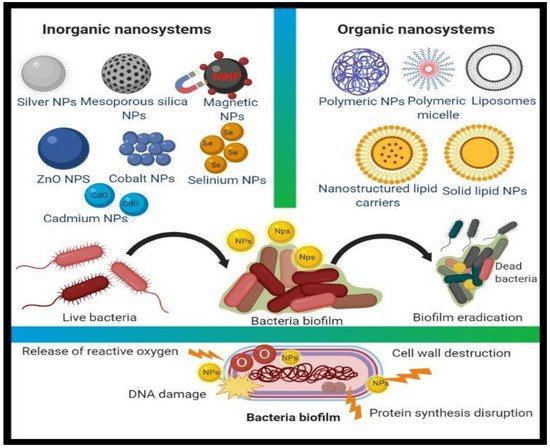
[3][29]. According to Mamun et al. “nAbts exemplify a potential Trojan horse technique to overcome antibiotic resistance processes”. Metal-based nanomaterials are frequently found to exert non-specific negative bacterial effects, such as the fact that they do not bind to a specific receptor in the bacterial cell, making it more difficult for bacteria to develop resistance (
Figure 12) and extending the antibacterial field
[2][28]. A wide range of nanostructures, including Au, Ag, CuO, TiO
2, MgO, and ZnO, are projected to become antibacterial nanosystem alternatives
[4][5][6][30,31,32].
Figure 12. Different types of organic and inorganic nanomaterial for antimicrobial action
[2][28].
12.1. Synergistic Effect (Antibiotics within Nanoparticles)
The influence of synergism is associated with the formation of exceptionally active hydroxyl radicals, as well as alterations in defensive cellular functions and anti-biofilm efficacy. It is associated with the use of antibiotics in connection with nanostructures, which is notably effective in increasing antibiotic functionality; this contrasts with the impact of the use of antibiotics in clinical practice, which minimizes antibiotic doses, exposure time, and bacterial resistance
[7][33]. The majority of harmful microorganisms are resistant to conventional antibiotics. Nanoparticles are affixed to antibiotics to augment their potency. NPs fight pathogenic germs through many pathways, which are activated concurrently by nanoparticles and antibiotics. The significance of these contemporary approaches is that even if microorganisms have a large number of mutant genes, NPs may help to minimize bacterial resistance
[6][32]. Research indicates that employing silver nanoparticles into certain drugs successfully increases the cumulative effects of antibiotics such as cefuroxime, azithromycin, fosfomycin, cefoxime, and chloramphenicol against
Escherichia coli. Although the antibacterial efficacy of silver nanoparticles in conjunction with oxacillin and neomycin was reported to be lower against
Staphylococcus aureus when contrasted with antibiotics alone, the conjunction of zinc oxide nanoparticles within antibiotics improved the antimicrobial efficacy
[5][31]. On the other hand, following green-route synthesis employing
Allium sativum extract to develop silver nanoparticles was also studied. The results demonstrated that the combined antibacterial activity of Ag NPs and cephalothin, cephem omycin, and cefazolin is an innovative and remarkable technique for drug carrier enhancement, which can be used in biomedical nanodevices. Cephem drugs are more expensive than other antibiotics; therefore, employing these nanoparticle combinations could also decrease the use of antibiotics, lowering their cost and their detrimental consequences
[7][33]. Bankier et al.
[8][34] performed research to evaluate the potential impact of several metallic nanoparticles on
Staphylococcus aureus and
Pseudomonas aeruginosa, either with or without antibiotics. The nanoparticles included tungsten carbide (WC), silver nanoparticles (Ag NPs), and copper nanoparticles (Cu NPs). Essentially, when the nanoparticles were mixed with antibiotics, the antimicrobial effects significantly improved compared to individual nanomaterials. This has also been reported by several other researchers in the field of nanomedicine and drug delivery.
12.2. Metal Nanoparticles (Inorganic Nanoparticles)
Metal and metal-oxide nanomaterials appear to be good inorganic nanostructures that have performed admirably as antibiotic resistance therapies. Nanomaterials act in different ways to antibiotics, indicating their efficacy against pathogens that have already acquired immunity. Furthermore, nanoparticles target a wide range of biomolecules, which affects the genesis of antibiotic strains
[9][10][35,36].
12.2.1. Antimicrobial Role of Silver Nanoparticles
Ag NPs have antibacterial, antifungal, and antiviral properties. Ag NPs have the ability to pass through bacterial cell walls, modifying the cellular structure and, evidently, inflicting cell injury. Due to the larger exposed sites and narrow diameters of nanomaterials
[10][36], when Ag NPs interact with bacteria, they accumulate at the membrane and form complexes, creating abnormalities that lead to cell death
[11][37]. It is thought that Ag NPs release silver ions perpetually. Pei et al.
[12][38] created Ag nanostructures with a nano range of 6–45 nm using
Coptis chinensis (CC). The investigators assessed AgNPs against
Bacillus subtilis,
Staphylococcus aureus,
Pseudomonas aeruginosa, Klebsiella pneumonia, and
Aspergillus niger. The results revealed that the Ag NPs were exceptionally reactive with the
B. subtilis and had a notably weaker impact on the
A. niger at increasing concentrations (25, 50, 75, and 100 L/mL). Throughout the incubation phase, the AgNPs were found to carry a rapid generation of diffusible suppressive species from bacterial membranes.
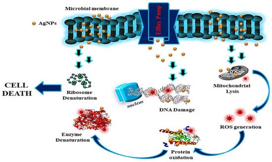
Ag NPs of varied sizes (1–100 nm) interact with bioactive components to limit microorganism proliferation. By generating superoxide radicals, Ag NPs inhibit the growth of bacterial enzymes and induce cytotoxicity (ROS). Silver has also been found to serve as a buffer between thiol-containing molecules, causing permanent agglomeration.
(Figure 3) [39,40] Many other substances, particularly DNA, peptides, and bio-molecules, have been identified as targets of the ions identified in the lethal bacterium. Antibiotics attempt to regulate particular elements of the bacterial life cycle, but silver ions can attach to any high-affinity component, implying that several bacterial cell mechanisms are disrupted, leading to cellular death. Further, silver ions are believed to bind non-specifically to a wide variety of sites and disrupt several aspects of cell metabolism simultaneously, eventually resulting in cell death
[7][33].
1
Figure 3. Mode of action of AgNPs Reprinted with permission from ref. [40]. Copyright 2018 Springer.
2.2.2. Gold Nanoparticles (Au NPs)
Gold nanoparticles have been used in a variety of applications, ranging from engineering to medical. Since gold nanoparticles are biocompatible, they have a greater potential for use in anticancer
[15][41] and antibacterial drugs. MacDonald et al.
[16][42] used a simple procedure followed by a thiol-functionalized AuNPs nanosystem to adjust the antibacterial surface structures to disrupt the bacterial cell in lighter and darker settings. The approach was examined and found to be significantly more effective against bacteria and other noxious microbiological entities. Gnanamoorthy et al. (2022) demonstrated the synthesis of 15-nanometer, average-sized AuNPs employing leaf extracts of Bauhinia tomentosa Linn. The synthesized AuNPs were assessed for their antimicrobial activity against
E. coli and
Staphylococcus aureus by the disk diffusion method. The disc diffusion method was used to test the antibacterial activity of the synthesized AuNPs against
E. coli and
Staphylococcus aureus. The results demonstrated that the plant-mediated AuNPs had greater antibacterial efficacy against both microorganisms
[17][43].
12.3. Copper Nanoparticles (Cu NPs)
Copper is a ubiquitous metal that is also a fundamentally important mineral in the majority of life forms. Copper nanoparticles are employed in a multitude of scenarios, notably electrochemical sensors, optoelectronic devices, solar panels, and paints and varnishes
[18][44]. Parikh et al.
[5][31] employed Datura leaf extract to synthesize copper nanoparticles. The copper NPs outperformed standard chloramphenicol in antibacterial activity versus
E. coli,
Bacillus megaterium, and
Bacillus subtilis, demonstrating that copper NPs can be used as antimicrobial agents instead of antibiotics. Chitosan was used as a stabilizer to inhibit the deposition and fast oxidation of copper nanoparticles with diameters ranging from 2–350 nm that were synthesized chemically. The antibacterial and antifungal activity of chitosan-copper NPs with different stoichiometric ratios (0.05 wt%, 0.1 wt%, 0.2 wt%, and 0.5 wt%, and chitosan) versus methicillin-resistant
Staphylococcus aureus, Bacillus subtilis,
Pseudomonas aeruginosa, and
Salmonella choleraesuis has been studied. The 0.5 wt% combination demonstrated the greatest zone inhibition
[19][45]. Kruk et al.
[20][46] utilized hydrazine in an aqueous SDS solution to form Cu nanomaterials accompanied by Cu salt reduction. The Cu NPs were tested for antibacterial activity versus Gram-positive bacteria, including both conventional and clinical strains, namely methicillin-resistant
Staphylococcus aureus, and antifungal activity towards
Candida species. The results demonstrated that the as-prepared Cu NPs were more efficient as antimicrobial compounds than Ag nanoparticles, as well as some antibiotics
[9][35].
12.4. Zinc Oxide Nanoparticles (ZnO NPs)
Transitional metals, such as zinc and iron, as well as their derivatives, are vital elements that play a pivotal role in the catalytic performances of a range of enzymes in the body and are widely dispersed across its tissues
[21][22][24,47]. Gram-positive bacteria include
Staphylococcus aureus, Staphylococcus epidermis, Bacillus subtilis, Bacillus cereus, Listeria monocytogenes, and
Escherichia faecium. Gram-negative bacteria include
Pseudomonas aeruginosa, Escherichia coli, Klebsiella pneumoniae, and
Salmonella sp.
[23][48]. Furthermore,
Acinetobacter baumannii is a multi-drug-resistant opportunistic bacteria that primarily induces respiratory and urinary tract infections. Carbapenems, which belong to the beta-lactam group of antibiotics, have been found to be the most efficient medicines against
A. baumannii so far; however, the progression of bacterial resistance to this antibiotic could result in extreme levels of fatality. To address this serious issue, scientists developed metal-based oxides, such as ZnO NPs, and observed them as prospective compounds to generate rapid reactive oxygen species, in order to increase membrane lipid peroxidation, resulting in the membrane leakage of reducing sugars, DNA, and proteins, as well as limiting cell survival. Researchers also proved that ZnO-NPs could be manufactured as novel anti-
A. baumannii drugs
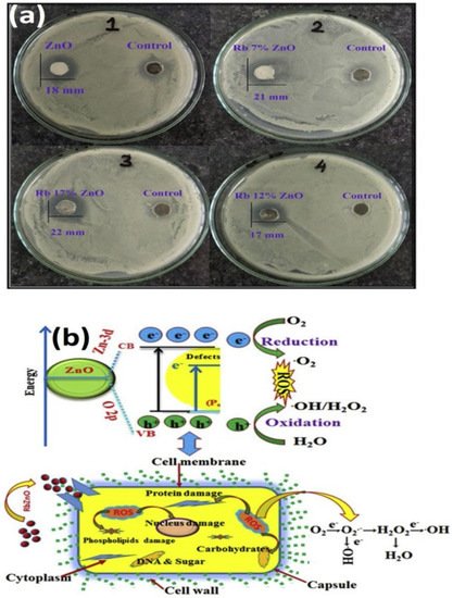
[24][49]. Since semiconducting nanomaterials have the electrical properties necessary to function as antibacterial agents, they are under investigation as both pure and doped semiconductors. Inwati et al.
[21][24] developed rare ion (Rb)-doped ZnO and utilized it as an antibacterial agent
(Figure 4).
Figure 4. (a) Growth inhibition zone of Bacillus subtilis of pure ZnO and ZnO: Rb samples; (b) Schematic representation of cell death mechanism of S. Bacillus under the influence of ZnO: Rb. Antibacterial activities of Rb-doped ZnO NPs Reprinted with permission from ref. [24]. Copyright 2020 Elsevier.
The antibacterial activity of synthesized nanostructures can be defined in terms of microbe cell destruction or plasma depletion after ion implantation. Furthermore, nanoparticle strikes on cellular components and cell dissociation in grown cells may affect bacterial routine cellular metabolism, leading to cytoplasmic discharge and bacterial cell death, with an unexpected rise in the inhibiting system. The more Rb were available, the more irregularities, such as oxygen vacancies, were incorporated into the ZnO frameworks. ZnO NPs with Rb deficiency generated pairs of electron-holes (e
−–h
+) during the VB-to-CB transition. A bandgap of 3.5 eV was detected in the ZnO NPs, yielding into holes and free electrons on the VB and CB, respectively. This hole-and-electron pair had a considerable impact on the antibacterial tests
[21][24].
12.5. Titanium Dioxide Nanoparticles (TiO2 NPs)
Titanium dioxide (TiO
2) NPs are among the most frequently researched nanomaterials for antimicrobial applications due to their unique properties, including bactericidal photocatalytic activity, security, and self-cleaning characteristics
[25][50]. They offer significant potential as bactericidal and fungicidal agents in food packaging and containers
[24][49]. When exposed to light, TiO
2 NPs generate reactive oxygen species (ROS) with high oxidizing potential, leading to a shift in band-gap in the open atmosphere (O
2)
[26][51]. Arora et al.
[27][52] investigated the role of TiO
2 NPs in controlling the growth rate of
Pseudomonas aeruginosa isolated by the endotracheal tract, pus, alveolar lavage, and sputum. They reported that exposing TiO
2 nanoparticles to UV light for 60 min dramatically improved their antibacterial activity against
P. aeruginosa with multiple drug resistance (MDR). Cefoxime’s potency was also demonstrated to be increased when coupled with UV-irradiated TiO
2 NPs for 60 min. Combinational studies on the activity of TiO
2 against pathogens emphasize its wider implications in clinical diagnostics as a strategy to combat the increasing problem of antibiotic resistance.
Semiconducting materials, such as TiO
2, have holes that split H
2O molecules into OH
− and H
+ free ions, with freed electrons interacting with soluble oxygen ions. Molecular oxygen’s anionic superoxide (O
2) radicals eventually caused the formation of hydrogen peroxide anions (HO
2−) and H
2O
2. The H
2O
2 produced penetrated the cell membrane and caused damage to it. Consequently, the rate of H
2O
2 formation increased as the number of electron-hole pairs increased
[28][53].
12.6. CuO Nanoparticles (CuO NPs)
CuO has recently been established as a robust p-type nanomaterial (1.2-electronvolt bandgap) for different applications due to its easy distribution and manufacture, better heat resistance, chemical inertness, and wider optical absorptivity. The morphological features of CuO (cupric oxide) or Cu
2O (cuprous oxide) nanomaterials can be modified by adjusting their size or shape, such as cylindrical, pyramidal, 1-D or 2-D, nanowires, and so on
[28][29][16,53]. Many professionals have modified and investigated the interfacial, electrical, physical, and chemical properties of pure CuO by applying alkali, transitional, and lanthanide ions. CuONPs are among the most frequently studied nanomaterials for antibacterial effects due to the fact that their appropriate ionic radii involve numerous ions such as Zn, Cd, Ag, Ce, Ni, and Co dopants
[30][31][54,55]. When a metal ion is introduced into a CuO substrate, the structure and morphology of pure CuO crystalline solids changes. It has been observed that the ionic radii of the dopant play an essential part in the atomic interaction between both the dopant and the host environment. Ultimately, during the dispersion of the NPs into the deionized water, the electric charge chemically reacts with the active hole (h
+) and free electrons (e
−). BY contrast, superoxide ions are formed when free electrons contact with soluble oxygen atoms
[6][32]. Furthermore, the hole at the top of the valence band reacts with aqueous ions to produce OH entities, which subsequently interact with superoxide ions to form hydrogen peroxide through H
+ ions. As a response, the hydrogen superoxide disrupts the usual metabolism of the bacterial cell nucleus, and causes cell death.
Gnanamoorthy et al. (2021) synthesized copper aminophosphate (X-CuAP) nanoparticles with improved electrochemical, photocatalytic, and biological characteristics. The researchers observed that the (en)-CuAP nanocrystals had greater reusability and enhanced photocatalytic and antibacterial properties
[32][56].
In addition, Gnanamoorthy et al. (2020) described the hydrothermal synthesis of (Cr)-CuSnO3 nanoparticles and their application for photocatalytic activity and antibacterial characteristics. The synthesized NPs were evaluated and their specific characteristics were examined using modern instruments
[33][57].
Gnanamoorthy et al. (2021) synthesized morphologically diverse varieties of SnO
2 rods and examined them for antifungal and antibacterial activity. The SnO
2 rods were found to have antimicrobial effects on
Enterococcus fecalis and antifungal activity against
Candida albicans [34][2].
23. Carbon Nanotubes as Antimicrobial Agents
Carbon nanotubes (CNTs) are a class of antibacterial agents
[35][84]. The literature is rich with accounts of CNTs being employed as drug carriers. CNTs are desirable agents in the biomedical industry due to their high mechanical strength, substantial photoluminescent property, and high surface-area-to-volume ratio. CNTs have been extensively used as effective and harmless drug delivery agents for both metallic and organic antimicrobial agents. Furthermore, there are multiple examples of CNTs being employed alone or surface-functionalized with various antimicrobial agents. Furthermore, in various cases, CNTs have been coupled with a polymeric substance, which may have antibacterial properties. Such CNT-based composites were shown to retain the toxic properties of CNTs to microbes whilst promoting cell–CNT interaction(
Table 13).
Table 13. Organic, inorganic, and carbon-containing nanomaterials for targeted medicinal uses
[35][84].
CNTs have a wide variety of antimicrobial activities. In comparison with commercial antibiotics, functionalized multi-walled carbon nanotubes (F-MWNTs) were produced as antimicrobial nanomaterials. The generated F-MWNTs were tested for antimicrobial properties against Gram-positive
S.aureus and Gram-negative
E.coli. For
E.coli and
S.aureus, the optimal concentrations for maximal inhibition and antibacterial function were observed to be 80 and 60 g/mL, respectively. F-MWNTs were reported to be 85% more effective against E.coli and 57% more effective against
S.aureus compared to conventional antibiotics
[36][85]. Some researchers have documented CNT antibacterial activity. For example, Maas et al. (2016) demonstrated that when anchored to the surface, CNTs may block the adhesion of microorganisms, particularly bacteria, and prevent the development of bacterial biofilm
[40][86]. CNTs’ antibacterial activity has also been reported by Malek et al. (2016) and Yick et al. (2015). Kang et al. (2007) revealed that single-walled carbon nanotubes were employed directly for antimicrobial purposes against
E. coli, which injured the bacterial cell membrane. Furthermore, multiple studies have shown that multiwall CNTs, not just SWCNTs, are powerful antimicrobial agents. MWCNTs with small diameters promote partitioning and partial penetration into the cell wall. Longer CNTs offer better antimicrobial activities due to their exceptional agglomeration with the bacterial cell
[35][41][42][84,87,88]. CNTs have antimicrobial activity for the purification of water and pathogen control, according to Liu et al. (2018). CNTs, in general, cause mechanical stress to microbial or bacterial cells, resulting in further cell disruption and, eventually, the release of intracellular substances. Furthermore, these CNTs cause oxidative stress in microorganisms
[43][89]. The main disadvantage of using CNTs as antimicrobial agents is that they cannot compete with traditional techniques targeting biofilm owing to the lesser-known toxicity profile of human beings
[44][90]. CNT anti-microbial properties are affected by a variety of parameters, including the CNT diameter and length, the residual catalyst, the electronic structure, the surface functional groups, the surface chemistry, the microbe type and shape, as well as the microbe growth state
[45][46][91,92]. Fullerenes are made up of carbon units organized in spherical clusters. They exhibit antimicrobial properties against a number of pathogens, including
Salmonella, Streptococcus spp., and
E. coli. It is believed that when the nanostructures are ingested by the microorganisms, the decrease in energy metabolism improves their anti-bacterial activity. Fullerene compounds are suspected to inhibit bacterial growth by interfering with respiratory systems.
Notably, new pharmaceutical items must pass through all the phases of clinical trials, and they must be approved by the relevant authorizing agencies prior to the final phase. The progression from phase I to phase V, as well as its approval, applies to all clinical products, whether conventional or nanomaterial. Since nanomaterials are still under development, the majority of nanocosmeceuticals, nanopharmaceuticals, and other clinically important nanoformulations are still under clinical study. Numerous instances of nanoformulations with antimicrobial abilities under clinical study are listed below. Bruinenberg et al. (2010)
[47][93] stated that ciprofloxacin-loaded liposomes might be employed to treat Pseudomonas aeruginosa respiratory functions. The current medication is in clinical phase III and further developments are required before it can be approved. Ciprofloxacin-loaded liposomes are also in phase IIa and III trials for respiratory infections and cystic fibrosis, respectively. Furthermore, Amikacin is being tested in stage II and III clinical trials
[48][94]. Other antimicrobial peptides are yet to be commercialized and are currently undergoing clinical trials. Antimicrobial peptides, such as mutacin 1140 (MU1140), lipohexapeptides 1345 (HB1345), avidocin and purocin, arenicin (AP139), arenicin (AP138), arenicin (AP114), and no-varifyn are in the preclinical phase of development against various Gram-positive, Gram-negative, and antibiotic-resistant bacteria
[49][50][51][95,96,97]. Despite the fact that there is a long list of clinical studies, the main issue is their high cost and time frame. After all the clinical trials and approvals are concluded, it is hoped that nanoformulations or nanotherapeutics could soon gain in market share.