Chitin and Chitosan: Comparison
Please note this is a comparison between Version 2 by Rita Xu and Version 1 by Azizur Rahman.

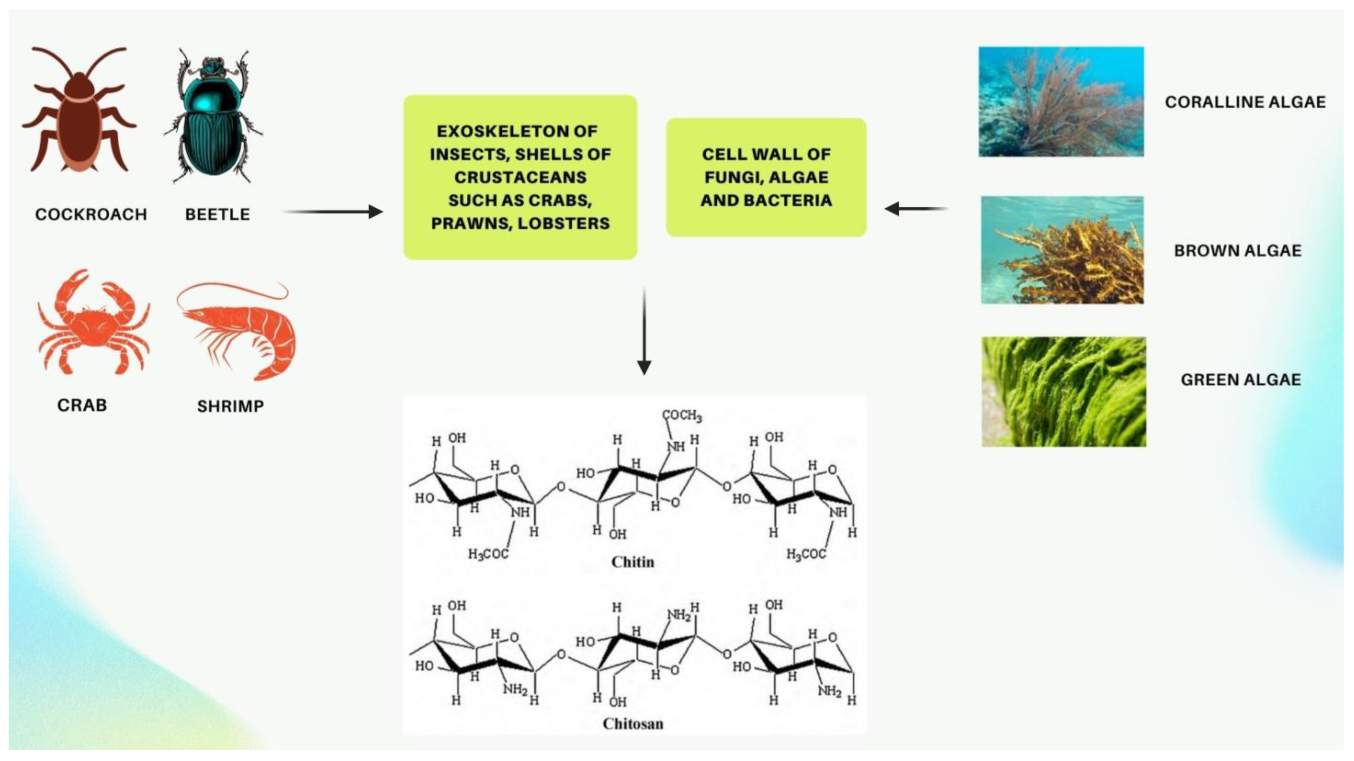
Chitin and its derivative chitosan are highly abundant polymers in nature, appearing in both the shells and exoskeletons of various marine and non-marine species. Since they possess favorable properties, such as biocompatibility, biodegradability, non-toxicity, and non-immunogenicity, they have gained recent attention due to their enormous potential biomedical applications. The polycationic surface of chitosan enables it to form hydrogenic and ionic bonds with drug molecules, which is one of its most useful properties. Because chitosan is biocompatible, it can therefore be used in drug delivery systems. The development of chitosan-based nanoparticles has also contributed to the significance of chitin as a drug delivery system that can deliver drugs topically. Furthermore, chitin can be used in cancer treatment as a vehicle for delivering cancer drugs to a specific site and has an antiproliferative effect by reducing the viability of cells. Finally, chitosan can be used as a wound dressing in order to promote the faster regeneration of skin epithelial cells and collagen production by fibroblasts.

  • chitin
  • chitosan
  • wound healing
  • drug delivery

1. Introduction

Chitin is a polysaccharide composed of crystallized N-Acetyl D-glucosamine monomers and 1–4 glycosidic bonds [1][2]. This structural polymer is highly abundant in the shells of marine crustaceans, cell walls of various organisms such as fungi, coralline algae, green and brown algae, and bacteria, as well as the exoskeleton of crustaceans, molluscs, and insects (Figure 1) [3][4][5][6][7][8][9]. In 2014, the first evidence of chitin inside the cell walls of the coralline algae Clathromorphum compactum was observed, and chitin was found to play an important role in the calcification process of this marine species [4]. Initially named fungine in 1811, it was subsequently renamed chitin in 1823 [10]. It occurs in about 19 animal phyla, bacteria, algae, and fungi, with a production rate of 1011–1014 tons per year [10]. Although chitin is present in nature, multiple tons of it transform into marine shell wastes or other types of industrial wastes [11]. Chitin has recently gained much attention from scientists due to its important properties, which include nontoxicity, biocompatibility, and biodegradability, the ability to degrade by enzymes, enabling it to be used in many biomedical applications, including drug delivery, tissues engineering, wound healing, and cancer therapy. [11]. In spite of being the second most abundant polymer after cellulose, chitin is still largely unutilized because its semi-crystalline structure with a large number of intermolecular hydrogen bonds causes it to be insoluble in most solvents [6][12]. With the advent of new technologies, however, the partial N-deacetylation of chitin can now be converted into a soluble substance called chitosan. The deacetylation of extracted chitin is largely carried out in strong alkaline media, resulting in chitosan becoming soluble in acidic environments [13]. Hence, chitosan is a synthetically deacetylated form of chitin that can be used in the food industry as an edible and biodegradable film for packaging food, food preservations, pharmaceuticals, water and wastewater treatment, cosmetics, wound dressings, and many other applications [1][6][11][14][15].
Figure 1. Sources of chitin and the structure of chitin and chitosan. [Created with BioRender.com accessed on 10 May 2022].
As mentioned earlier, the shells of crustaceans and shrimps, insect exoskeletons, and the cell walls of fungi such as aspergillus and mucor are the main sources of chitin and chitosan [16]. A large scale of industrial chitin production is derived from crab shells and shrimp shells, so recycling these wastes will yield valuable byproducts that can be used in medical and pharmaceutical applications [16]. To prepare chitin, the first step is to extract it from shellfish wastes. Shellfish wastes contain chitin, lipids, inorganic salts, and proteins as major components. Hence, chitin is extracted from these structural components using two common methods: chemical and biological processes (Figure 2) [17][18][19]. The purpose of these processes is to isolate chitin from minerals such as calcium carbonate and calcium phosphate and proteins and lipids or other macromolecules embedded in shells and cell walls [17]. In strong alkaline (>1 M NaOH) and acetic (>3 M HCl) environments combined with heat, chitin can be transformed into its deacetylated form, chitosan, or even partially fragmented [20]. Therefore, sulfuric acid, nitric acid, formic acid, acetic acid, and in particular, dilute hydrochloric acid are commonly used in the demineralization process of chitin [17][18][21]. The separation of proteins and pigments such as carotenoids is performed using dilute sodium hydroxide and acetone (or other organic solvents), respectively [17][18]. As an alternative method, biological processes may provide more desirable results due to their relatively low cost and environmental friendliness, low energy consumption, and reproducibility. They can also produce chitin with a higher molecular weight and better crystal structure [17]. Biological processes rely largely on enzymatic reactions and microbial fermentation [17][22]. However, the basis of these reactions, such as with the chemical process, is the production of acidic and alkaline products but from microorganism fermentation and enzymatic metabolites [17].
Figure 2. Chemical and biological processes to isolate chitin from animal sources. [Created with BioRender.com accessed on 10 May 2022].
Though chemical processes make chitin extraction easier and more efficient, they have some drawbacks when used on an industrial scale with industrial protocols. There are two main concerns about conventional methods of chitin extraction: the use of strong chemicals such as HCl, and the lower quality of raw extracted chitin due to contamination [15]. Therefore, obtaining high-quality chitin involves sophisticated instrumentation such as the use of ultrafiltration and molecular sieving. However, these techniques are costly, which creates significant financial barriers, especially for developing countries. Recent efforts are underway to develop novel methods for chitin extraction. As an example, Tissera et al. introduced a new process called “pretreatment” for the extraction of chitin from blue swimmer crab shell waste, proposing that this procedure helped improve the quality of extracted chitin for large-scale production [15]. They soaked crushed crab shell pieces in acetic acid (with concentrations of 0.05, 0.10, and 0.50 M) and citric acid with concentrations of 0.05, 0.10, and 0.50 M and found that acetic acid at 0.5 M resulted in the better cleaning of crab shells. They found that this pre-acid treatment helped the rigid structure of shells to become more flexible as well as slightly beginning the process of demineralization [15]. The results showed that this treatment with 0.5 M acetic acid helped remove any remaining muscle tissue that remained, which led to a lower concentration of NaOH being required for the deproteinization of crab shells. Furthermore, this pretreatment led to a lower concentration of acid required for the demineralization step since acetic acid aids in calcium carbonate removal [15].
Traditional chitin extraction methods have been thought of as time-consuming and expensive, with production times greater than one day. Therefore, other methods aimed at speeding up the process of chitin extraction have been studied in recent years. Kaya et al. proposed to treat the shells of crustaceans such as crabs, crayfish, and shrimp with two NaClO treatments (10 min each) before the main processes of demineralization and deproteinization to ensure the complete disintegration and removal of pigments [23]. They found that by introducing NaClO, they were able to extract chitin in a shorter amount of time while the yield of chitin was similar to conventional extraction, and the color of extracted chitin was white, as in commercially available chitin [23]. The elemental analysis, FT-IR, and XRD of extracted chitin suggested that it has a high similarity to commercial chitin; consequently, they suggested that this new treatment of chitin should be considered for extracting chitin from crab shells, crayfish shells, and shrimp shells [23].
In another innovative study, Machałowski et al. isolated chitin from Caribena versicolor spider molt cuticles [24]. Arachnoids have a mineral-free cuticle, so demineralization (such as decalcification) can be skipped, and chitin isolation can begin with deproteinization and depigmentation [24]. Their study included removing pigments as well as chitin from spider cuticles using microwave-assisted methods (MWI) [24]. In the first step, which involved removing lipids and waxes from the cuticle, the cuticles were exposed to microwave radiation while being treated with chloroform and ethanol in a ratio of 2 to 1 [24]. In the second step, they used microwave radiation alongside NaOH treatment to remove other proteins and pigments from the cuticle [24]. The final step was to treat the cuticles with H2O2 under microwave radiation in order to remove any residual pigments and obtain tubular chitin [24]. The bodies of C. versicolor spiders are covered with a strong exoskeleton (cuticle) consisting of an inner layer with chitin and proteins and an outer layer (without chitin) [24]. Due to this, these spiders lose a large number of chitin-rich cuticles during their molting cycle; therefore, tubular chitin from their cuticles could be used as a scaffold-based catalyst and tissue engineering material [24].

2. Chitosan as a Possible Drug Delivery Agent

The term “drug delivery systems” (DDS) refers to products that combine an active pharmaceutical agent with a suitable carrier—typically a polymer—and have been used in the pharmaceutical industry to improve the therapeutic potential and bioavailability of drugs [1][25]. By using hydrogels, scaffolds, micro- and nano-particles, and organic or inorganic matrices as carriers of active drug agents, this system delivers drugs in a sustained and controlled manner [1][10]. Among these, hydrogels, due to their chemical stability, are able to protect and maintain the encapsulated drug in extreme environmental conditions, and by dissolving in body fluids, lead to the release of the drug at the desired site and with the required concentration [1]. Controlled delivery is a basic property of the ideal DDS, which is used to maintain the plasma concentration of the active drug at a constant rate and within the therapeutic window [25]. Another important feature of DDS is targeted drug delivery or directing the active substance to a specific site [25]. In DDS, different types of vehicles such as tablets, capsules, and hydrogels are commonly used, depending on the target site and the route of administration [1]. The delivery of drugs can be performed either through invasive or non-invasive routes, such as parenteral and mucosal administration, respectively [25]. Mucosal drug delivery includes oral, buccal, nasal, pulmonary, and rectal routes, and most DDS use this route of administration to enhance mucoadhesiveness and, consequently, increase drug release [25].
A number of studies have been conducted on chitin and chitosan to determine their potential as drug delivery agents. Chitosan, for example, is significantly utilized in the production of hydrogels for drug delivery due to its valuable properties, including bioadhesion, having a polycationic surface that facilitates the creation of hydrogenic and ionic bonds, and biocompatibility, meaning that in contact with living tissue or body fluids, it does not produce any toxins or elicit an immune response [1][2].
Marine sponges provide a good source of naturally occurring chitin that can be used for drug delivery by extracting chitin from their skeletons [26]. Kovalchuk et al. examined the potential application of fabric-like chitin scaffolds from the demosponge Ianthella flabelliformis for the first time [26]. The researchers found that these chitin scaffolds possess three-dimensional (3D) matrices that retain antiseptic decamethoxine absorbed from a 0.1% ethanol solution [26]. Moreover, they demonstrated that decamethoxine diffused from these mesh-like scaffolds into an agar diffusion assay and inhibited the growth of Staphylococcus aureus, confirming the potential use of pure chitinous scaffolds in drug delivery [26]. In another study, Zheng et al. prepared 10–100 μm carboxymethyl chitin microspheres (CMCH-Ms) and then encapsulated them in thermosensitive hydroxypropyl chitin hydrogels (HPCH) [27]. To maintain the spherical structure of microspheres with porous microstructures, they used physical cross-linking procedures and increased the temperature in an aqueous two-phase system without using cross-linking agents. Using the physical cross-linking method, carboxymethyl chitins are able to form microspheres at high temperatures because of their high acetylation levels, which make them temperature sensitive [27]. Similarly, hydroxypropyl chitin (HPCH) is also temperature sensitive, and it is capable of reversibly converting into solid hydrogels at elevated temperatures, enabling the in situ formation of hydrogels [27]. Two drugs, naproxen (NPX) and ropivacaine (Ropivacaine), were analyzed in vitro for their loading and release capacities into CMCH-Ms and HPCH gel scaffolds [27]. They found that CMCH-Ms/HPCH gel scaffolds sustained in vitro drug release over a longer period and caused fewer bursts of drug release. Therefore, they determined that drug-loaded CMCH-Ms/HPCH gel scaffolds could be used as a means of delivering local drugs [27].
Another technology that has attracted attention in recent years in the drug delivery industry is the use of nanomaterials, which include objects with a size of 1–100 nm [2][28]. Due to the size of these particles, they have a special feature as they follow the rules of quantum mechanics [28]. Nanotechnology can be used for accurate and precise imaging and drug delivery [2][29]. One method of drug delivery is the use of chitosan-based nanoparticles, which are used greatly in the mucosal route to transport the drug to the brain and eyes, as well as to treat diseases such as cancer and gastrointestinal and pulmonary diseases [29][30]. The solubility, diffusion, and size of chitosan-based nanoparticles determine the rate of drug release [31]. Most chitosan-based nanoparticles have a size between 100 and 400 nanometers. Photon correlation spectroscopy can be used to determine the size of the nanoparticles by measuring their Brownian motion [31]. Chitosan nanoparticles are stabilized based on several physical and chemical factors, including agglomeration, coagulation, temperature, the molecular weight of the polymer, and the pH of the medium [31].
In many eye diseases, drugs are used superficially on the ocular surface. However, due to defense mechanisms, less than 5% of drugs used topically are able to cross the cornea and reach intraocular tissue [32][33]. As a result, chitosan-based nanoparticles could be considered a possible solution to increase bioavailability and prolong drug bioavailability in the ocular tissue [32][33][34]. In a study by Silva et al., using nanotechnology, eye drops containing polymer-based nanoparticles consisting of the antibiotic ceftazidime were used to treat ocular infection with pseudomonas aeruginosa, and the degree of mucoadhesion or the interaction between nanoparticles delivered through the eye and ocular mucosal tissue was later analyzed [32]. The results of their study demonstrated that the use of polymeric-based nanoparticles facilitated the transport of antibiotics through the mucosal barrier of the eye as it enhanced the bioavailability of the drug and protected against degradation [32]. In addition, they were able to show that chitosan, due to its structural properties, increases the contact time between antibiotics and ocular mucosal tissue, so that a strong electrostatic interaction between the amine group of chitosan and salicylic acid, which is a main component of mucin, causes the drug to stay inside the eye longer. Other properties of chitosan have demonstrated its potential for drug delivery systems, including hydrophilicity, biodegradability, and antimicrobial activity [32].
In an innovative study, Cánepa et al. investigated the efficacy of interferon-alpha (IFNα) delivery using chitosan-based nanoparticles through oral administration [35]. It has been known that the use of drugs via the oral route could reduce the absorption of drugs into the gastrointestinal tract due to the degradation by brush border enzymes and hepatic metabolism [35][36][37]. As a result, many pharmaceutical companies are trying to increase the bioavailability of prescribed orally-administered drugs [37]. IFNα is used as a drug to treat cancer and viral infections; however, due to its short half-life, it usually needs to be taken regularly by patients, and this regime could have its own side effects and drawbacks [38][39][40][41][42]. Cánepa et al. showed that the antiviral effect of encapsulated IFNα by chitosan-based nanoparticles was comparable to commercial IFNα on the market [35]. Furthermore, the bioavailability of the drug improved within the gastrointestinal tract due to the electrostatic interaction between the chemical groups in chitosan and salicylic acid in mucin, which led to the increased size of nanoparticles following the adsorption of mucin, as well as decreased surface charge of nanoparticles [35].

References

  1. Parhi, R. Chitin and Chitosan in Drug Delivery. In Sustainable Agriculture Reviews 36: Chitin and Chitosan: Applications in Food, Agriculture, Pharmacy, Medicine and Wastewater Treatment; Crini, G., Lichtfouse, E., Eds.; Sustainable Agriculture Reviews; Springer International Publishing: Cham, Switzerland, 2019; pp. 175–239. ISBN 978-3-030-16581-9.
  2. Ahmad, A.; Mubarak, N.M.; Naseem, K.; Tabassum, H.; Rizwan, M.; Najda, A.; Kashif, M.; Bin-Jumah, M.; Hussain, A.; Shaheen, A.; et al. Recent Advancement and Development of Chitin and Chitosan-Based Nanocomposite for Drug Delivery: Critical Approach to Clinical Research. Arab. J. Chem. 2020, 13, 8935–8964.
  3. Dave, U.; Somanader, E.; Baharlouei, P.; Pham, L.; Rahman, M.A. Applications of Chitin in Medical, Environmental, and Agricultural Industries. J. Mar. Sci. Eng. 2021, 9, 1173.
  4. Rahman, M.A.; Halfar, J. First Evidence of Chitin in Calcified Coralline Algae: New Insights into the Calcification Process of Clathromorphum Compactum. Sci. Rep. 2015, 4, 6162.
  5. Rahman, M.A.; Halfar, J.; Adey, W.H.; Nash, M.; Paulo, C.; Dittrich, M. The Role of Chitin-Rich Skeletal Organic Matrix on the Crystallization of Calcium Carbonate in the Crustose Coralline Alga Leptophytum Foecundum. Sci. Rep. 2019, 9, 11869.
  6. Ahmad, S.I.; Ahmad, R.; Shoeb Khan, M.; Kant, R.; Shahid, S.; Gautam, L.; Hasan, G.M.; Hassan, M.I. Chitin and Its Derivatives: Structural Properties and Biomedical Applications. Int. J. Biol. Macromol. 2020, 164, 526–539.
  7. Singh, S.K. Solubility of Lignin and Chitin in Ionic Liquids and Their Biomedical Applications. Int. J. Biol. Macromol. 2019, 132, 265–277.
  8. Silva, S.S.; Mano, J.F.; Reis, R.L. Ionic Liquids in the Processing and Chemical Modification of Chitin and Chitosan for Biomedical Applications. Green Chem. 2017, 19, 1208–1220.
  9. Elieh-Ali-Komi, D.; Hamblin, M.R. Chitin and Chitosan: Production and Application of Versatile Biomedical Nanomaterials. Int. J. Adv. Res. 2016, 4, 411–427.
  10. Broek, L.A.; Boeriu, C.G.; Stevens, C.V. Chitin and Chitosan: Properties and Applications; Wiley series in renewable resources; Wiley: Hoboken, NJ, USA, 2020; ISBN 978-1-119-45046-7.
  11. Sivanesan, I.; Gopal, J.; Muthu, M.; Shin, J.; Oh, J.-W. Reviewing Chitin/Chitosan Nanofibers and Associated Nanocomposites and Their Attained Medical Milestones. Polymers 2021, 13, 2330.
  12. Joseph, S.M.; Krishnamoorthy, S.; Paranthaman, R.; Moses, J.A.; Anandharamakrishnan, C. A Review on Source-Specific Chemistry, Functionality, and Applications of Chitin and Chitosan. Carbohydr. Polym. Technol. Appl. 2021, 2, 100036.
  13. Rasti, H.; Parivar, K.; Baharara, J.; Iranshahi, M.; Namvar, F. Chitin from the Mollusc Chiton: Extraction, Characterization and Chitosan Preparation. Iran. J. Pharm. Res. IJPR 2017, 16, 366–379.
  14. Anraku, M.; Ifuku, S.; Iohara, D.; Hirayama, F.; Otagiri, M.; Gebicki, J.M. Future Aspects of Biomedical Applications of Chitin and Chitosan in Diseases Associated with Oxidative Stress. In Handbook of Chitin and Chitosan; Elsevier: Amsterdam, The Netherlands, 2020; pp. 589–608. ISBN 978-0-12-817966-6.
  15. Tissera, W.M.J.C.; Rathnayake, S.I.; Abeyrathne, E.D.N.S.; Nam, K.-C. An Improved Extraction and Purification Method for Obtaining High-Quality Chitin and Chitosan from Blue Swimmer (Portunus Pelagicus) Crab Shell Waste. Food Sci. Biotechnol. 2021, 30, 1645–1655.
  16. Islam, S.; Bhuiyan, M.A.R.; Islam, M.N. Chitin and Chitosan: Structure, Properties and Applications in Biomedical Engineering. J. Polym. Environ. 2017, 25, 854–866.
  17. Özel, N.; Elibol, M. A Review on the Potential Uses of Deep Eutectic Solvents in Chitin and Chitosan Related Processes. Carbohydr. Polym. 2021, 262, 117942.
  18. Younes, I.; Rinaudo, M. Chitin and Chitosan Preparation from Marine Sources. Structure, Properties and Applications. Mar. Drugs 2015, 13, 1133–1174.
  19. Pachapur, V.L.; Guemiza, K.; Rouissi, T.; Sarma, S.J.; Brar, S.K. Novel Biological and Chemical Methods of Chitin Extraction from Crustacean Waste Using Saline Water: Novel Biological and Chemical Methods of Chitin Extraction. J. Chem. Technol. Biotechnol. 2016, 91, 2331–2339.
  20. Tsurkan, M.V.; Voronkina, A.; Khrunyk, Y.; Wysokowski, M.; Petrenko, I.; Ehrlich, H. Progress in Chitin Analytics. Carbohydr. Polym. 2021, 252, 117204.
  21. Kumari, S.; Rath, P.; Sri Hari Kumar, A.; Tiwari, T.N. Extraction and Characterization of Chitin and Chitosan from Fishery Waste by Chemical Method. Environ. Technol. Innov. 2015, 3, 77–85.
  22. Arbia, W.; Arbia, L.; Adour, L.; Amrane, A. Chitin Extraction from Crustacean Shells Using Biological Methods—A Review. Food Technol. Biotechnol. 2012, 51, 12.
  23. Kaya, M.; Baran, T.; Karaarslan, M. A New Method for Fast Chitin Extraction from Shells of Crab, Crayfish and Shrimp. Nat. Prod. Res. 2015, 29, 1477–1480.
  24. Machałowski, T.; Wysokowski, M.; Tsurkan, M.V.; Galli, R.; Schimpf, C.; Rafaja, D.; Brendler, E.; Viehweger, C.; Żółtowska-Aksamitowska, S.; Petrenko, I.; et al. Spider Chitin: An Ultrafast Microwave-Assisted Method for Chitin Isolation from Caribena Versicolor Spider Molt Cuticle. Molecules 2019, 24, 3736.
  25. Parhi, R. Drug Delivery Applications of Chitin and Chitosan: A Review. Environ. Chem. Lett. 2020, 18, 577–594.
  26. Kovalchuk, V.; Voronkina, A.; Binnewerg, B.; Schubert, M.; Muzychka, L.; Wysokowski, M.; Tsurkan, M.V.; Bechmann, N.; Petrenko, I.; Fursov, A.; et al. Naturally Drug-Loaded Chitin: Isolation and Applications. Mar. Drugs 2019, 17, 574.
  27. Zheng, J.; Lv, S.; Zhong, Y.; Jiang, X. Injectable Hydroxypropyl Chitin Hydrogels Embedded with Carboxymethyl Chitin Microspheres Prepared via a Solvent-Free Process for Drug Delivery. J. Biomater. Sci. Polym. Ed. 2021, 32, 1564–1583.
  28. Sim, S.; Wong, N.K. Nanotechnology and Its Use in Imaging and Drug Delivery (Review). Biomed. Rep. 2021, 14, 1–9.
  29. Mohammed, M.A.; Syeda, J.T.M.; Wasan, K.M.; Wasan, E.K. An Overview of Chitosan Nanoparticles and Its Application in Non-Parenteral Drug Delivery. Pharmaceutics 2017, 9, 53.
  30. Garg, U.; Chauhan, S.; Nagaich, U.; Jain, N. Current Advances in Chitosan Nanoparticles Based Drug Delivery and Targeting. Adv. Pharm. Bull. 2019, 9, 195–204.
  31. Naskar, S.; Sharma, S.; Kuotsu, K. Chitosan-Based Nanoparticles: An Overview of Biomedical Applications and Its Preparation. J. Drug Deliv. Sci. Technol. 2019, 49, 66–81.
  32. Silva, M.M.; Calado, R.; Marto, J.; Bettencourt, A.; Almeida, A.J.; Gonçalves, L.M.D. Chitosan Nanoparticles as a Mucoadhesive Drug Delivery System for Ocular Administration. Mar. Drugs 2017, 15, 370.
  33. Shinde, U.A.; Joshi, P.N.; Jain, D.D.; Singh, K. Preparation and Evaluation of N-Trimethyl Chitosan Nanoparticles of Flurbiprofen for Ocular Delivery. Curr. Eye Res. 2019, 44, 575–582.
  34. Zhao, R.; Li, J.; Wang, J.; Yin, Z.; Zhu, Y.; Liu, W. Development of Timolol-Loaded Galactosylated Chitosan Nanoparticles and Evaluation of Their Potential for Ocular Drug Delivery. AAPS PharmSciTech 2017, 18, 997–1008.
  35. Cánepa, C.; Imperiale, J.C.; Berini, C.A.; Lewicki, M.; Sosnik, A.; Biglione, M.M. Development of a Drug Delivery System Based on Chitosan Nanoparticles for Oral Administration of Interferon-α. Biomacromolecules 2017, 18, 3302–3309.
  36. Peterson, B.; Weyers, M.; Steenekamp, J.H.; Steyn, J.D.; Gouws, C.; Hamman, J.H. Drug Bioavailability Enhancing Agents of Natural Origin (Bioenhancers) That Modulate Drug Membrane Permeation and Pre-Systemic Metabolism. Pharmaceutics 2019, 11, 33.
  37. Maderuelo, C.; Lanao, J.M.; Zarzuelo, A. Enteric Coating of Oral Solid Dosage Forms as a Tool to Improve Drug Bioavailability. Eur. J. Pharm. Sci. 2019, 138, 105019.
  38. Hasselbalch, H.C.; Holmström, M.O. Perspectives on Interferon-Alpha in the Treatment of Polycythemia Vera and Related Myeloproliferative Neoplasms: Minimal Residual Disease and Cure? Semin. Immunopathol. 2019, 41, 5–19.
  39. Lombardi, A.; Tsomos, E.; Hammerstad, S.S.; Tomer, Y. Interferon Alpha: The Key Trigger of Type 1 Diabetes. J. Autoimmun. 2018, 94, 7–15.
  40. Talpaz, M.; Mercer, J.; Hehlmann, R. The Interferon-Alpha Revival in CML. In Chronic Myeloid Leukemia; Hehlmann, R., Ed.; Hematologic Malignancies; Springer International Publishing: Cham, Switzerland, 2021; pp. 179–226. ISBN 978-3-030-71913-5.
  41. Pires de Mello, C.P.; Tao, X.; Kim, T.H.; Bulitta, J.B.; Rodriquez, J.L.; Pomeroy, J.J.; Brown, A.N. Zika Virus Replication Is Substantially Inhibited by Novel Favipiravir and Interferon Alpha Combination Regimens. Antimicrob. Agents Chemother. 2018, 62, e01983-17.
  42. Fleischmann, W.R.; Koren, S.; Fleischmann, C.M. Orally Administered Interferons Exert Their White Blood Cell Suppressive Effects via a Novel Mechanism. Proc. Soc. Exp. Biol. Med. 1992, 201, 200–207.
More