1. The Neurexin–Neuroligin Mediated Trans-Synaptic Modulation
1.1. The Dynamic Synaptic Regulation by Neurexins
Early evidence suggested that these Nrxs and their binding partners can modulate synapse numbers and distributions, thereby contributing to the recruitment of pre-and postsynaptic machinery
[1][2][8,20]. These findings, together with data from recent studies, strongly suggest that Nrxs are key regulators of the overall functionality of synapses, shaping synaptic processes such as transmission and plasticity
[1][3][8,21].
Several mechanisms govern the roles of Nrxs in organizing diverse synaptic properties. Primarily, Nrxs influence components of the presynaptic machinery and synaptic functional efficiency. For example, Nrx deletion results in the loss of presynaptic active-zone GABARs, decreasing the sensitivity of neurotransmitter release to GABAR activation at both excitatory and inhibitory synapses
[4][22]. Given the importance of presynaptic GABARs in the nucleation of signaling complexes that control the release of neurotransmitters, the ability of Nrxs to regulate these receptors enables them to further govern synaptic transmission and plasticity from the presynaptic perspective
[5][6][7][23,24,25]. Additionally, conditional knockout mice deficient for all three β-Nrxs exhibit impaired presynaptic release probability. However, this phenotype is not due to a direct presynaptic effect but is attributed to a trans-synaptic regulatory loop in which presynaptic β-Nrxs regulate postsynaptic tonic endocannabinoid signaling
[8][26].
The critical roles played by Nrxs in the synapse are not limited to presynaptic modulation. Nrxs can also orchestrate postsynaptic properties, thereby shaping the input/output relations of their resident trans-synaptic circuits. For instance, the expression levels of the N-methyl-D-aspartate receptor (NMDAR) and the α-amino-3-hydroxy-5-methyl-4-isoxazolepropionic acid receptor (AMPAR) are affected by the constitutive inclusion of SS4 in hippocampal presynaptic Nrx-1 and Nrx-3, respectively
[9][27]. Accordingly, conditional control of insertions at Nrx-3 SS4 suppresses the responsivity of glutamate receptor responses mediated by AMPARs, whereas the same manipulation in Nrx-1 enhances NMDAR-mediated glutamate receptor postsynaptic strength. Collectively, these findings suggest an economical molecular mechanism whereby Nrxs and their alternative splicing can contribute to the regulation of AMPAR- and NMDAR-mediated excitatory postsynaptic strength and plasticity
[10][28].
Likewise, GABARs require inhibitory presynaptic terminals for their postsynaptic localization. Specifically, ectopic expression of Nrx-3 in presynaptic neurons could recruit GABARs to postsynaptic sites, thus establishing a trans-synaptic interaction
[11][29]. Consistent with this finding, the Nrx-3 knockout shows altered inhibitory postsynaptic strength, with a pronounced impact on inhibitory postsynaptic current (IPSC) amplitudes in males. In contrast, the same change results in enhanced IPSC amplitudes in females
[12][30]. Moreover, circumstantial evidence indicates that GABAergic synapse specification is influenced by the expression of the highly selective Nrxs SS4 splicing factor sam68-like molecule 2
[13][31]. These data link the Nrx expression levels and their alternative splicing modulations with the functional regulation of excitatory and inhibitory postsynaptic transmission and plasticity.
Another important mechanism that deserves special attention in relation to Nrxs in synaptic efficiency is the process of proteolytic cleavage. Physiologically, Nrx-1 is cleaved by a disintegrin and metalloproteinase-10 (ADAM-10), resulting in 4–6% of Nrx-1 in the adult brain existing as a soluble ectodomain protein. Blocking ADAM10-mediated Nrx-1 cleavage dramatically increases the synaptic Nrx-1 content, thereby elevating the percentage of excitatory synapses containing Nrx-1 nanoclusters
[14][32]. In fact, the ectodomain cleavage might be critical for the synaptic activity mediated by Nrxs since blocking ectodomain cleavage by metalloproteases reduces β-Nrxs mobility and enhances glutamate release
[15][33]. Moreover, a loss of presenilins that mediate Nrx cleavage induces a drastic accumulation of Nrx C-terminal fragments in cultured rat hippocampal neurons and mouse brains, which coincides with synaptic and memory impairments. These findings suggest that impaired Nrx proteolytic processing may be an early event in the development of dysfunctional synaptic plasticity
[16][34].
Glycosylation of Nrxs may also contribute to their functions in synaptic transmission and plasticity. Nrxs are HS proteoglycans, and the HS component plays a critical role in high-affinity Nrx interaction with Nlgs, LRRTMs, and novel ligands. In line with this notion, reductions in the frequency and amplitude of miniature excitatory postsynaptic currents (EPSCs) recorded from hippocampal neurons have been reported in mice harboring a mutation that interferes with Nrx-1 HS modification, indicating that the HS modification is required for the regulation of synaptic transmission
[17][35]. Given that HS modification of Nrxs is tightly regulated by an activity-dependent mechanism, further research aimed at clarifying how Nrx glycosylation participates in synaptic functional modulation is necessary to shine additional light on this dynamic regulatory process. Overall, the available evidence is consistent with a model in which Nrxs exhibit a high degree of plasticity in the context of synaptic activation, allowing them to shape synaptic transmission and plasticity.
1.2. The Dynamic Synaptic Regulation by Neuroligins
Nlgs aid in organizing and orchestrating several aspects of synaptic function
[18][36]. While early experimental evidence suggests that Nlgs can induce the recruitment of presynaptic specializations in an Nrx-dependent manner, further analyses of mice constitutively lacking Nlg1/2/3 expression suggest that these Nlgs are dispensable in the context of initial synaptic formation
[19][37]. Instead, Nlgs appear to serve as mediators of synaptic transmission and plasticity that are modulated by neural activity, leading to activity-induced synaptic circuit functional reshaping, as emphasized in several recent studies
[20][38].
Firstly, the expression profiles of Nlgs are highly specific, with Nlg-1 localizing mainly to excitatory synapses, Nlg-2 localizing primarily to inhibitory synapses, Nlg-3 localizing to both of these synaptic types, and Nlg-4 localizing primarily to the glycinergic synapses of the retinal system
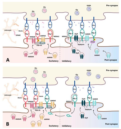
[21][39]. In agreement with their subcellular distributions, Nlg isoforms contribute differently to the function of glutamatergic vs. GABAergic synapses through their capacity to assemble appropriate scaffolds and functional receptors in the postsynaptic membrane opposing the presynaptic terminals. Specifically, Nlg-1 favors the functional modulation of glutamatergic synapses by recruiting NMDARs via the PSD-95 scaffold proteins and trapping surface-diffusing AMPARs by binding with PSD-95 and stargazin
[22][40]. In contrast, Nlg-2 recruits GABARs or glycine receptors through a specific interaction with gephyrin, driving the functional properties of inhibitory synapses
[23][41]. Thus, Nlgs are confined to excitatory synapses or inhibitory synapses, positioning them to influence the excitation/inhibition ratio, the imbalance of which leads to synaptic dysfunction-associated pathologies (
Figure 12A). Indeed, manipulating Nlg expression levels in vitro and in vivo has demonstrated their isoform-specific modulation of synaptic functions. The overexpression of Nlg-1 can specifically enhance AMPAR- and NMDAR-mediated EPSCs in an NMDAR-dependent fashion. In contrast, Nlg-1-knockout mice exhibit reduced NMDAR-mediated EPSC amplitudes and a loss of NMDAR-dependent long-term potential (LTP)
[24][25][26][42,43,44]. Of note, NMDAR-dependent LTP is not dependent on the binding of Nlg-1 to PDZ-domain-containing proteins, such as PSD-95. Rather, it requires Nlg-1 binding to Nrxs, as the rescue of LTP by Nlg-1 can be prevented by the mutation of residues critical for Nrx binding. This finding demonstrates the specific and pronounced regulatory role played by Nlg-1 in the context of excitatory synaptic transmission in an Nrx-dependent manner. However, the rescue of basal NMDAR-mediated synaptic transmission after Nlg deletion requires the Nlg-1 intracellular domain but not Nrx binding
[27][45]. This molecular dissociation of Nlg-1 domains required for LTP versus those required for the maintenance of basal NMDAR-mediated synaptic transmission indicates the complexity of the molecular architecture responsible for regulating synaptic strength at excitatory synapses.
Figure 12. Schematic diagram of trans-synaptic interaction between neurexins and neuroligins in normal and pathologic pain. (A) In normal conditions, Nlg-1 and Nlg-2 contribute differently to the function of excitatory and inhibitory transmissions, respectively, in front of corresponding presynaptic Nrxs. Nlg-1 favors the functional modulation of glutamatergic synapses by recruiting NMDARs via the PSD-95 scaffold and trapping surface-diffusing AMPARs by binding with PSD-95 and stargazing. Nlg-2 recruits GABARs or glycine receptors through a specific interaction with gephyrin. The mechanistic importance of Nrx–Nlg is associated with a range of proteins capable of modulating their interaction at the synaptic cleft, in which hevin and SPARC directly interact with Nrx and Nlg, while MDGAs occupy the interaction site between Nrx and Nlg and, thereby, block Nrx–Nlg interaction. (B) In pathologic pain, Nlg-1, as well as the recruitment of corresponding excitatory glutamatergic receptors, is upregulated in the context of pathological pain. Meanwhile, traditionally inhibitory Nlg-2 undergoes a fundamental shift in functionality from inhibition towards excitation, with an increase in co-localization with PSD-95 and subsequent AMPAR subunit targeting under the same condition.
Research on Nlg-2 has fundamentally shaped our understanding of the molecular architecture of Nlg-2 as a central organizer of inhibitory synapses. The protein components of the GABAergic postsynaptic complex, GABARs and gephyrin, are reduced in Nlg-2-knockout mice
[23][41], accompanied by a general reduction in inhibitory synaptic transmission. In contrast, overexpression of Nlg-2 reportedly results in a specific increase in IPSC amplitude, underscoring the role of Nlg-2 as a mediator and regulator of inhibitory synaptic transmission
[28][29][46,47].
Nlg-3 can either upregulate or downregulate inhibitory synaptic transmission in a splice-variant-dependent manner, suggesting that the specific subcellular localization of the Nlg-3 isoforms may contribute to the functional differences observed between them
[30][31][48,49]. However, when overexpressed, Nlg-3 enhances excitatory transmission and presynaptic vesicular glutamate transporter 1 expression, irrespective of the Nlg-3 splice variant. Together, these data indicate that alterations in Nlg expression can modulate both excitatory and inhibitory synaptic transmission, thus contributing to synaptic plasticity.
It is also noteworthy that Nlgs function as mediators of synaptic transmission and plasticity in a phosphorylation-dependent manner. For example, calcium/calmodulin-dependent protein kinase II (CaMKII)-mediated phosphorylation of T739 in the Nlg-1 C-terminal domain results in increased surface trafficking, while the mutant protein that could not be phosphorylated leads to reduced Nlg-mediated excitatory synaptic potentiation
[32][50]. Nlg phosphorylation also plays a role in governing postsynaptic protein recruitment, including both surface receptors and scaffold proteins. For instance, protein kinase A can phosphorylate Nlg-1 at S839 and dynamically regulate PSD-95 binding since a phosphomimetic mutation of Nlg-1 at S839 significantly reduces PSD-95 binding
[33][51]. Nevertheless, Nlg-1 phosphorylation at Y782 inhibits gephyrin binding rather than promoting PSD-95 recruitment
[34][52]. Proline-directed Nlg-2 phosphorylation at the S714 residue can ablate gephyrin binding, even though the residue does not fall in the gephyrin-binding domain. A previous study shows that S714 phosphorylation promotes the recruitment of the peptidyl-proly cis-trans isomerase (Pin1) enzyme, which mediates Nlg-2 isomerization and disrupts gephyrin interaction
[35][53].
Nlg phosphorylation also influences the recruitment of functional surface receptors, with phosphorylation of Nlg-1 at Y782 recruiting cell-surface AMPARs and thus augmenting AMPAR-mediated EPSCs
[36][54]. On the other hand, endogenous Nlg-2 phosphorylation is associated with reductions in spontaneous GABAR-mediated postsynaptic current amplitudes due to reduced GABAR and gephyrin retention at inhibitory synapses
[35][53]. As Nlg phosphorylation is controlled in an activity-dependent fashion, this may represent another distinct mechanism whereby these proteins can dynamically modulate synaptic transmission and plasticity.
Indeed, Nlgs have been found to undergo activity-dependent proteolytic cleavage, which can alter their activity and/or expression. For example, proteolytic cleavage of Nlg-1 prevents its surface expression, leading to the destabilization of trans-synaptic Nrx–Nlg complexes
[37][38][55,56]. Furthermore, the extracellular fragments of Nlg-1 generated through its proteolytic cleavage can bind to presynaptic metabotropic glutamate receptors, ultimately leading to their activation and the suppression of glutamate release, thereby reducing synaptic strength
[39][57]. Likewise, the induction of Nlg-3 proteolytic cleavage by protein kinase C results in reduced synaptic strength that can be counteracted by the prevention of cleavage. It should be noted that the proteolytic cleavage of Nlg-1 and Nlg-2, indeed, depends on the presence of Nlg-3 through Nlg-1/Nlg-3 or Nlg-2/Nlg-3 heterodimers, indicating the potential role of Nlg-3 as a key regulator of other Nlg cleavage events
[40][58].
2. The Regulators of Neurexin–Neuroligin Interaction
2.1. Hevin
Hevin, also known as SPARC-like protein 1, is a member of the SPARC family that is highly expressed in developing astrocytes, even after reaching adulthood
[41][59]. Structurally, hevin is a cysteine-rich glycoprotein containing a flexible acidic N-terminal region and a globular C-terminal region, which contains a follistatin-like (FS) domain and an extracellular calcium-binding (EC) domain. The FS–EC tandem domains exist as a monomer in solution, maintaining an elongated structure where the FS and EC domains do not interact, suggesting their independent functions. Further studies using surface plasmon resonance have shown that the FS domain contains residues responsible for the hevin–Nlg interaction. It has also been found that the FS domain is sufficient for Nrx binding and that the interaction is calcium-dependent
[42][60].
Interestingly, these structural findings correspond with previous results showing that hevin forms a trans-synaptic bridge between Nrx-1α and Nlg-1, which otherwise do not directly interact with one another under physiological conditions, thus regulating the formation and refinement of thalamocortical glutamatergic synapses
[43][61]. It is found that expression of Nlg-1, PSD-95, homer-1, and the NMDAR subunits are significantly reduced in the cortical postsynaptic membranes of hevin-knockout mice compared with wild-type mice
[43][61]. Of note, in addition to the recruitment of NMDARs, the treatment of cortical neurons with hevin can also raise the number of AMPARs on the cell surface, increasing both the amplitude and frequency of AMPAR-mediated EPSCs
[44][62]. Furthermore, increased co-localization of hevin with the excitatory synaptic markers vesicular glutamate transporter 1, AMPAR subunit, and NMDAR subunit is observed in epileptogenesis, while no co-localization is seen with the inhibitory synaptic markers vesicular GABA transporter and GABAR subunit, suggesting an association of hevin with the modulation of excitatory synapses
[45][63]. Therefore, the trans-synaptic interaction mediated by hevin might be exploited to modulate synaptic reorganization under various neurological conditions via the stabilization of the Nrx–Nlg trans-synaptic bridges.
2.2. SPARC
SPARC, also called osteonectin, is a prototypical member of the SPARC family of biologically active glycoproteins expressed largely in microglia and some subcortical astrocytes in the central nervous system
[46][64]. Examination of the protein structure shows that SPARC is highly homologous to hevin, with three identical structural domains, including the N-terminal, FS, and EC domains
[47][65]. Notably, the SPARC FS domain shares 56% sequence identity with the FS domain of hevin, and they are structurally similar. This intriguing feature suggests that SPARC may interact directly with Nrxs and Nlgs, and, indeed, it has been recently demonstrated that SPARC can bind both Nrxs and Nlgs with a similar affinity as hevin. In particular, SPARC resembles hevin in binding Nlg-2 with comparable affinity and in binding Nrx-1α in a similar calcium-dependent manner, suggesting that it may compete with hevin for binding to both Nrxs and Nlgs
[42][60]. In short, both SPARC and hevin can stabilize the Nrx–Nlg trans-synaptic bridge, and the ratio of SPARC versus hevin regulates Nrx–Nlg interaction and determines the net effect on synaptic function.
It is possible that the actions of one regulator might oppose those of another. Characteristically, SPARC antagonizes the action of hevin in synaptogenesis, with SPARC specifically inhibiting hevin-induced excitatory synaptogenesis; SPARC-null mice show increased synapse formation
[47][65]. Strikingly, SPARC may play multiple roles in synaptic function by regulating the postsynaptic glutamate receptors. During development, SPARC acts more like a “molecular brake” to prevent the over-accumulation of AMPARs, thereby altering both the amplitude and frequency of AMPAR-mediated mEPSCs and decreasing synaptic strength
[48][66]. However, under injury or disease in the mature nervous system, SPARC expression is increased, accompanied by the upregulation of AMPAR substrates at synapses and enhanced synaptic function
[49][67]. Specifically, SPARC treatment reduces the loss of AMPAR subunits in the synapses of hippocampal neurons following oxygen-glucose deprivation, and SPARC may play a novel role in regulating neuronal health and recovery following central nervous system injury. In addition, pretreatment with SPARC is accompanied by a significant increase in synaptic NDMAR subunit levels under the same condition, which could, in turn, enhance synaptic strength and plasticity
[49][67]. Taken together, it is conceivable that SPARC has differential effects on synaptic signaling pathways during development and following injury/disease. It would be important to test the effect of SPARC on the trans-synaptic Nrx–Nlg connection and determine whether this trans-synaptic bridge influences the activities of functional receptors.
2.3. MDGA
MDGAs are vertebrate-specific proteins belonging to the immunoglobulin superfamily and are specifically expressed in the nervous system. The two homologous MDGA proteins, MDGA1 and MDGA2, have a characteristic domain organization composed of an N-terminal signal peptide followed by six immunoglobulin domains, a MAM domain, and a C-terminal domain containing a cleavage site for GPI for anchorage in the cell membrane
[50][68]. Structural determination of the MDGA extracellular region shows a compact, approximately triangular structure that is stabilized by extensive interdomain contacts
[51][69].
Unlike hevin and SPARC, MDGAs interact with Nlgs and, thereby, block sites for Nrx binding, thus suppressing trans-synaptic bridge formation and disrupting synaptic activity
[51][52][69,70]. The affinity profiles of different MDGAs are Nlg isoform-specific, providing unique modes for regulating different neuronal populations during synaptic development and transmission. Specifically, MDGAs bind in a cis configuration with a lower affinity to Nlg-3 and Nlg-4 than to Nlg-1 and Nlg-2. In addition, MDGA1 selectively forms complexes with Nlg-2, and MDGA2 selectively forms complexes with Nlg-1
[53][71]. Analysis of mouse mutants shows that targeted mutations of MDGA1 and MDGA2 promote the activities of inhibitory and excitatory synapses, respectively, suggesting functional divergence between the proteins
[51][69]. In line with this notion, RNAi-mediated knockdown of MDGA1 increases the number of inhibitory synapses in cultured rat hippocampal neurons without affecting the number of excitatory synapses
[54][72]. Mutation of MDGA2 increases excitatory synapse numbers and elevates excitatory transmission by blocking Nlg-1 interaction with Nrxs, with no influence on inhibitory synapses
[55][73]. Intriguingly, MDGAs and hevin compete for the same Nlg binding sites and such competition may shape the excitatory and inhibitory balance mediated by molecular crosstalk between different modifiers of the Nrx–Nlg interaction
[42][60]. Thus, examining how different MDGAs modulate Nrx–Nlg interaction will enhance our understanding of the mechanisms underlying synapse plasticity and provide new insights into the etiology of neurological disorders.