Nanobioengineered-based hybrid electrochemical biosensors exploit the synergistic properties of hybrid systems that connect biomolecules with nanomaterials to engineer highly sensitive biosensing platforms for the specific electrochemical detection of different target analytes.
- hybrid
- nanobiomaterial
- bioreceptor
- bioaffinity
- biocatalytic
- biosensor
- cytosensor
- genosensor
- immunosensor
- electrochemical
Graphical abstract
11. Introduction
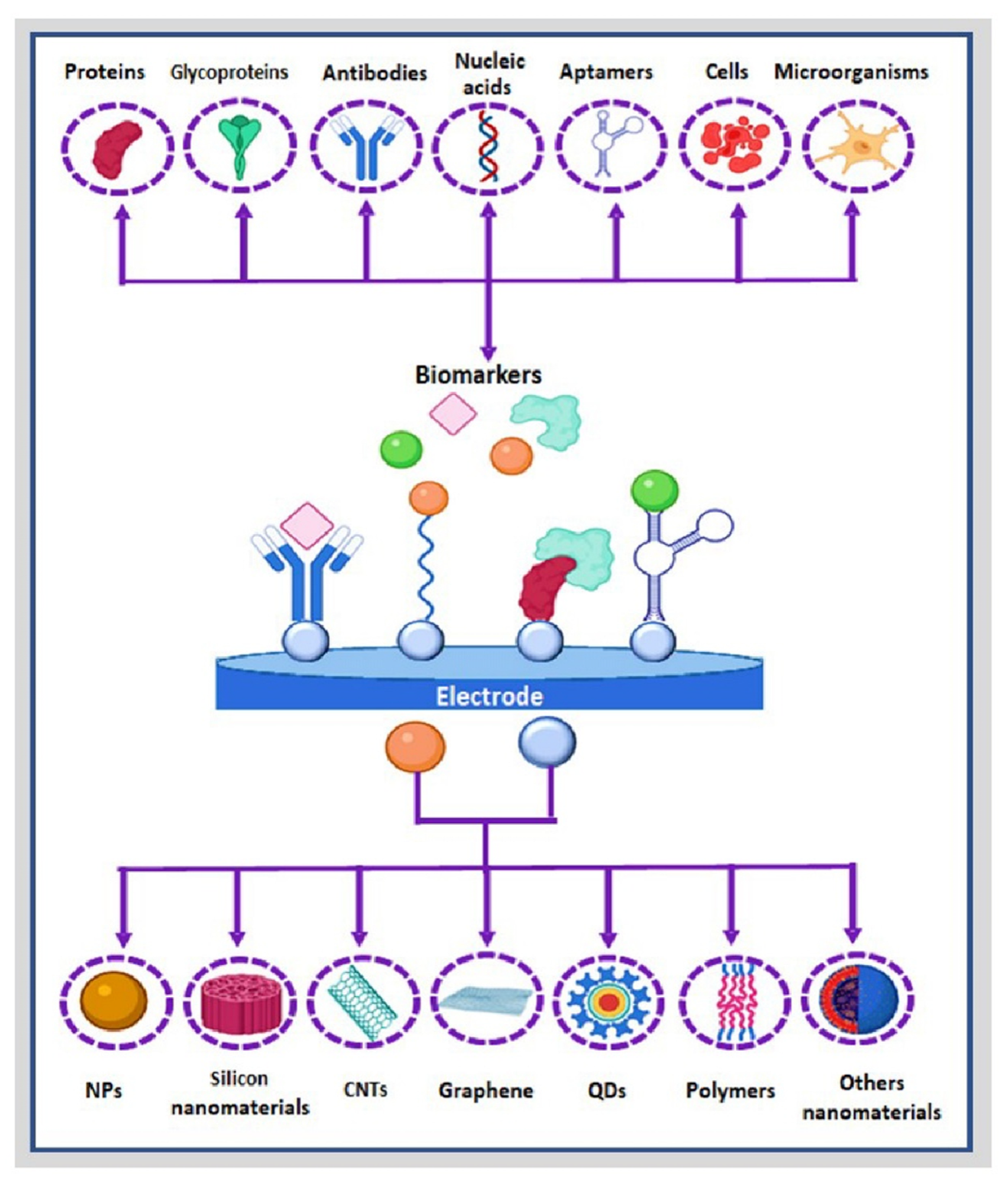
2. Nanohybrids and Nanocomposites
Nanostructured nanomaterials, both nanohybrids and nanocomposites, have been increasingly exploited in developing electrochemical biosensors [16] and functional interfaces [17][18][19][20][21] with enhanced properties in terms of sensitivity, selectivity, robustness, and simplicity [22]. Nanocomposite materials are prepared by combining two or more different materials with different physicochemical properties, where one of the constituents has dimensions at the nanoscale or, instead, the nanocomposite structure exhibits a nanometric phase separation of the individual components. In preparing nanocomposites, one of the constituent materials acts as a support matrix in which other materials called reinforcement agents are incorporated [23][24]. Nanocomposites present mixed properties based on the original properties of each constituent nanomaterial, not modified during the preparation process [25]. Similarly, a hybrid nanomaterial combines organic and inorganic building blocks [4], which present a continuous interface between the structural components [25], and new, improved physicochemical properties emerge that are distinct from the specific properties of the components alone [26]. Hybrid nanomaterials can function as novel electrode materials, signal amplifiers, and catalysts of the electrochemical reaction of the product generated in situ during the biorecognition event. To date, the most common hybrid nanomaterials applicable to electrochemical biosensing include metallic nanostructures [27][28], silicon nanomaterials [29][30][31][32], carbon nanostructures [17][33][34], and semiconductor polymers [35][36][37][38][39], with great potential for the development of electrochemical nanobiosensors with enhanced performance [40][41][42], as commented. This section will comment on the main examples of the last ten years (Table 1), focused on nanostructured nanomaterials employed in developing nanohybrids for their implementation in electrochemical biosensing.|
Nanomaterial |
Hybrid a |
Target b |
Analytical Characteristics |
Comments |
References |
|
|---|---|---|---|---|---|---|
|
Linear Range |
LOD |
|||||
|
Metallic nanostructures |
3D hybrid graphene–GNR. |
H2O2 |
0 to 50 mM |
2.9 µM |
Metallic nanostructures have high catalytic activity, easy preparation, and relatively low cost. However, this kind of nanomaterial can change its oxidation state due to variations in conditions of the medium, such as pH, ionic strength, and temperature upon time. |
[43] |
|
TiO2 nanoparticles encapsulated ZIF-8 |
Glucose |
2 to 10 mM |
80 nM |
[44] |
||
|
Nanohybrid of VS2/AuNP and CoFe2O4 nanozyme |
Kana |
1 pM to 1 μM |
0.5 pM |
[45] |
||
|
Ag and hybrid Ag–Fe3O4 metallic nanoparticles. |
AA |
0.2–60 μM |
74 nM |
[46] |
||
|
Silicon nanomaterials |
mSiO2@MWCNT. |
Thrombin |
0.0001 nM and 80 nM |
50 fM |
These nanomaterials have high mechanical resistance, thermal stability, long functional life, and versatility; nonetheless, they require long synthetic processes, and their application is limited to certain analytes. |
[31] |
|
MSF/APTES/AgNP |
STR |
1 to 6.2 ng/mL |
0.33 fg/mL |
[47] |
||
|
Ap–GA–NH2MCM-41–GCE |
hemin and Hb |
1.0 × 10⁻¹⁹ to 1.0 × 10⁻⁶ M |
7.5 × 10⁻²⁰ M and 6.5 × 10⁻²⁰ M |
[48] |
||
|
AuNPs loaded in functionalized MSNPs |
CEA |
1.0 × 10⁻³ to 100 ng/mL |
9.8 × 10⁻⁴ ng/mL |
[49] |
||
|
Carbon nanostructures |
MWCNTs and GQDs. |
IL-13Rα2 |
2.7 to 100 ng/mL |
0.8 ng/mL |
These nanomaterials enjoy thermal stability, large surface area, and a wide range of nanostructures and functional groups. They are the main nanomaterials used in the preparation of electrochemical biosensors. |
[50] |
|
GQDs/AuNPs. |
P53 |
0.000592–1.296 pM |
0.065 fM |
[51] |
||
|
CQDs/AuNps |
Glucose |
0.05 mM to 2.85 mM |
17 μM |
[52] |
||
|
CoCu-ZIF@CDs |
B16-F10 cells |
1 × 10² to 1 × 10⁵ cells/mL |
33 cells/mL |
[53] |
||
|
Polymers |
(Chi-Py) mixture, AuNPs, and MWCNT |
Escherichia coli |
3 × 101 to 3 × 10⁷ cfu/mL |
~30 CFU/mL |
These have high biocompatibility, high affinity, strong adsorption ability, low molecular permeability, physical rigidity, and chemical inertness in biological processes. However, functionalizing their surface is necessary for the anchorage of bioreceptors, and some polymers oxidize due to changes in medium conditions. |
[54] |
|
PANI/active carbon and n-TiO2 |
Glucose |
0.02 mM to 6.0 mM |
18 μM |
[55] |
||
|
PEG/AuNPs/PANI |
alpha-fetoprotein |
10−14 to 10−6 mg/mL |
0.007 pg/mL |
[56] |
||
|
Other nanostructured nanomaterials |
WSe2 and AuNPs |
Thrombin |
0–1 ng/mL |
190 fg/mL |
Other hybrid nanostructures have a large specific surface area, excellent electrical conductivity, and electrocatalytic properties. |
[57] |
|
MoS2/Ti3C2 nanohybrids |
miRNA |
1 fM to 0.1 nM |
0.43 fM |
[58] |
||
|
AuNPs/Ti3C2 MXene 3D |
miRNA155 |
1.0 fM to 10 nM |
0.35 fM |
[59] |
||
a GNR, graphene–gold nanorod; AuNPs, gold nanoparticles; Ap, aptamer; GA, glutaraldehyde; GCE, glassy carbon electrode; MSNPs, mesoporous silica nanoparticles; MWCNTs, multiwalled carbon nanotube; MSF, mesoporous silica thin film; APTES, (3-aminopropyl) triethoxysilane; AgNP, silver nanoparticles; CDs, carbon-dots; Chi-Py, pyrrole branched chitosan; PEG, polyethylene glycols; PANI, polyaniline. b AA, ascorbic acid; STR, streptomycin; miRNA; micro-RNA.
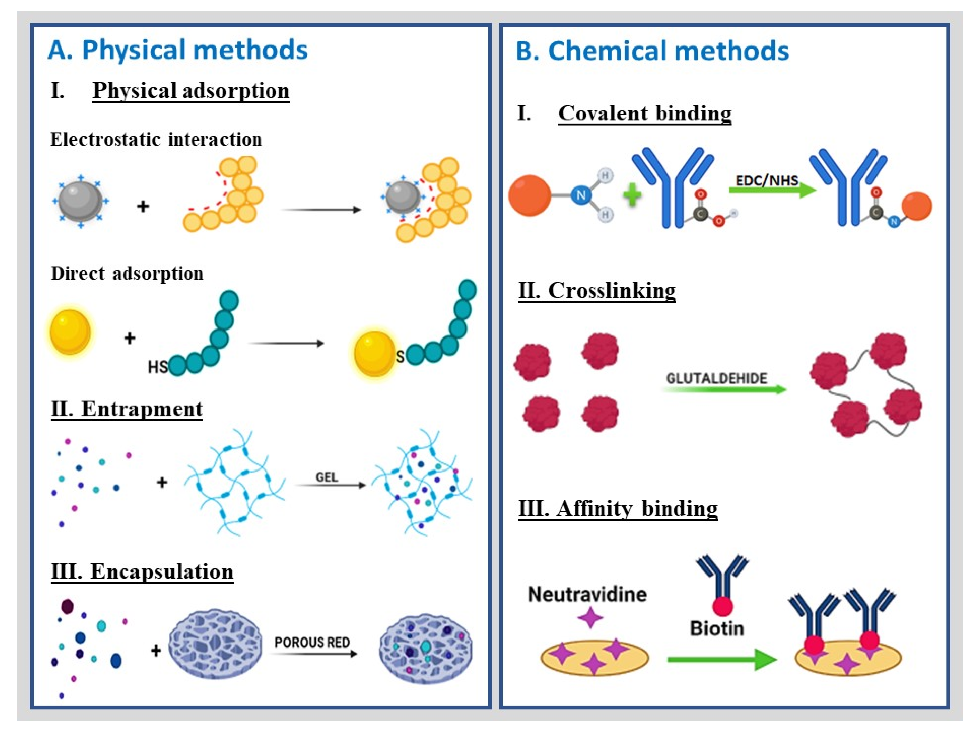
3. Conjugation of Nanohybrid Materials with Biomolecules
Biosensors can be label-free and label-based. Briefly, in a label-free mode, the detected signal is generated directly by the interaction of the analyzed (bio)material with the transducer. In contrast, label-based sensing involves chemical or biological compounds that act as labels, generating a detectable signal by analytical techniques such as colorimetry, fluorescence, and electrochemistry [60].3.1. Bioreceptor
3.1. Bioreceptors
s
Biological receptors are biomolecules that bind to a specific ligand with a defined structure, commonly through bioaffinity interactions. These biological components are used when assembling nanobiosensors due to their high specificity, differentiating the target molecule from analogous counterparts and even isomers of the same molecule (Figure 1). Different bioreceptors can be anchored at electrochemical transducers to confer specificity to nanobioengineered devices. They can generally be classified into five major categories, i.e., enzymes, antibody/antigens, nucleic acids, cellular structures/cells, and biomimetic entities, as depicted in Figure 1.
4. Functional Groups and Conjugation Chemistry

5. Characterization of Nanobioengineered Platforms
|
Techniques |
Physicochemical Characteristics Analyzed |
|---|---|
|
Fourier transform infrared spectroscopy (FTIR). |
This technique characterizes the functional groups, surface properties, structure, and conformation of hybrid nanomaterials and nanobioconjugates. |
|
Thermogravimetric analysis (TGA). |
Thermogravimetric analysis of nanohybrids determines their thermal stability by estimating organic and inorganic material extent. |
|
Ultraviolet spectroscopy (UV-Vis). |
This technique can be used to estimate variables such as Km and Vmax in enzyme nanobioconjugates. |
|
Dynamic light scattering (DLS). |
This technique can estimate the hydrodynamic size distribution of nanostructures. |
|
Electrophoretic light scattering. |
The stability of nanomaterials is highly dependent on the surface charge, among other factors. |
|
X-ray diffraction (XRD). |
These techniques characterize hybrid nanomaterials’ size, shape, and crystalline structure. |
|
X-ray photoelectron spectroscopy (XPS). |
|
|
Transmission electron microscopy (TEM). |
Imaging techniques study size, size distribution, aggregation, dispersion, heterogeneity, morphological characteristics, and compositional analysis of the hybrid nanomaterials and nanobioconjugates. |
|
Scanning electron microscopy (SEM). |
|
|
Electrochemical techniques. |
Electrochemical techniques such as CV and EIS are used to evaluate electron transfer before, during, and after the bioreceptors attach to the surface of hybrid nanomaterials. They are also used to characterize the analytical properties of the resultant biosensors. |
6. Examples of Nanobioengineered Platforms for Electrochemical Biosensing in the Last Five Years
The significant advance in developing nanobioengineered platforms for electrochemical biosensing has been remarkable in the last five years. However, new 2D and 3D nanomaterials emerge year by year with various improved properties ranging from quantum tunneling, excellent stability, and high conductivity and versatility, which provide new opportunities to develop electrochemical biosensors with high selectivity and extremely low LODs. Furthermore, the appearance of these novel nanostructured materials has led to the implementation of advanced and ultrasensitive biodetection tools (Table 3).
Table 3. Examples of nanobioengineered biosensors, indicating the nanobiohybrid (nanomaterial and biomolecules) and analytic characteristics
|
Biosensor |
Application a |
Nanobiohybrid: Nanomaterial and Biomolecules b |
Characterization c |
Analytical Performance (Linear Range and LOD) d |
Reference |
|
Immunosensor |
PSA |
Antibody/HP5@AuNPs@g-C3N4 bioconjugated with PSA-Ab2 |
CV, EIS, and DPV |
0.0005 to 0.00 ng/mL with LOD of 0.12 pg/mL |
[71] |
|
HER2 |
Ab/g-C3N4/AuNPs/Cu-MOF |
CV and EIS |
1.00 to 100.00 ng/mL with LOD of 3.00 fg/mL |
[72] |
|
|
AXL |
Ab/fGQDs |
XRD, FTIR, UV-Vis, TEM, EIS, DPV |
1.7 to 1000 pg/mL with LOD of 0.5 pg/mL |
[73] |
|
|
CEA |
CdSe-QD-melamine and Ab1-TiO2-AuNP-ITO |
DPV |
0.005 - 1000 ng/mL with a LOD of 5 pg/mL |
[74] |
|
|
CA19-9 |
CeO2/FeOx@mC |
XPS, TEM, EIS, CV |
0.1 mU/mL to 10 U/mL with a LOD of 10 μU/mL |
[75] |
|
|
NMP-22 |
Co-MOFs/CuAu NWs/Ab |
SEM, XPS, CV, and chronoamperometry |
0.1 pg/mL to 1 ng/mL with a LOD of 33 fg/mL |
[76] |
|
|
Genosensor |
Zika |
Anti-Dig-HRP |
Chronoamperometry, CV, EIS |
5 to 300 pmol/L with LOD of 0.7 pM |
[77] |
|
Zika genes |
AuNPs/ssDNA |
SEM, CV, DPV, and chronoamperometry |
10 to 600 fM with LOD of 0.2 fM |
[78] |
|
|
CaMV35S gen |
Fe3O4-Au@Ag-sDNA on MWCNT/AuNPs/SH-sDNA |
TEM, XRD, UV-Vis, CV, and DPV |
1 ×10⁻¹⁶ M to 1 ×10⁻¹⁰ M with LOD of 1.26 × 10⁻¹⁷ M |
[79] |
|
|
mi-R21 |
3-(trimethoxysilyl)propyl methacrylate/ITO/PET/Fc-hybrid DNA hydrogel |
DPV |
10 nM to 50 μM with a LOD of 5 nM |
[80] |
|
|
miRNA-122 |
rGO/Au/DNA |
XRD, TEM, Raman, XPS, CV, and DPV |
10 μM to 10 pM with a LOD of 1.73 pM |
[81] |
|
|
OVA |
SiO2@Au/dsDNA/CeO2 |
DPV |
1 pg/mL to 1000 ng/mL with a LOD of 0.87 pg/mL |
[82] |
|
|
Enzymatic |
Glucose |
GOx/n-TiO2/PANI |
CV and chronoamperometry |
0.02 to 6.0 mM with LOD of 18 μM |
[83] |
|
Glucose |
Cu-nanoflowers-Gox-HRP/AuNPs-GO-PVA nanofibers |
UV-Vis, SEM, TEM, XDR, CV, and chronoamperometry |
0.001 to 0.1 mM with a LOD of 0.018 μM |
[84] |
|
|
Organophosphate pesticides |
acetylcholinesterase/chitosan-transition metals/graphene/GCE |
SEM, TEM, XPS, XRD, CV, DPV and EIS |
11.31 μM to 22.6 nM with LOD of 14.45 nM |
[85] |
|
|
β-hydroxybutyric acid |
Ti3C2Tx nanosheets conjugated with β-hydroxybutyrate dehydrogenase |
SEM, CV, and chronoamperometry |
0.36 to 17.9 mM with a LOD of 45 μM |
[86] |
|
|
Based on peptides |
norovirus |
Cys/peptide/gold layer |
CV and EIS |
The LOD was 99.8 nM and 7.8 copies/mL for rP2 and human norovirus, respectively. |
[87] |
|
PSA |
MXene-Au-MB nanohybrid/peptide |
DPV |
5 pg/mL to 10 ng/mL with a LOD of 0.83 pg/mL |
[88] |
|
|
PKA and CK2 |
Peptide/MSF/ITO |
Chronoamperometry |
The LODs were 0.083 and 0.095 U/mL, for PKA and CK2, respectively |
[89] |
|
|
NHE |
Cys -PEG-QRRMIEEPA-MB |
DPV and SWV |
10 and 150 nM with a LOD of 250 pM |
[90] |
|
|
Based on glycoproteins |
Toxoplasma gondii |
Ab glycosylphosphatidylinositol/SPAuE |
CV, EIS |
1.0 to 10.0 IU/mL, with a LOD of 0.31 IU/mL |
[91] |
|
MIPs/glycoproteins |
Fc/MPBA/AuNPs-SiO2 nanobioconjugate |
FTIR, CV, EIS, DPV, and chronoamperometry |
1 pg/mL to 100 ng/mL and reached a LOD of 0.57 pg/mL |
[92] |
|
|
Based on aptamers |
tumor exosomes extracted from lymph node carcinoma of a prostate cells line |
MNPs/aptamer-DNA/double-stranded DNA/GCE |
DPV |
The LOD was 70 particles/μL |
[93] |
|
miRNA |
DSN/AuNPS/HRP |
CV, EIS and chronoamperometry |
The LOD was 43.3 aM |
[94] |
|
|
CA125 and living MCF-7 cells |
Tb-MOF-on-Fe-MOF |
SEM, TEM, XPS, CV, and EIS |
100 μU/mL to 200 U/mL with a LOD of 58 μU/mL towards CA125. Moreover, biosensor detecting MCF-7 cells with a LOD of 19 cells/mL |
[95] |
|
|
CEA and NSE |
Paper-electrode functionalized with amino-modified graphene-Thi-AuNPs and PB-PEDOT |
DPV |
0.01 to 500 ng/mL for CEA and 0.05 - 500 ng/mL for NSE with a LOD of 2 pg/mL for CEA and 10 pg/mL for NSE, respectively |
[96] |
|
|
Other types of biosensors (based on cells or mimicking biosensors) |
Impedimetric biosensor/Escherichia coli B. |
CNT/PEI-T2 virus/GCE |
EIS |
103 to 107 CFU/mL with LOD of 1.5 × 10³ CFU/mL |
[97] |
|
Nonenzymatic biosensor/glucose |
GS/GNR/Ni |
Chronoamperometry |
5 nM to 5 mM with a LOD of 2.5 nM. |
[98] |
|
|
Mimicking biosensor/H2O2 released from H9C2 cardiac cells |
AuNFs/Fe3O4@ZIF-8-MoS2 |
SEM, fluorescence, CV, EIS, and chronoamperometry |
5 μM - 120 mM and a LOD of 0.9 μM |
[99] |
|
|
Electrochemical/glucose |
CuOx@Co3O4 core-shell nanowires/ZIF-67 |
SEM, TEM, XRD, XPS, CV, and chronoamperometry |
0.1 to 1300.0 μM with a LOD of 36 nM |
[100] |
|
|
Mimicking/L-tyrosinase |
UT-g-C3N4/Ag hybrids |
TEM, XPS, XRD, AFM, EIS, CV, and DPV |
1.00× 10⁻⁶ to 1.50 × 10⁻⁴ mol/L with a LOD of 1.40 × 10⁻⁷ mol/L |
[101] |
|
|
Biomimetic biosensor/glucose |
Fe3O4@PNE-GOx |
Chronoamperometry |
0.24 to 24 mM with a LOD of 6.1 µM |
[102] |
|
|
PAD/creatinine |
CuO/IL/ERGO/SPCE |
Chronoamperometry |
0.01 to 2.0 mM and a LOD of 0.22 μM |
[103] |
|
|
3D paper-based microfluidic electrochemical biosensor/glucose
|
rGO-TEPA/PB |
SEM, Raman, CV, and chronoamperometry |
0.1 mM - 25 mM with a LOD of 25 μM |
[104] |
.
|
Biosensor |
Application a |
Nanobiohybrid: Nanomaterial and Biomolecules b |
Characterization c |
Analytical Performance (Linear Range and LOD) d |
Reference |
|
Immunosensor |
PSA |
Antibody/HP5@AuNPs@g-C3N4 bioconjugated with PSA-Ab2 |
CV, EIS, and DPV |
0.0005 to 0.00 ng/mL with LOD of 0.12 pg/mL |
[71] |
|
HER2 |
Ab/g-C3N4/AuNPs/Cu-MOF |
CV and EIS |
1.00 to 100.00 ng/mL with LOD of 3.00 fg/mL |
[72] |
|
|
AXL |
Ab/fGQDs |
XRD, FTIR, UV-Vis, TEM, EIS, DPV |
1.7 to 1000 pg/mL with LOD of 0.5 pg/mL |
[73] |
|
|
CEA |
CdSe-QD-melamine and Ab1-TiO2-AuNP-ITO |
DPV |
0.005 - 1000 ng/mL with a LOD of 5 pg/mL |
[74] |
|
|
CA19-9 |
CeO2/FeOx@mC |
XPS, TEM, EIS, CV |
0.1 mU/mL to 10 U/mL with a LOD of 10 μU/mL |
[75] |
|
|
NMP-22 |
Co-MOFs/CuAu NWs/Ab |
SEM, XPS, CV, and chronoamperometry |
0.1 pg/mL to 1 ng/mL with a LOD of 33 fg/mL |
[76] |
|
|
Genosensor |
Zika |
Anti-Dig-HRP |
Chronoamperometry, CV, EIS |
5 to 300 pmol/L with LOD of 0.7 pM |
[77] |
|
Zika genes |
AuNPs/ssDNA |
SEM, CV, DPV, and chronoamperometry |
10 to 600 fM with LOD of 0.2 fM |
[78] |
|
|
CaMV35S gen |
Fe3O4-Au@Ag-sDNA on MWCNT/AuNPs/SH-sDNA |
TEM, XRD, UV-Vis, CV, and DPV |
1 ×10⁻¹⁶ M to 1 ×10⁻¹⁰ M with LOD of 1.26 × 10⁻¹⁷ M |
[79] |
|
|
mi-R21 |
3-(trimethoxysilyl)propyl methacrylate/ITO/PET/Fc-hybrid DNA hydrogel |
DPV |
10 nM to 50 μM with a LOD of 5 nM |
[80] |
|
|
miRNA-122 |
rGO/Au/DNA |
XRD, TEM, Raman, XPS, CV, and DPV |
10 μM to 10 pM with a LOD of 1.73 pM |
[81] |
|
|
OVA |
SiO2@Au/dsDNA/CeO2 |
DPV |
1 pg/mL to 1000 ng/mL with a LOD of 0.87 pg/mL |
[82] |
|
|
Enzymatic |
Glucose |
GOx/n-TiO2/PANI |
CV and chronoamperometry |
0.02 to 6.0 mM with LOD of 18 μM |
[83] |
|
Glucose |
Cu-nanoflowers-Gox-HRP/AuNPs-GO-PVA nanofibers |
UV-Vis, SEM, TEM, XDR, CV, and chronoamperometry |
0.001 to 0.1 mM with a LOD of 0.018 μM |
[84] |
|
|
Organophosphate pesticides |
acetylcholinesterase/chitosan-transition metals/graphene/GCE |
SEM, TEM, XPS, XRD, CV, DPV and EIS |
11.31 μM to 22.6 nM with LOD of 14.45 nM |
[85] |
|
|
β-hydroxybutyric acid |
Ti3C2Tx nanosheets conjugated with β-hydroxybutyrate dehydrogenase |
SEM, CV, and chronoamperometry |
0.36 to 17.9 mM with a LOD of 45 μM |
[86] |
|
|
Based on peptides |
norovirus |
Cys/peptide/gold layer |
CV and EIS |
The LOD was 99.8 nM and 7.8 copies/mL for rP2 and human norovirus, respectively. |
[87] |
|
PSA |
MXene-Au-MB nanohybrid/peptide |
DPV |
5 pg/mL to 10 ng/mL with a LOD of 0.83 pg/mL |
[88] |
|
|
PKA and CK2 |
Peptide/MSF/ITO |
Chronoamperometry |
The LODs were 0.083 and 0.095 U/mL, for PKA and CK2, respectively |
[89] |
|
|
NHE |
Cys -PEG-QRRMIEEPA-MB |
DPV and SWV |
10 and 150 nM with a LOD of 250 pM |
[90] |
|
|
Based on glycoproteins |
Toxoplasma gondii |
Ab glycosylphosphatidylinositol/SPAuE |
CV, EIS |
1.0 to 10.0 IU/mL, with a LOD of 0.31 IU/mL |
[91] |
|
MIPs/glycoproteins |
Fc/MPBA/AuNPs-SiO2 nanobioconjugate |
FTIR, CV, EIS, DPV, and chronoamperometry |
1 pg/mL to 100 ng/mL and reached a LOD of 0.57 pg/mL |
[92] |
|
|
Based on aptamers |
tumor exosomes extracted from lymph node carcinoma of a prostate cells line |
MNPs/aptamer-DNA/double-stranded DNA/GCE |
DPV |
The LOD was 70 particles/μL |
[93] |
|
miRNA |
DSN/AuNPS/HRP |
CV, EIS and chronoamperometry |
The LOD was 43.3 aM |
[94] |
|
|
CA125 and living MCF-7 cells |
Tb-MOF-on-Fe-MOF |
SEM, TEM, XPS, CV, and EIS |
100 μU/mL to 200 U/mL with a LOD of 58 μU/mL towards CA125. Moreover, biosensor detecting MCF-7 cells with a LOD of 19 cells/mL |
[95] |
|
|
CEA and NSE |
Paper-electrode functionalized with amino-modified graphene-Thi-AuNPs and PB-PEDOT |
DPV |
0.01 to 500 ng/mL for CEA and 0.05 - 500 ng/mL for NSE with a LOD of 2 pg/mL for CEA and 10 pg/mL for NSE, respectively |
[96] |
|
|
Other types of biosensors (based on cells or mimicking biosensors) |
Impedimetric biosensor/Escherichia coli B. |
CNT/PEI-T2 virus/GCE |
EIS |
103 to 107 CFU/mL with LOD of 1.5 × 10³ CFU/mL |
[97] |
|
Nonenzymatic biosensor/glucose |
GS/GNR/Ni |
Chronoamperometry |
5 nM to 5 mM with a LOD of 2.5 nM. |
[98] |
|
|
Mimicking biosensor/H2O2 released from H9C2 cardiac cells |
AuNFs/Fe3O4@ZIF-8-MoS2 |
SEM, fluorescence, CV, EIS, and chronoamperometry |
5 μM - 120 mM and a LOD of 0.9 μM |
[99] |
|
|
Electrochemical/glucose |
CuOx@Co3O4 core-shell nanowires/ZIF-67 |
SEM, TEM, XRD, XPS, CV, and chronoamperometry |
0.1 to 1300.0 μM with a LOD of 36 nM |
[100] |
|
|
Mimicking/L-tyrosinase |
UT-g-C3N4/Ag hybrids |
TEM, XPS, XRD, AFM, EIS, CV, and DPV |
1.00× 10⁻⁶ to 1.50 × 10⁻⁴ mol/L with a LOD of 1.40 × 10⁻⁷ mol/L |
[101] |
|
|
Biomimetic biosensor/glucose |
Fe3O4@PNE-GOx |
Chronoamperometry |
0.24 to 24 mM with a LOD of 6.1 µM |
[102] |
|
|
PAD/creatinine |
CuO/IL/ERGO/SPCE |
Chronoamperometry |
0.01 to 2.0 mM and a LOD of 0.22 μM |
[103] |
|
|
3D paper-based microfluidic electrochemical biosensor/glucose
|
rGO-TEPA/PB |
SEM, Raman, CV, and chronoamperometry |
0.1 mM - 25 mM with a LOD of 25 μM |
[104] |
aPSA, prostate-specific antigen; HER2, human epidermal growth factor receptor 2; AXL, tyrosine kinase; CaMV35S, cauliflower mosaic virus 35S; SAMs, self-assembled monolayers; MIPs, molecular imprinted polymers; miRNA, micro-ribonucleic acid; CA19-9, carbohydrate antigen 19-9; CA125, carbohydrate antigen 125; NMP-22, nuclear matrix protein-22; PKA, protein kinase A; CK2, casein kinase II; PAD, paper-based analytical devices; OVA, ovalbumin; CEA, carcinoembryonic antigen; NHE, human neutrophil elastase; NSE, neuron-specific enolase.
bHP5, hydroxylpillar[5]arene; AuNPs, gold nanoparticles; Ab, antibody; Gox, glucose oxidase; PANI, polyaniline; Cys, cysteine; MOFs, metal-organic framework; fGQDs, functionalized graphene quantum dots; anti-Dig-HRP, antibody-digoxigenin-horseradish peroxidase; ssDNA, single-strand DNA; MWCNT, multiwalled carbon nanotube; CNT, carbon nanotube; PEI, polyethyleneimine; GCE, glassy carbon electrode; SPAuE, screen-printed gold electrode; MNPs, metallic nanoparticles; Fc, ferrocene; MPBA, 4-mercaptophenylboronic acid; QD, quantum dots; ITO, indium tin oxide; DSN, duplex-specific nuclease; PET, polyethylene terephthalate; GO, graphene oxide; PVA, poly(vinyl alcohol); GS, graphene sheet; GNR, graphene-gold nanorod; rGO, reduced graphene oxide; UT, ultrathin; PEG, polyethylene glycol; MB, methylene blue; THI, electron-mediating thionin; PB, Prussian blue; PEDOT, poly(3,4-ethylenedioxythiophene), SPCE, screen-printed carbon electrode; MSF, mesoporous silica thin film; PNE, polynorepinephrine; IL, ionic liquid; ERGO, electrochemically reduced graphene.
cCV, cyclic voltammetry; EIS, electrochemical impedance spectroscopy; DPV, differential pulse voltammetry; XRD, X-ray diffraction; XPS, X-ray photoelectron spectroscopy; FTIR, infrared spectroscopy; UV-Vis, ultraviolet visible spectroscopy; SEM, scanning electron microscopy; TEM, transmission electron microscopy; SWV, square wave voltammetry.
dLOD, limit of detection.
7. Limitations, Opportunities, and Concluding Remarks
Biosensor technology based on nanobiohybrid materials represents a vast field that significantly impacts healthcare, the environment, and food quality control. These functional platforms promote target molecule detection with high specificity and sensitivity, particularly in the biomedical field [105][106][107][108][71][109]. Furthermore, the rational design of the nanobiohybrids has been demonstrated to enhance the response and long-term stability of the resultant devices due to the incorporation of nanomaterials with improved properties that promote a favorable nanoenvironment for bioreceptors anchoring. Besides, the versatility of nanomaterials facilitates the conjugation with molecules by multiple conjugation chemistry, opening options to detect numerous target molecules.
Electrochemical-based nanohybrid biosensors have the potential to solve most of the limitations and concerns of bioanalysis and diagnostic tests while maintaining the required sensitivity, selectivity, and LOD to face real needs. Besides, integrating sample preparation into the device allows the possibility of direct analysis within a sample matrix and offers opportunities for new strategies of long-term analysis in vivo, among many other exciting applications. Electrochemical nanohybrid biosensors are particularly suitable for miniaturization and integration in microfluidic devices, thus reducing the consumption of reagents and samples {Formatting Citation}. Applications include detecting whole cells, cell components, proteins, and small molecules to address diagnostics and food and environmental control tasks online and in real-time, but still require more sophisticated platforms with additional elements, such as sample preparation. Although nanobioengineered biosensors are an affordable analytical strategy relative to gold standard detection methods, the development of large-scale electrochemical nanobiosensors is still challenging because they require state-of-the-art technologies for their production in a reproducible and stable manner, directly influencing the cost of the sensing device [109][110]. This apparent drawback could be overcome by scaling, automation, and mass manufacturing to lower costs through advanced methods in elaborating cost-affordable and disposable electrochemical nanobiosensors based on additive manufacturing, including screen inkjet 3D printing or microfabrication technologies [111][110][112][113].
Overall, this revisearcwh exemplified nanobiosensors mainly based on screen-printed electrodes modified with nanohybrids conjugated with highly specific bioreceptors for enhanced biosensing. Yet, the richness in the art of biosensors deserves deeper exploration and support of exciting new ideas. Overall, nanobiohybrids are paving the way in the pioneering development of highly sensitive and selective electrochemical nanobiosensors and represent remarkable research advances that are a step forward in increasing the impact of this exciting, cutting-edge technology in the field of biomarker detection of clinical interest [110]
.
References
- Cajigas, S.; Orozco, J. Nanobioconjugates for Signal Amplification in Electrochemical Biosensing. Molecules 2020, 25, 3542.
- Kumar, A.; Purohit, B.; Maurya, P.K.; Pandey, L.M.; Chandra, P. Engineered nanomaterial assisted signal-amplification strategies for enhancing analytical performance of electrochemical biosensors. Electroanalysis 2019, 31, 1615–1629.
- Low, S.S.; Ji, D.; Chai, W.S.; Liu, J.; Khoo, K.S.; Salmanpour, S.; Karimi, F.; Deepanraj, B.; Show, P.L. Recent progress in nanomaterials modified electrochemical biosensors for the detection of MicroRNA. Micromachines 2021, 12, 1409.
- Jones, R.G.; Kahovec, J.; Stepto, R.; Wilks, E.S.; Hess, M.; Kitayama, T.; Metanomski, W.V. Definitions of terms relating to the structure and processing of sols, gels, networks, and inorganic-organic hybrid materials (IUPAC Recommendations 2007). Pure Appl. Chem. 2007, 79, 1801–1829.
- Suni, I.I. Substrate materials for biomolecular immobilization within electrochemical biosensors. Biosensors 2021, 11, 239.
- Singh, A.; Sharma, A.; Ahmed, A.; Sundramoorthy, A.K.; Furukawa, H.; Arya, S.; Khosla, A. Recent advances in electrochemical biosensors: Applications, challenges, and future scope. Biosensors 2021, 11, 336.
- Yüce, M.; Kurt, H. How to make nanobiosensors: Surface modification and characterisation of nanomaterials for biosensing applications. RSC Adv. 2017, 7, 49386–49403.
- Rocchitta, G.; Spanu, A.; Babudieri, S.; Latte, G.; Madeddu, G.; Galleri, G.; Nuvoli, S.; Bagella, P.; Demartis, M.; Fiore, V.; et al. Enzyme biosensors for biomedical applications: Strategies for safeguarding analytical performances in biological fluids. Sensors 2016, 16, 780.
- Taylor-Pashow, K.M.L.; Della Rocca, J.; Huxford, R.C.; Lin, W. Hybrid nanomaterials for biomedical applications. Chem. Commun. 2010, 46, 5832–5849.
- Hu, Y.; Huang, Y.; Tan, C.; Zhang, X.; Lu, Q.; Sindoro, M.; Huang, X.; Huang, W.; Wang, L.; Zhang, H. Two-dimensional transition metal dichalcogenide nanomaterials for biosensing applications. Mater. Chem. Front. 2016, 1, 24–36.
- Toyos-Rodríguez, C.; García-Alonso, F.J.; Escosura-Muñiz, A. de la Electrochemical biosensors based on nanomaterials for early detection of Alzheimer’s disease. Sensors 2020, 20, 4748.
- Holzinger, M.; Le Goff, A.; Cosnier, S. Synergetic effects of combined nanomaterials for biosensing applications. Sensors 2017, 17, 1010.
- Holzinger, M.; Le Goff, A.; Cosnier, S. Nanomaterials for biosensing applications: A review. Front. Chem. 2014, 2, 1–10.
- Aziz, M.A.; Oyama, M. Nanomaterials in Electrochemical Biosensor. Adv. Mater. Res. 2014, 995, 125–143.
- Saxena, U.; Das, A.B. Nanomaterials towards fabrication of cholesterol biosensors: Key roles and design approaches. Biosens. Bioelectron. 2016, 75, 196–205.
- Zhang, S.; Geryak, R.; Geldmeier, J.; Kim, S.; Tsukruk, V.V. Synthesis, assembly, and applications of hybrid nanostructures for biosensing. Chem. Rev. 2017, 117, 12942–13038.
- Soto, D.; Alzate, M.; Gallego, J.; Orozco, J. Hybrid nanomaterial/catalase-modified electrode for hydrogen peroxide sensing. J. Electroanal. Chem. 2020, 880, 114826.
- Liu, H.; Fu, Z.-e; Song, F.; Liu, Q.; Chen, L. The controllable construction and properties characterization of organic-inorganic hybrid materials based on benzoxazine-bridged polysilsesquioxanes. RSC Adv. 2017, 7, 3136–3144.
- Zhao, X.; Zhang, P.; Chen, Y.; Su, Z.; Wei, G. Recent advances in the fabrication and structure-specific applications of graphene-based inorganic hybrid membranes. Nanoscale 2015, 7, 5080–5093.
- Gong, Y.; Chen, X.; Lu, Y.; Yang, W. Self-assembled dipeptide–gold nanoparticle hybrid spheres for highly sensitive amperometric hydrogen peroxide biosensors. Biosens. Bioelectron. 2015, 66, 392–398.
- Yin, P.T.; Shah, S.; Chhowalla, M.; Lee, K.-B. Design, synthesis, and characterization of graphene-nanoparticle hybrid materials for bioapplications. Chem. Rev. 2015, 115, 2483–2531.
- Wang, Z.; Dai, Z. Carbon nanomaterial-based electrochemical biosensors: An overview. Nanoscale 2015, 7, 6420–6431.
- Fernández-Sánchez, C.; Pellicer, E.; Orozco, J.; Jiménez-Jorquera, C.; Lechuga, L.M.; Mendoza, E. Plasma-activated multi-walled carbon nanotube–polystyrene composite substrates for biosensing. Nanotechnology 2009, 20, 335501.
- Mendoza, E.; Orozco, J.; Jiménez-Jorquera, C.; González-Guerrero, A.B.; Calle, A.; Lechuga, L.M.; Fernández-Sánchez, C. Scalable fabrication of immunosensors based on carbon nanotube polymer composites. Nanotechnology 2008, 19, 075102.
- Ashby, M.; Cope, E.; Cebon, D. Materials selection for engineering design. In Informatics for Materials Science and Engineering; Elsevier: Amsterdam, The Netherland, 2013; pp. 219–244.
- Gu, H.; Liu, C.; Zhu, J.; Gu, J.; Wujcik, E.K.; Shao, L.; Wang, N.; Wei, H.; Scaffaro, R.; Zhang, J.; et al. Introducing advanced composites and hybrid materials. Adv. Compos. Hybrid Mater. 2018, 1, 1–5.
- Arduini, F.; Micheli, L.; Moscone, D.; Palleschi, G.; Piermarini, S.; Ricci, F.; Volpe, G. Electrochemical biosensors based on nanomodified screen-printed electrodes: Recent applications in clinical analysis. TrAC Trends Anal. Chem. 2016, 79, 114–126.
- Orozco, J.; Villa, E.; Manes, C.L.; Medlin, L.K.; Guillebault, D. Electrochemical RNA genosensors for toxic algal species: Enhancing selectivity and sensitivity. Talanta 2016, 161, 560–566.
- You, M.; Yang, S.; An, Y.; Zhang, F.; He, P. A novel electrochemical biosensor with molecularly imprinted polymers and aptamer-based sandwich assay for determining amyloid-β oligomer. J. Electroanal. Chem. 2020, 862, 114017.
- Ahmed, J.; Rashed, M.A.; Faisal, M.; Harraz, F.A.; Jalalah, M.; Alsareii, S.A. Novel SWCNTs-mesoporous silicon nanocomposite as efficient non-nzymatic glucose biosensor. Appl. Surf. Sci. 2021, 552, 149477.
- Zhang, J.; Chai, Y.; Yuan, R.; Yuan, Y.; Bai, L.; Xie, S. A highly sensitive electrochemical aptasensor for thrombin detection using functionalized mesoporous carbon nanotubes as signal tags and DNAzyme signal amplification. Analyst 2013, 138, 6938–6945.
- Hasanzadeh, M.; Shadjou, N.; de la Guardia, M.; Eskandani, M.; Sheikhzadeh, P. Mesoporous silica-based materials for use in biosensors. TrAC Trends Anal. Chem. 2012, 33, 117–129.
- Hasanzadeh, M.; Tagi, S.; Solhi, E.; Mokhtarzadeh, A.; Shadjou, N.; Eftekhari, A.; Mahboob, S. An innovative immunosensor for ultrasensitive detection of breast cancer specific carbohydrate (CA 15-3) in unprocessed human plasma and MCF-7 breast cancer cell lysates using gold nanospear electrochemically assembled onto thiolated graphene quantum dots. Int. J. Biol. Macromol. 2018, 114, 1008–1017.
- Henry, P.A.; Raut, A.S.; Ubnoske, S.M.; Parker, C.B.; Glass, J.T. Enhanced electron transfer kinetics through hybrid graphene-carbon nanotube films. Electrochem. Commun. 2014, 48, 103–106.
- Promphet, N.; Rattanarat, P.; Rangkupan, R.; Chailapakul, O.; Rodthongkum, N. An electrochemical sensor based on graphene/polyaniline/polystyrene nanoporous fibers modified electrode for simultaneous determination of lead and cadmium. Sens. Actuators B Chem. 2015, 207, 526–534.
- Barsan, M.M.; Ghica, M.E.; Brett, C.M.A. Electrochemical sensors and biosensors based on redox polymer/carbon nanotube modified electrodes: A review. Anal. Chim. Acta 2015, 881, 1–23.
- Duan, T.; Chen, Y.; Wen, Q.; Yin, J.; Wang, Y. Three-dimensional macroporous CNT–SnO2 composite monolith for electricity generation and energy storage in microbial fuel cells. RSC Adv. 2016, 6, 59610–59618.
- Noreña-Caro, D.; Álvarez-Láinez, M. Functionalization of polyacrylonitrile nanofibers with β-cyclodextrin for the capture of formaldehyde. Mater. Des. 2016, 95, 632–640.
- Yuan, L.; Jiang, L.; Hui, T.; Jie, L.; Bingbin, X.; Feng, Y.; Yingchun, L. Fabrication of highly sensitive and selective electrochemical sensor by using optimized molecularly imprinted polymers on multi-walled carbon nanotubes for metronidazole measurement. Sens. Actuators B Chem. 2015, 206, 647–652.
- Liu, J.; Bo, X.; Yang, J.; Yin, D.; Guo, L. One-step synthesis of porphyrinic iron-based metal-organic framework/ordered mesoporous carbon for electrochemical detection of hydrogen peroxide in living cells. Sens. Actuators B Chem. 2017, 248, 207–213.
- Azzouzi, S.; Rotariu, L.; Benito, A.M.; Maser, W.K.; Ben Ali, M.; Bala, C. A novel amperometric biosensor based on gold nanoparticles anchored on reduced graphene oxide for sensitive detection of l-lactate tumor biomarker. Biosens. Bioelectron. 2015, 69, 280–286.
- Lawal, A.T. Synthesis and utilization of carbon nanotubes for fabrication of electrochemical biosensors. Mater. Res. Bull. 2016, 73, 308–350.
- Xue, C.; Kung, C.C.; Gao, M.; Liu, C.C.; Dai, L.; Urbas, A.; Li, Q. Facile fabrication of 3D layer-by-layer graphene-gold nanorod hybrid architecture for hydrogen peroxide based electrochemical biosensor. Sens. Bio-Sens. Res. 2015, 3, 7–11.
- Paul, A.; Srivastava, D.N. Amperometric glucose sensing at nanomolar level using MOF-encapsulated TiO2 platform. ACS Omega 2018, 3, 14634–14640.
- Tian, L.; Zhang, Y.; Wang, L.; Geng, Q.; Liu, D.; Duan, L.; Wang, Y.; Cui, J. Ratiometric dual signal-enhancing-based electrochemical biosensor for ultrasensitive Kanamycin detection. ACS Appl. Mater. Interfaces 2020, 12, 52713–52720.
- Hashemi, S.A.; Mousavi, S.M.; Bahrani, S.; Ramakrishna, S.; Babapoor, A.; Chiang, W.H. Coupled graphene oxide with hybrid metallic nanoparticles as potential electrochemical biosensors for precise detection of ascorbic acid within blood. Anal. Chim. Acta 2020, 1107, 183–192.
- Roushani, M.; Ghanbari, K. An electrochemical aptasensor for streptomycin based on covalent attachment of the aptamer onto a mesoporous silica thin film-coated gold electrode. Microchim. Acta 2019, 185, 1–9.
- Shekari, Z.; Zare, H.R.; Falahati, A. An ultrasensitive aptasensor for hemin and hemoglobin based on signal amplification via electrocatalytic oxygen reduction. Anal. Biochem. 2017, 518, 102–109.
- Shekari, Z.; Zare, H.R.; Falahati, A. Developing an impedimetric aptasensor for selective label–free detection of CEA as a cancer biomarker based on gold nanoparticles loaded in functionalized mesoporous silica films. J. Electrochem. Soc. 2017, 164, 739–745.
- Serafín, V.; Valverde, A.; Martínez-García, G.; Martínez-Periñán, E.; Comba, F.; Garranzo-Asensio, M.; Barderas, R.; Yáñez-Sedeño, P.; Campuzano, S.; Pingarrón, J.M. Graphene quantum dots-functionalized multi-walled carbon nanotubes as nanocarriers in electrochemical immunosensing. Determination of IL-13 receptor α2 in colorectal cells and tumor tissues with different metastatic potential. Sens. Actuators B Chem. 2019, 284, 711–722.
- Hasanzadeh, M.; Baghban, H.N.; Shadjou, N.; Mokhtarzadeh, A. Ultrasensitive electrochemical immunosensing of tumor suppressor protein p53 in unprocessed human plasma and cell lysates using a novel nanocomposite based on poly-cysteine/graphene quantum dots/gold nanoparticle. Int. J. Biol. Macromol. 2018, 107, 1348–1363.
- Buk, V.; Pemble, M.E.; Twomey, K. Fabrication and evaluation of a carbon quantum dot/gold nanoparticle nanohybrid material integrated onto planar micro gold electrodes for potential bioelectrochemical sensing applications. Electrochim. Acta 2019, 293, 307–317.
- Liu, Y.; Huang, S.; Li, J.; Wang, M.; Wang, C.; Hu, B.; Zhou, N.; Zhang, Z. 0D/2D heteronanostructure–integrated bimetallic CoCu-ZIF nanosheets and MXene-derived carbon dots for impedimetric cytosensing of melanoma B16-F10 cells. Microchim. Acta 2021, 188, 1–12.
- Güner, A.; Çevik, E.; Şenel, M.; Alpsoy, L. An electrochemical immunosensor for sensitive detection of Escherichia coli O157:H7 by using chitosan, MWCNT, polypyrrole with gold nanoparticles hybrid sensing platform. Food Chem. 2017, 229, 358–365.
- Tang, W.; Li, L.; Zeng, X. A glucose biosensor based on the synergistic action of nanometer-sized TiO2 and polyaniline. Talanta 2015, 131, 417–423.
- Hui, N.; Sun, X.; Song, Z.; Niu, S.; Luo, X. Gold nanoparticles and polyethylene glycols functionalized conducting polyaniline nanowires for ultrasensitive and low fouling immunosensing of alpha-fetoprotein. Biosens. Bioelectron. 2016, 86, 143–149.
- Wang, Y.H.; Xia, H.; Huang, K.J.; Wu, X.; Ma, Y.Y.; Deng, R.; Lu, Y.F.; Han, Z.W. Ultrasensitive determination of thrombin by using an electrode modified with WSe2 and gold nanoparticles, aptamer-thrombin-aptamer sandwiching, redox cycling, and signal enhancement by alkaline phosphatase. Mikrochim. Acta 2018, 185, 502.
- Liu, L.; Wei, Y.; Jiao, S.; Zhu, S.; Liu, X. A novel label-free strategy for the ultrasensitive miRNA-182 detection based on MoS2/Ti3C2 nanohybrids. Biosens. Bioelectron. 2019, 137, 45–51.
- Yang, X.; Feng, M.; Xia, J.; Zhang, F.; Wang, Z. An electrochemical biosensor based on AuNPs/Ti3C2 MXene three-dimensional nanocomposite for microRNA-155 detection by exonuclease III-aided cascade target recycling. J. Electroanal. Chem. 2020, 878, 114669.
- Damborský, P.; Švitel, J.; Katrlík, J. Optical biosensors. Essays Biochem. 2016, 60, 91–100.
- Orozco, J.; Baudart, J.; Medlin, L.K. Evaluation of probe orientation and effect of the digoxigenin-enzymatic label in a sandwich hybridization format to develop toxic algae biosensors. Harmful Algae 2011, 10, 489–494.
- Datta, S.; Christena, L.R.; Rajaram, Y.R.S. Enzyme immobilization: An overview on techniques and support materials. Biotech 2013, 3, 1–9.
- Zehani, N.; Fortgang, P.; Saddek Lachgar, M.; Baraket, A.; Arab, M.; Dzyadevych, S.V.; Kherrat, R.; Jaffrezic-Renault, N. Highly sensitive electrochemical biosensor for bisphenol A detection based on a diazonium-functionalized boron-doped diamond electrode modified with a multi-walled carbon nanotube-tyrosinase hybrid film. Biosens. Bioelectron. 2015, 74, 830–835.
- Alzate, D.; Cajigas, S.; Robledo, S.; Muskus, C.; Orozco, J. Genosensors for differential detection of Zika virus. Talanta 2020, 210, 1–8.
- Ricci, F.; Zari, N.; Caprio, F.; Recine, S.; Amine, A.; Moscone, D.; Palleschi, G.; Plaxco, K.W. Surface chemistry effects on the performance of an electrochemical DNA sensor. Bioelectrochemistry 2009, 76, 208–213.
- Orozco, J.; Jiménez-Jorquera, C.; Fernández-Sánchez, C. Gold nanoparticle-modified ultramicroelectrode arrays for biosensing: A comparative assessment. Bioelectrochemistry 2009, 75, 176–181.
- Jungseung, K.; Desch, R.J.; Thiel, S.W.; Guliants, V.V.; Pinto, N.G.; Kim, J.; Desch, R.J.; Thiel, S.W.; Guliants, V.V.; Pinto, N.G. Energetics of protein adsorption on amine-functionalized mesostructured cellular foam silica. J. Chromatogr. A 2011, 1218, 7796–7803.
- Zhou, Z.; Piepenbreier, F.; Marthala, V.R.R.; Karbacher, K.; Hartmann, M. Immobilization of lipase in cage-type mesoporous organosilicas via covalent bonding and crosslinking. Catal. Today 2015, 243, 173–183.
- Dugas, V.; Elaissari, A.; Chevalier, Y. Surface sensitization techniques and recognition receptors immobilization on biosensors and microarrays. In Recognition Receptors in Biosensors; Springer: Berlin/Heidelberg, Germany, 2010; pp. 47–134.
- Wang, J.; Zeng, H. Recent advances in electrochemical techniques for characterizing surface properties of minerals. Adv. Colloid Interface Sci. 2021, 288, 102346.
- Zhou, X.; Yang, L.; Tan, X.; Zhao, G.; Xie, X.; Du, G. A robust electrochemical immunosensor based on hydroxyl pillar[5]arene@AuNPs@g-C3N4 hybrid nanomaterial for ultrasensitive detection of prostate specific antigen. Biosens. Bioelectron. 2018, 112, 31–39.
- Yola, M.L. Sensitive sandwich-type voltammetric immunosensor for breast cancer biomarker HER2 detection based on gold nanoparticles decorated Cu-MOF and Cu2ZnSnS4 NPs/Pt/g-C3N4 composite. Microchim. Acta 2021, 188, 1–13.
- Mollarasouli, F.; Serafín, V.; Campuzano, S.; Yáñez-Sedeño, P.; Pingarrón, J.M.; Asadpour-Zeynali, K. Ultrasensitive determination of receptor tyrosine kinase with a label-free electrochemical immunosensor using graphene quantum dots-modified screen-printed electrodes. Anal. Chim. Acta 2018, 1011, 28–34.
- Li, J.; Zhang, Y.; Kuang, X.; Wang, Z.; Wei, Q. A network signal amplification strategy of ultrasensitive photoelectrochemical immunosensing carcinoembryonic antigen based on CdSe/melamine network as label. Biosens. Bioelectron. 2016, 85, 764–770.
- Wang, M.; Hu, M.; Hu, B.; Guo, C.; Song, Y.; Jia, Q.; He, L.; Zhang, Z.; Fang, S. Bimetallic cerium and ferric oxides nanoparticles embedded within mesoporous carbon matrix: Electrochemical immunosensor for sensitive detection of carbohydrate antigen 19-9. Biosens. Bioelectron. 2019, 135, 22–29.
- Li, S.; Yue, S.; Yu, C.; Chen, Y.; Yuan, D.; Yu, Q. A label-free immunosensor for the detection of nuclear matrix protein-22 based on a chrysanthemum-like Co-MOFs/CuAu NWs nanocomposite. Analyst 2019, 144, 649–655.
- Alzate, D.; Cajigas, S.; Robledo, S.; Muskus, C.; Orozco, J. Genosensors for differential detection of Zika virus. Talanta 2020, 210, 1–8.
- Cajigas, S.; Alzate, D.; Orozco, J. Gold nanoparticle/DNA-based nanobioconjugate for electrochemical detection of Zika virus. Microchim. Acta 2020, 187, 1–10.
- Ye, Y.; Mao, S.; He, S.; Xu, X.; Cao, X.; Wei, Z.; Gunasekaran, S. Ultrasensitive electrochemical genosensor for detection of CaMV35S gene with Fe3O4-Au@Ag nanoprobe. Talanta 2020, 206, 120205.
- Liu, S.; Su, W.; Li, Y.; Zhang, L.; Ding, X. Manufacturing of an electrochemical biosensing platform based on hybrid DNA hydrogel: Taking lung cancer-specific miR-21 as an example. Biosens. Bioelectron. 2018, 103, 1–5.
- Kasturi, S.; Eom, Y.; Torati, S.R.; Kim, C.G. Highly sensitive electrochemical biosensor based on naturally reduced rGO/Au nanocomposite for the detection of miRNA-122 biomarker. J. Ind. Eng. Chem. 2021, 93, 186–195.
- Sun, X.; Jian, Y.; Wang, H.; Ge, S.; Yan, M.; Yu, J. Ultrasensitive microfluidic paper-based electrochemical biosensor based on molecularly imprinted film and boronate affinity sandwich assay for glycoprotein detection. ACS Appl. Mater. Interfaces 2019, 11, 16198–16206.
- Tang, W.; Li, L.; Zeng, X. A glucose biosensor based on the synergistic action of nanometer-sized TiO2 and polyaniline. Talanta 2015, 131, 417–423.
- Baek, S.H.; Roh, J.; Park, C.Y.; Kim, M.W.; Shi, R.; Kailasa, S.K.; Park, T.J. Cu-nanoflower decorated gold nanoparticles-graphene oxide nanofiber as electrochemical biosensor for glucose detection. Mater. Sci. Eng. C 2020, 107, 110273.
- Wang, B.; Li, Y.; Hu, H.; Shu, W.; Yang, L.; Zhang, J. Acetylcholinesterase electrochemical biosensors with graphene-transition metal carbides nanocomposites modified for detection of organophosphate pesticides. PLoS ONE 2020, 15, e0231981.
- Koyappayil, A.; Chavan, S.G.; Mohammadniaei, M.; Go, A.; Hwang, S.Y.; Lee, M.H. β-Hydroxybutyrate dehydrogenase decorated MXene nanosheets for the amperometric determination of β-hydroxybutyrate. Microchim. Acta 2020, 187, 1–7.
- Hwang, H.J.; Ryu, M.Y.; Park, C.Y.; Ahn, J.; Park, H.G.; Choi, C.; Ha, S. Do; Park, T.J.; Park, J.P. High sensitive and selective electrochemical biosensor: Label-free detection of human norovirus using affinity peptide as molecular binder. Biosens. Bioelectron. 2017, 87, 164–170.
- Xu, Y.; Wang, X.; Ding, C.; Luo, X. Ratiometric antifouling electrochemical biosensors based on multifunctional peptides and MXene loaded with Au nanoparticles and methylene blue. ACS Appl. Mater. Interfaces 2021, 13, 20388–20396.
- Liu, J.; Cheng, H.; He, D.; He, X.; Wang, K.; Liu, Q.; Zhao, S.; Yang, X. Label-free homogeneous electrochemical sensing platform for protein Kinase assay based on Carboxypeptidase Y-assisted peptide cleavage and vertically ordered mesoporous silica films. Anal. Chem. 2017, 89, 9062–9068.
- González-Fernández, E.; Staderini, M.; Yussof, A.; Scholefield, E.; Murray, A.F.; Mount, A.R.; Bradley, M. Electrochemical sensing of human neutrophil elastase and polymorphonuclear neutrophil activity. Biosens. Bioelectron. 2018, 119, 209–214.
- Echeverri, D.; Garg, M.; Varón Silva, D.; Orozco, J. Phosphoglycan-sensitized platform for specific detection of anti-glycan IgG and IgM antibodies in serum. Talanta 2020, 217, 121117.
- You, M.; Yang, S.; Tang, W.; Zhang, F.; He, P.G. Ultrasensitive electrochemical detection of Ggycoprotein based on boronate affinity sandwich assay and signal amplification with functionalized SiO2@Au nanocomposites. ACS Appl. Mater. Interfaces 2017, 9, 13855–13864.
- Dong, H.; Chen, H.; Jiang, J.; Zhang, H.; Cai, C.; Shen, Q. Highly sensitive electrochemical detection of tumor exosomes based on aptamer recognition-induced multi-DNA release and cyclic enzymatic amplification. Anal. Chem. 2018, 90, 4507–4513.
- Zhang, H.; Fan, M.; Jiang, J.; Shen, Q.; Cai, C.; Shen, J. Sensitive electrochemical biosensor for MicroRNAs based on duplex-specific nuclease-assisted target recycling followed with gold nanoparticles and enzymatic signal amplification. Anal. Chim. Acta 2019, 1064, 33–39.
- Wang, M.; Hu, M.; Li, Z.; He, L.; Song, Y.; Jia, Q.; Zhang, Z.; Du, M. Construction of Tb-MOF-on-Fe-MOF conjugate as a novel platform for ultrasensitive detection of carbohydrate antigen 125 and living cancer cells. Biosens. Bioelectron. 2019, 142, 111536.
- Wang, Y.; Luo, J.; Liu, J.; Sun, S.; Xiong, Y.; Ma, Y.; Yan, S.; Yang, Y.; Yin, H.; Cai, X. Label-free microfluidic paper-based electrochemical aptasensor for ultrasensitive and simultaneous multiplexed detection of cancer biomarkers. Biosens. Bioelectron. 2019, 136, 84–90.
- Zhou, Y.; Marar, A.; Kner, P.; Ramasamy, R.P. Charge-directed immobilization of bacteriophage on nanostructured electrode for whole-cell electrochemical biosensors. Anal. Chem. 2017, 89, 5734–5741.
- Jothi, L.; Jayakumar, N.; Jaganathan, S.K.; Nageswaran, G. Ultrasensitive and selective non-enzymatic electrochemical glucose sensor based on hybrid material of graphene nanosheets/graphene nanoribbons/nickel nanoparticle. Mater. Res. Bull. 2018, 98, 300–307.
- Lu, J.; Hu, Y.; Wang, P.; Liu, P.; Chen, Z.; Sun, D. Electrochemical biosensor based on gold nanoflowers-encapsulated magnetic metal-organic framework nanozymes for drug evaluation with in-situ monitoring of H2O2 released from H9C2 cardiac cells. Sensors Actuators B Chem. 2020, 311, 127909.
- Ding, J.; Zhong, L.; Wang, X.; Chai, L.; Wang, Y.; Jiang, M.; Li, T.T.; Hu, Y.; Qian, J.; Huang, S. General approach to MOF-derived core-shell bimetallic oxide nanowires for fast response to glucose oxidation. Sensors Actuators B Chem. 2020, 306, 127551.
- Zou, J.; Mao, D.; Wee, A.T.S.; Jiang, J. Micro/nano-structured ultrathin g-C3N4/Ag nanoparticle hybrids as efficient electrochemical biosensors for l-tyrosine. Appl. Surf. Sci. 2019, 467–468, 608–618.
- Jędrzak, A.; Kuznowicz, M.; Rębiś, T.; Jesionowski, T. Portable glucose biosensor based on polynorepinephrine@magnetite nanomaterial integrated with a smartphone analyzer for point-of-care application. Bioelectrochemistry 2022, 145, 108071.
- Boobphahom, S.; Ruecha, N.; Rodthongkum, N.; Chailapakul, O.; Remcho, V.T. A copper oxide-ionic liquid/reduced graphene oxide composite sensor enabled by digital dispensing: Non-enzymatic paper-based microfluidic determination of creatinine in human blood serum. Anal. Chim. Acta 2019, 1083, 110–118.
- Cao, L.; Han, G.C.; Xiao, H.; Chen, Z.; Fang, C. A novel 3D paper-based microfluidic electrochemical glucose biosensor based on rGO-TEPA/PB sensitive film. Anal. Chim. Acta 2020, 1096, 34–43.
- Cajigas, S.; Orozco, J. Nanobioconjugates for Signal Amplification in Electrochemical Biosensing. Molecules 2020, 25, 3542.
- Hasanzadeh, M.; Tagi, S.; Solhi, E.; Mokhtarzadeh, A.; Shadjou, N.; Eftekhari, A.; Mahboob, S. An innovative immunosensor for ultrasensitive detection of breast cancer specific carbohydrate (CA 15-3) in unprocessed human plasma and MCF-7 breast cancer cell lysates using gold nanospear electrochemically assembled onto thiolated graphene quantum dots. Int. J. Biol. Macromol. 2018, 114, 1008–1017.
- Li, Z.; Liu, C.; Sarpong, V.; Gu, Z. Multisegment nanowire/nanoparticle hybrid arrays as electrochemical biosensors for simultaneous detection of antibiotics. Biosens. Bioelectron. 2019, 126, 632–639.
- Hossain, M.F.; Slaughter, G. PtNPs decorated chemically derived graphene and carbon nanotubes for sensitive and selective glucose biosensing. J. Electroanal. Chem. 2020, 861, 113990.
- Yoon, J.; Shin, M.; Lee, T.; Choi, J.W. Highly sensitive biosensors based on biomolecules and functional nanomaterials depending on the types of nanomaterials: A perspective review. Materials 2020, 13, 299.
- Agrahari, S.; Kumar Gautam, R.; Kumar Singh, A.; Tiwari, I. Nanoscale materials-based hybrid frameworks modified electrochemical biosensors for early cancer diagnostics: An overview of current trends and challenges. Microchem. J. 2022, 172, 106980.
- Park, W.; Shin, H.; Choi, B.; Rhim, W.K.; Na, K.; Keun Han, D. Advanced hybrid nanomaterials for biomedical applications. Prog. Mater. Sci. 2020, 114, 100686.
- Zhang, Y.; Zhou, N. Electrochemical biosensors based on micro-fabricated devices for point-of-care testing: A review. Electroanalysis 2022, 34, 168–183.
- Fahmy, H.M.; Samy, E.; Serea, A.; Reem, O.; Salah-Eldin, E.; Al-Hafiry, S.A.; Ali, K.; Shalan, A.E.; Lanceros-Méndez, S. Recent progress in graphene and related carbon nanomaterial based electrochemical biosensors for early disease detection. ACS Biomater. Sci. Eng. 2022, 8, 964–1000.
- Fahmy, H.M.; Samy, E.; Serea, A.; Reem, O.; Salah-Eldin, E.; Al-Hafiry, S.A.; Ali, K.; Shalan, A.E.; Lanceros-Méndez, S. Recent progress in graphene and related carbon nanomaterial based electrochemical biosensors for early disease detection. ACS Biomater. Sci. Eng. 2022, 8, 964–1000.