Your browser does not fully support modern features. Please upgrade for a smoother experience.
Please note this is a comparison between Version 2 by Lindsay Dong and Version 1 by Hongyan Yang.
Graphene, a novel form of the hexagonal honeycomb two-dimensional carbon-based structural material with a zero-band gap and ultra-high specific surface area, has unique optoelectronic capabilities, promising a suitable basis for its application in the field of optical fiber sensing. Graphene optical fiber sensing has also been a hotspot in cross-research in biology, materials, medicine, and micro-nano devices in recent years, owing to prospective benefits, such as high sensitivity, small size, and strong anti-electromagnetic interference capability and so on.
- graphene
- optical fiber
- biochemical sensor
1. Introduction
Graphene is a two-dimensional single-crystal functional material with a hexagonal honeycomb lattice composed of carbon atoms with sp2 hybrid orbitals, and has been considered a hypothetical structure for a long time. In 2004, graphene was separated by the 2010 Nobel Prize winners Andre Geim and Konstantin Novoselov [1], who made it become a distinct entity. Three in-plane σ bonds and one out-of-plane π bond exist in each carbon atom of graphene and are responsible for out-of-plane bonding, such as the stacking of graphene sheets. [2]. The carbons inside the monolayer graphene sheet are connected by covalent bonds, while the different graphene layers are connected by van der Waals bonds. Monolayer graphene has an extremely high surface-to-volume ratio due to the ultra-thin thickness (~0.3 nm). [3]. Moreover, graphene has a very active surface area, as all the carbon atoms are located on the surface. In addition, it also has outstanding mechanical, chemical and op-tical properties. [4]. Graphene is quite sensitive to the external environment because each atom of graphene is a surface atom; therefore, graphene-based nanostructures have great potential for developing various types of sensors [5]. Because of these characteristics, in less than 20 years, graphene has been widely studied in many different fields, such as field emission displays, transparent conductors, field effect transistors (FETs), optical biochemical sensors and composite reinforcement of material, etc. [6,7,8,9,10,11,12,13,14,15,16][6][7][8][9][10][11][12][13][14][15][16].
Optical biochemical sensors have broad development prospects because of their advantages of non-destructive detection, high sensitivity and fast detection speed, etc. [17]. The optical fiber sensor, comprised of an important branch of optical sensors and integrating signal acquisition and transmission, has become a research hotspot in the field of sensing. In the past ten years, optical fiber-based biochemical sensors have attracted people’s interest due to their attractive features such as compact size, flexibility, relatively low cost, and biocompatibility [18]. Additionally, their magnetic resonance compatibility and distant and multiplexed detection capabilities [19] allow them to be used in a variety of industries [20]. The physical properties of optical fibers make it possible to measure the level of biomarkers and chemical markers in biological and chemical liquids, non-liquid media, and hard-to-reach environments for minimally invasive in vivo medical diagnosis [21]. The sensing mechanism of the fiber-optic biochemical sensor modulates the physical signal of the transmitted light in the fiber, such as refractive index (RI), intensity, amplitude and phase by using the biochemical information, which is generated by the selective interaction (i.e., specific binding of antigen-antibody or receptor-ligand; nucleic acid molecular base complementary pairing; specificity of enzyme to substrate, etc.) between the measured object and the biological sensor. Among them, the biochemical sensitive element is the key component of the sensor, and the general biochemical sensitive element is comprised of antigens, antibodies, enzymes, or oligonucleotides [22,23,24,25][22][23][24][25]. In order to improve the sensitivity of biochemical detection, the contact area between light and the biochemical sensitive element is increased by changing the optical fiber structure at a specific detection point or covering the sensitive medium material at the end of the optical fiber. Then, the transmission spectrum or reflection spectrum caused by the change of the surrounding biochemical sensitive mediumis detected by a spectral analyzer or vector analyzer [26].
2. The Sensing Properties and Mechanism of Graphene-Based Optical Fiber
Graphene has been considered as a multifunctional material because of its 2D planar structure. It has excellent potential applications in wide range of fields, such as photonics, optoelectronics, sensors, flexible electronics and nanocomposites, etc. [57][27]. In particular, the integration of graphene and its derivatives with biochemical sensor modules also can also be applied to detect different samples, such as cells, proteins, and small molecules [58,59][28][29]. The chemical derivatives of graphene, including graphene-oxide (GO), reduced-GO (rGO), few-layer graphene (FLG), wrinkled graphene (WG), hydrogenated graphene (HG), and nano-size GO etc. are also broadly applied as a components for biosensors and cancer diagnosis and treatment, etc. [60,61][30][31].2.1. Sensing Properties of Graphene
The various properties of graphene are excellent: its strength is about 200 times that of steel, its electrical and thermal conductivity are higher than copper, and its weight per square meter is less than 1 milligram. The reason why graphene has high electrical conductivity and thermal conductivityis that its room temperature electron mobility is as high as 15,000 cm2/(Vs). Because of its excellent carrier mobility and mechanical properties, graphene is widely favored by researchers in condensed matter physics and materials science. Graphene has been widely used in the field of biochemical sensing based on its good photoelectric characteristics and excellent high-energy transfer efficiency, large surface area, and biocompatibility [62][32].2.1.1. Optical Absorption Characteristics
Graphene has excellent optical absorption characteristics. The absorption rate of monolayer graphene to visible light is only 2.3% [63][33], and the reflectivity is negligible. The light absorptivity of graphene increases linearly with the graphene layers. The theoretical research indicated that monolayer, double-layer, and few-layer graphene absorb the TE (transversal electric wave) mode more than TM (transversal magnetic wave) mode in the total internal reflection structure [64][34]. The different light absorptivity of graphene leads to the reflectivity of the TM mode, which is greater than the TE mode [65][35]. Therefore, the resolution and sensitivity of the sensing system can be greatly improved by measuring the difference in the reflected light intensity between the TM mode and TE mode. Graphene exhibits strong polarization-dependent optical absorption under total internal reflection [66][36]. All of the extinction ratios are higher than 36 dB, which means that the graphene polarizer has an excellent polarization performance. For optical fiber sensors, the light absorption performance of graphene can greatly improve the sensing resolution. Therefore, improving and enhancing the light absorption properties of graphene by adopting various new technological methods has become a pursuit of researchers. [68,69,70,71,72][37][38][39][40][41].2.1.2. Photoluminescence Characteristics
Photoluminescence (PL) is an optical phenomenon that semiconductors launch light emissions by absorbing incident light, in which semiconductors emit light by absorbing incident light with energy higher than the energy band gap of the semiconductor. In the mechanism of PL, the excited electrons generated by optical excitation will return to the ground state with the emission of photons [73][42]. Graphene is a photoluminescent material. Graphene quantum dots (GQDs) are generally regarded as a new material with graphene sheet sizes less than 100 nm, the number of graphene sheet layers is less than 10. GQDs can produce a photoluminescence effect, which is the most important and most widely studied performance of graphene, and also makes graphene close to practical application [74][43].32.1.3. Optical Conductivity
Graphene’s unique structure enables it to have special properties, such as the perfect quantum tunneling effect and quantum Hall effect, etc. The optical conductivity of graphene depends on electron density, layer number and width at low temperatures. When electrons are transmitted in the graphene layer, the interference is very small and it is not easy to be scattered. The mobility can reach 2 × 105 cm2/(V·s), which is about 100 times that of the electron in silicon and the resistivity is about 10−6 Ω·cm, which is lower than that of metallic copper or silver. Therefore, it is the material with the lowest resistance at room temperature, and its conductive density is one million times that of copper. As the number of graphene layers increases, its conductivity gradually decreases, and then tends to remain unchanged at eight monolayers [79][44]. The optical conductivity of monolayer graphene is proportional to the number of layers when the light energy is larger than twice the kinetic energy of the Fermi level. Doped graphene will also increase its optical conductivity.32.1.4. Surface Plasmon Properties
Graphene is an excellent plasmon material [77][45], which is applied to optical fiber sensors by the surface plasmon effect to increase the sensitivity of biochemical sensors. The graphene-coated fiber forms a surface plasmon resonance (SPR) structure as surface plasmon. The resonant wavelength of SPR will change with the refractive index of biochemical analytes. The common materials to excite surface plasmon are noble metals, such as gold or silver. However, due to the existence of noble metal resistance, its large loss in visible light band and the complexity of metal coating process make it very easy to be oxidized and worn; therefore, the sensitivity of its sensing is greatly limited. Graphene has stable chemical properties, is not easily oxidized, and can prevent metal oxidation, increase the elastic modulus of metal surface, and promote the effective propagation of surface plasma in the infrared band. Therefore, surface plasmon-graphene can improve the sensitivity of fiber optic biochemical sensors, and has been widely used in the field of biochemical sensing [77,80][45][46].2.2. The Sensing Mechanism of Graphene-Based Optical Fiber
2.2.1. Kubo Model of Graphene
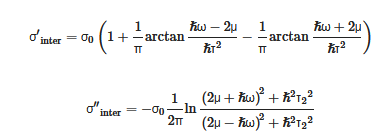
Graphene is a two-dimensional honeycomb lattice structure material that is tightly packed with a monolayer of carbon atoms on the sp2 hybrid orbital. Kx and Ky are the components of the wave vector k. As k and k’ are symmetric, and the conduction and valence bands at two points in the Brillouin zone are degenerate, the linear dispersion relationship of the energy bands of graphene is obtained. Hence, electrons can be considered to be massless relativistic particles, namely Dirac fermions, and the dispersion relation of the two-dimensional electron energy is isotropic, and is called Dirac cones. [81,82][47][48]. Graphene is a semi-metallic material with zero-band gap. The conduction band (c band) and the valence band (v band) are symmetrically tapered and intersect at one point. This band structure satisfies the Dirac equation, rather than the Schrodinger equation satisfied by traditional metals or semiconductors, and the intersection is called the Dirac point. The unique zero bandgap, excellent mechanical properties, high thermal conductivity, large specific surface area, ultra-broad photo-response spectrum, and strong nonlinear optical properties have significant advantages in novel optical and optoelectronic-sensing technologies. Graphene can be transformed from semi-metallic to metallic behavior by chemical doping or electrical gating, which depends heavily on µ (chemical potential). [83,84,85,86][49][50][51][52]. From the complex Kubo equation, a random phase approximation of the dynamic optical response of graphene can be obtained, including inter-band and intra-band contributions [87,88][53][54].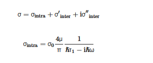
 where τ2 is the inter-band transitions rate of relaxation.
From the above formular, it can be observed that the intra-band and inter-band optical conductivities of graphene are concerned with μ and ω. The μ of doped graphene is controlled by the carrier concentration n0 = (μ/hυ)2/π, which can be controlled by chemical doping or applied voltage. There is no intra-band contribution in the µ = 0 of pristine graphene. It can be observed from the theoretical expression and experimental results of optical absorption that the intra-band photoconductivity σ in the terahertz and far-infrared bands dominates due to the appearance of the zero-band gap, while the total conductivity variation in the near-infrared and visible light bands mainly depends on the transition process between the bands. Particularly, the propagation of surface plasmons in graphene has relations to the intra-band contribution.
where τ2 is the inter-band transitions rate of relaxation.
From the above formular, it can be observed that the intra-band and inter-band optical conductivities of graphene are concerned with μ and ω. The μ of doped graphene is controlled by the carrier concentration n0 = (μ/hυ)2/π, which can be controlled by chemical doping or applied voltage. There is no intra-band contribution in the µ = 0 of pristine graphene. It can be observed from the theoretical expression and experimental results of optical absorption that the intra-band photoconductivity σ in the terahertz and far-infrared bands dominates due to the appearance of the zero-band gap, while the total conductivity variation in the near-infrared and visible light bands mainly depends on the transition process between the bands. Particularly, the propagation of surface plasmons in graphene has relations to the intra-band contribution.
32.2.2. Sensing Mechanism-SPR and Evanescent Field
The evanescent wave generated by the total reflection enters the metal layer through the interface between the metal and the medium, and interacts with free electrons to excite surface plasmon waves (SPWs) that propagate on the surface of the metal layer. SPR is a type of optical physical phenomenon. When the incident wavelength reaches a certain value (resonance), most of the incident light is converted into the energy of SPW, resulting in a sharp drop in the reflected light energy and a resonance peak in the reflection spectrum. The incident wavelength at this time is called the resonance wavelength of the SPR. By measuring the shift of the resonance wavelength, the refractive index (RI) of the sample on the surface of the metal layer can be obtained. [89][55].3. Progress of Graphene Optical Fiber Biochemical Sensor
It is well known that biochemical sensors have been widely used in medical diagnosis, environmental monitoring, food safety, genetic engineering and other fields, and the accuracy and requirements of detection are becoming higher and higher with the development of artificial intelligence technology. Graphene and its derivatives (graphene oxide-GO), as new sensitive materials, are rapidly becoming a popular material for fiber-optic biochemical sensors. Based on the two-dimensional monolayer honeycomb lattice structure, GO is formed by enriching oxygen-containing functional groups (hydroxyl and carboxyl groups) on the surface of graphene through oxidation. Compared with graphene, the functional groups of GO are highly dispersed, hydrophilic and modifiable. Its unique two-dimensional structure and abundant oxy-gen-containing functional groups on the surface have presented unique advantages in biochemical-sensing applications.
Figure 4.
Diagrams of biochemical sensors for detecting important substances in different fields.
4.1. Graphene Fiber-Grating Sensor
3.1. Graphene Fiber-Grating Sensor
43.1.1. Graphene Long-Period Fiber-Grating Sensor
Long-period fiber gratings are also called transmission fiber gratings, and their period is relatively long, which is generally hundreds of microns. In 2017, Liu Chen’s research team studied biosensors based on graphene oxide nanosheets that functionalized dual-peak long-period grating (dLPG), and proposed a label-free biosensor based on GO-coated dLPG for the real-time detection of the biological affinity between the antibody and the antigen; this was through a new GO deposition technology that combines chemical bonding and physical adsorption, enabling GO to bind to dLPG firmlyand uniformly with good stability and high repeatability.
43.1.2. Graphene Bragg Fiber-Grating Sensor
Bragg fiber gratings are also called reflective or short-period fiber gratings, and the grating period is only a few hundred nanometers. In 2014, Sridevi et al. developed a graphene oxide-etched Bragg fiber-grating biosensor for the detection of the protein concanavalin A (Con A) [97][57]. In 2015, Yao et al. developed a graphene-based D-type Bragg fiber-grating biosensor to accurately detect the concentration of red blood cell solution [98][58]. Due to the addition of graphene, the sensor demonstrates high sensitivity for detecting surrounding biochemical parameters with a value higher than 1 pm/ppm. This biochemical sensor has a compact structure, which is clinically acceptable and provides good recoverability, providing an advanced sensing platform for high sensitivity in situ and live cell detection applications.4.2. Graphene No-Core Fiber Sensor
3.2. Graphene No-Core Fiber Sensor
The classic approach of improving the sensitivity of optical fiber sensors involve corroding the structure of fiber cladding [100][59]. However, this etching method is difficult to precisely control. This will not only increase the surface roughness of the fabricated sensing fiber, but also reduce the mechanical strength of the fiber, which is not conducive to subsequent fusion and sensing applications. Moreover, researchers began to ponder the use of surface plasmon resonance (SPR) method for optical fiber-sensing for solving the above problems. The performance of the sensor can be improved by the SPR method with localized surface enhancement effect, but the plasmonic sensor needs to introduce a metal with a negative dielectric constant as the excitation condition, which will greatly increase the internal loss of the sensor and affect the sensitivity and quality factor of the sensor. The single-multi-single-mode (SMS) sensors based on the step refractive index distribution can achieve multi-mode interference. Replacing multimode fiber with coreless fiber (NCF) can greatly overcome the above-mentioned shortcomings caused by chemical corrosion, and improve the sensitivity, and repeatability at the same time.4.3. Graphene Photonic Crystal Fiber Sensor
3.3. Graphene Photonic Crystal Fiber Sensor
Photonic crystal fiber (PCF) has been proven to be a good prism. Single-mode transmission, excellent birefringence, and tunable dispersion characteristics are all advantages of its unique structure and flexible design [102][60]. The combined action of graphene and metal on PCF has been proven in studies to increase the performance of traditional PCF sensors. At present, graphene photonic crystal fiber biochemical sensors based on plasmon resonance have been well researched, providing a new path for graphene-based fiber biochemical sensors. In 2014, Dash et al. proposed a birefringent PCF-SPR biochemical sensor based on a graphene-silver layer [80][46]; graphene helps prevent the oxidation of silver used as a plasma active metal, thereby improving the sensitivity of the sensor that is higher than the widely used bimetal configuration. In 2017, Yang et al. proposed a PCF-SPR biochemical sensor based on a graphene-silver layer with a wavelength sensitivity of 2520 nmRIU−1 and a resolution of 3.97 × 10⁻⁵ RIU [103][61]. Compared with circular-shaped PCF, D-shaped PCF has an asymmetric cross-sectional structure and a wide and flat side polishing surface, which is not only conducive to the transfer and deposition of graphene, but also make it easy to reprocess the graphene on the side-polishing surface [108][62]. Chemical etching, laser etching, and side polishing can be used to create D-shaped PCF from regular PCF. In recent years, researchers have been working on graphene D-shaped photonic crystal fiber biochemical sensors based on surface plasmon resonance; graphene/metal film/polymer composite materials have been employed to adjust the side-polishing surface to enhance the sensitivity of the sensor.4. Conclusions
For the past few years, graphene-based fiber-optic biochemical sensors have demonstrated excellent performance in genetic engineering, medical diagnosis, environmental monitoring, and food safety, etc. However, the widespread application of graphene-based commercial fiber biochemical sensors still faces challenges, and the following key issues need to be solved: Firstly, the current research on graphene is still limited to theories and laboratories-the existing preparation methods of graphene are not perfect enough. How to prepare high-quality graphene with controllable size and thickness, no defect, no pollution, and uniform distribution at a low cost is one of the urgent problems to be solved. Secondly, when graphene is transferred to the surface of the fiber, it is easy to be contaminated and damaged. How to realize the lossless and high-quality coupling between graphene and fiber is the second key problem to be solved. Thirdly, graphene’s flexible photoelectric characteristics make it difficult to find a perfect solution to the cross-sensitive detection. For example, temperature can interfere with the gas sensor when measuring gas concentrations. Finally, the repeatability and stability of graphene sensors are not optimistic either. When the external environment causes the electronic structure of graphene to change, it will result in graphene taking a certain time to recover. It is difficult to automatically restore it to the original state, which leads to the poisoning of the sensor when graphene adsorbs chemical molecules.References
- Novoselov, K.S.; Geim, A.K.; Morozov, S.V.; Jiang, D.; Zhang, Y.; Dubonos, S.V.; Grigorieva, I.V.; Firsov, A.A. Electric field effect in atomically thin carbon films. Science 2004, 306, 666–669.
- Choi, W.; Lahiri, I.; Seelaboyina, R.; Kang, Y.S. Synthesis of Graphene and Its Applications: A Review. Crit. Rev. Solid State Mater. Sci. 2010, 35, 52–71.
- Chen, K.; Lu, G.; Chang, J.; Mao, S.; Yu, K.; Cui, S.; Chen, J. Hg(II) Ion Detection Using Thermally Reduced Graphene Oxide Decorated with Functionalized Gold Nanoparticles. Anal. Chem. 2012, 84, 4057–4062.
- Novodchuk, I.; Bajcsy, M.; Yavuz, M. Graphene-based field effect transistor biosensors for breast cancer detection: A review on biosensing strategies. Carbon 2021, 172, 431–453.
- Geim, A.K. Graphene: Status and Prospects. Science 2009, 324, 1530–1534.
- Wassei, J.K.; Kaner, R.B. Graphene, a promising transparent conductor. Mater. Today 2010, 13, 52–59.
- Wang, Q.; Wang, B.-T. Surface plasmon resonance biosensor based on graphene oxide/silver coated polymer cladding silica fiber. Sens. Actuators B Chem. 2018, 275, 332–338.
- Rahman, M.S.; Anower, M.S.; Abdulrazak, L.F. Utilization of a phosphorene-graphene/TMDC heterostructure in a surface plasmon resonance-based fiber optic biosensor. Photonics Nanostruct. Fundam. Appl. 2019, 35, 100711.
- Rahman, M.S.; Noor, S.S.; Anower, M.S.; Abdulrazak, L.F.; Rahman, M.M.; Rikta, K.A. Design and numerical analysis of a graphene-coated fiber-optic SPR biosensor using tungsten disulfide. Photonics Nanostruct. Fundam. Appl. 2019, 33, 29–35.
- Hong, X.D.; Zheng, H.R.; Liang, D. Stable electron field emission from graphene/hexagonal boron nitride hybrid structure. Mater. Lett. 2020, 277, 128356.
- Hossain, M.B.; Islam, M.M.; Abdulrazak, L.F.; Rana, M.M.; Akib, T.B.A.; Hassan, M. Graphene-Coated Optical Fiber SPR Biosensor for BRCA1 and BRCA2 Breast Cancer Biomarker Detection: A Numerical Design-Based Analysis. Photonic Sens. 2020, 10, 67–79.
- Song, G.; Luo, S.; Zhang, J.; Zhang, M.; Qiu, G.; Meng, A.; Lin, Y.; Li, Z. Template-free one-step synthesis of the multi-layer carbon or stacked graphene sheets coessentially coating N-doped graphene tubes and their field emission and photoluminescence properties. J. Alloys Compd. 2020, 829, 154411.
- Dusane, P.R.; Gavhane, D.S.; Kolhe, P.S.; Bankar, P.K.; Thombare, B.R.; Lole, G.S.; Kale, B.B.; More, M.A.; Patil, S.I. Controlled decoration of palladium (Pd) nanoparticles on graphene nanosheets and its superior field emission behavior. Mater. Res. Bull. 2021, 140, 111335.
- Feng, D.; Niu, Z.; Yang, J.; Xu, W.; Liu, S.; Mao, X.; Li, X. Flexible artificial synapse with relearning function based on ion gel-graphene FET. Nano Energy 2021, 90, 106526.
- Xia, Y.; Sun, Y.; Li, H.; Chen, S.; Zhu, T.; Wang, G.; Man, B.; Pan, J.; Yang, C. Plasma treated graphene FET sensor for the DNA hybridization detection. Talanta 2021, 223, 121766.
- Singh, P.; Sohi, P.A.; Kahrizi, M. In silico design and analysis of Pt functionalized graphene-based FET sensor for COVID-19 biomarkers: A DFT coupled FEM study. Phys. E Low Dimens. Syst. Nanostruct. 2022, 135, 114972.
- Xu, Y.; Xiong, M.; Yan, H. A portable optical fiber biosensor for the detection of zearalenone based on the localized surface plasmon resonance. Sens. Actuators B Chem. 2021, 336, 129752.
- Yin, M.-J.; Gu, B.; An, Q.-F.; Yang, C.; Guan, Y.L.; Yong, K.-T. Recent development of fiber-optic chemical sensors and biosensors: Mechanisms, materials, micro/nano-fabrications and applications. Coord. Chem. Rev. 2018, 376, 348–392.
- Molardi, C.; Beisenova, A.; Issatayeva, A.; Korganbayev, S.; Blanc, W.; Tosi, D. Parallel multiplexing in Optical Backscatter Reflectometry by the use of nano-particles doped optical fiber. Opt. Fibers Sens. Med. Diagn. Treat. Appl. XIX 2019, 28, 10872.
- Sypabekova, M.; Korganbayev, S.; Gonzalez-Vila, A.; Caucheteur, C.; Shaimerdenova, M.; Ayupova, T.; Bekmurzayeva, A.; Vangelista, L.; Tosi, D. Functionalized etched tilted fiber Bragg grating aptasensor for label-free protein detection. Biosens. Bioelectron. 2019, 146, 111765.
- Ribaut, C.; Loyez, M.; Larrieu, J.-C.; Chevineau, S.; Lambert, P.; Remmelink, M.; Wattiez, R.; Caucheteur, C. Cancer biomarker sensing using packaged plasmonic optical fiber gratings: Towards in vivo diagnosis. Biosens. Bioelectron. 2017, 92, 449–456.
- Krishnan, Y.; Simmel, F.C. Nucleic Acid Based Molecular Devices. Angew. Chem. Int. Ed. 2011, 50, 3124–3156.
- Rodrigues, R.C.; Ortiz, C.; Berenguer-Murcia, A.; Torres, R.; Fernandez-Lafuente, R. Modifying enzyme activity and selectivity by immobilization. Chem. Soc. Rev. 2013, 42, 6290–6307.
- Carter, P.J.; Lazar, G.A. Next generation antibody drugs: Pursuit of the ‘high-hanging fruit’. Nat. Rev. Drug Discov. 2018, 17, 197–223.
- Jhunjhunwala, S.; Hammer, C.; Delamarre, L. Antigen presentation in cancer: Insights into tumour immunogenicity and immune evasion. Nat. Rev. Cancer 2021, 21, 298–312.
- Sypabekova, M.; Aitkulov, A.; Blanc, W.; Tosi, D. Reflector-less nanoparticles doped optical fiber biosensor for the detection of Case thrombin. Biosens. Bioelectron. 2020, 165, 112365.
- Chauhan, N.; Maekawa, T.; Kumar, D.N.S. Graphene based biosensors—Accelerating medical diagnostics to new-dimensions. J. Mater. Res. 2017, 32, 2860–2882.
- Deng, X.; Tang, H.; Jiang, J. Recent progress in graphene-material-based optical sensors. Anal. Bioanal. Chem. 2014, 406, 6903–6916.
- Ferrari, A.C.; Bonaccorso, F.; Fal’ko, V.; Novoselov, K.S.; Roche, S.; Boggild, P.; Borini, S.; Koppens, F.H.L.; Palermo, V.; Pugno, N.; et al. Science and technology roadmap for graphene, related two-dimensional crystals, and hybrid systems. Nanoscale 2015, 7, 4598–4810.
- Feng, L.; Wu, L.; Qu, X. New Horizons for Diagnostics and Therapeutic Applications of Graphene and Graphene Oxide. Adv. Mater. 2013, 25, 168–186.
- Morales-Narvaez, E.; Merkoci, A. Graphene Oxide as an Optical Biosensing Platform. Adv. Mater. 2012, 24, 3298–3308.
- Xing, F.; Meng, G.-X.; Zhang, Q.; Pan, L.-T.; Wang, P.; Liu, Z.-B.; Jiang, W.-S.; Chen, Y.; Tian, J.-G. Ultrasensitive Flow Sensing of a Single Cell Using Graphene-Based Optical Sensors. Nano Lett. 2014, 14, 3563–3569.
- Nair, R.R.; Blake, P.; Grigorenko, A.N.; Novoselov, K.S.; Booth, T.J.; Stauber, T.; Peres, N.M.R.; Geim, A.K. Fine structure constant defines visual transparency of graphene. Science 2008, 320, 1308.
- Ye, Q.; Wang, J.; Liu, Z.; Deng, Z.-C.; Kong, X.-T.; Xing, F.; Chen, X.-D.; Zhou, W.-Y.; Zhang, C.-P.; Tian, J.-G. Polarization-dependent optical absorption of graphene under total internal reflection. Appl. Phys. Lett. 2013, 102, 021912.
- Xing, F.; Liu, Z.-B.; Deng, Z.-C.; Kong, X.-T.; Yan, X.-Q.; Chen, X.-D.; Ye, Q.; Zhang, C.-P.; Chen, Y.-S.; Tian, J.-G. Sensitive Real-Time Monitoring of Refractive Indexes Using a Novel Graphene-Based Optical Sensor. Sci. Rep. 2012, 2, 908.
- Huynh Vinh, P.; Nguyen Ngoc, H. Nonlinear optical absorption in graphene via two-photon absorption process. Opt. Commun. 2015, 344, 12–16.
- Pirruccio, G.; Martin Moreno, L.; Lozano, G.; Rivas, J.G. Coherent and Broadband Enhanced Optical Absorption in Graphene. ACS Nano 2013, 7, 4810–4817.
- Cai, Y.; Zhu, J.; Liu, Q.H. Tunable enhanced optical absorption of graphene using plasmonic perfect absorbers. Appl. Phys. Lett. 2015, 106, 043105.
- Wang, J.; Cheng, Z.; Shu, C.; Tsang, H.K. Optical Absorption in Graphene-on-Silicon Nitride Microring Resonators. IEEE Photonics Technol. Lett. 2015, 27, 1765–1767.
- Selvakumar, N.; Biswas, A.; Krupanidhi, S.B.; Barshilia, H.C. Enhanced optical absorption of graphene-based heat mirror with tunable spectral selectivity. Sol. Energy Mater. Sol. Cells 2018, 186, 149–153.
- Liu, T.; Zhou, C.; Xiao, S. Gain-assisted critical coupling for enhanced optical absorption in graphene. Nanotechnology 2021, 32, 205202.
- Wang, F.; Liu, X.-K.; Gao, F. Chapter 1—Fundamentals of Solar Cells and Light-Emitting Diodes. In Advanced Nanomaterials for Solar Cells and Light Emitting Diodes; Gao, F., Ed.; Elsevier: Amsterdam, The Netherlands, 2019; pp. 1–35.
- Nemade, K.R.; Waghuley, S.A. in situ synthesis of graphene/SnO2 quantum dots composites for chemiresistive gas sensing. Mater. Sci. Semicond. Processing 2014, 24, 126–131.
- Nirmalraj, P.N.; Lutz, T.; Kumar, S.; Duesberg, G.S.; Boland, J.J. Nanoscale Mapping of Electrical Resistivity and Connectivity in Graphene Strips and Networks. Nano Lett. 2011, 11, 16–22.
- Zhao, Y.; Li, X.-g.; Zhou, X.; Zhang, Y.-n. Review on the graphene based optical fiber chemical and biological sensors. Sens. Actuators B Chem. 2016, 231, 324–340.
- Dash, J.N.; Jha, R. Graphene-Based Birefringent Photonic Crystal Fiber Sensor Using Surface Plasmon Resonance. IEEE Photonics Technol. Lett. 2014, 26, 1092–1095.
- Yang, H.; Liu, M.; Chen, Y.; Guo, L.; Xiao, G.; Liu, H.; Li, J.; Yuan, L. Highly Sensitive Graphene-Au Coated Plasmon Resonance PCF Sensor. Sensors 2021, 21, 818.
- Jiang, X.Q.; Liu, Z.B.; Tian, J.G. Progress in Optical Properties and Applications of Graphene. Prog. Phys. 2017, 37, 22–36.
- Li, Z.Q.; Henriksen, E.A.; Jiang, Z.; Hao, Z.; Martin, M.C.; Kim, P.; Stormer, H.L.; Basov, D.N. Dirac charge dynamics in graphene by infrared spectroscopy. Nat. Phys. 2008, 4, 532–535.
- Wang, F.; Zhang, Y.; Tian, C.; Girit, C.; Zettl, A.; Crommie, M.; Shen, Y.R. Gate-variable optical transitions in graphene. Science 2008, 320, 206–209.
- Bao, Q.; Loh, K.P. Graphene Photonics, Plasmonics, and Broadband Optoelectronic Devices. ACS Nano 2012, 6, 3677–3694.
- Jiao, S.; Gu, S.; Yang, H.; Fang, H.; Xu, S. High y sensitive dual-core photonic crystal fiber based on a surface plasmon resonance sensor with a silver nano-continuous grating. Appl. Opt. 2018, 57, 8350–8358.
- Gusynin, V.P.; Sharapov, S.G. Transport of Dirac quasiparticles in graphene: Hall and optical conductivities. Phys. Rev. 2006, 73, 245411.
- Falkovsky, L.A.; Pershoguba, S.S. Optical far-infrared properties of a graphene monolayer and multilayer. Phys. Rev. B 2007, 76, 153410.
- Xiao, G.; Xiuhua, H.; Yang, H.; Dou, W.; Xu, J.; Wei, Q.; Li, H.; Zhang, F.; Li, Q.; Chen, Y.; et al. Plasma Refractive Index Sensor with Tunable Cross Tie-Shaped Graphene Array Structure. Acta Opt. Sin. 2019, 39, 431–439.
- Xu, Y.; Cottenden, A.; Jones, N.B. A theoretical evaluation of fibre-optic evanescent wave absorption in spectroscopy and sensors. Opt. Lasers Eng. 2006, 44, 93–101.
- Sridevi, S.; Vasu, K.S.; Jayaraman, N.; Asokan, S.; Sood, A.K. Optical bio-sensing devices based on etched fiber Bragg gratings coated with carbon nanotubes and graphene oxide along with a specific dendrimer. Sens. Actuators B Chem. 2014, 195, 150–155.
- Yao, B.C.; Wu, Y.; Webb, D.J.; Zhou, J.H.; Rao, Y.J.; Pospori, A.; Yu, C.B.; Gong, Y.; Chen, Y.F.; Wang, Z.G. Graphene-Based D-Shaped Polymer FBG for Highly Sensitive Erythrocyte Detection. IEEE Photonics Technol. Lett. 2015, 27, 2399–2402.
- Wu, Q.; Semenova, Y.; Wang, P.; Farrell, G. High sensitivity SMS fiber structure based refractometer—Analysis and experiment. Opt. Express 2011, 19, 7937–7944.
- Ortigosa-Blanch, A.; Knight, J.C.; Wadsworth, W.J.; Arriaga, J.; Mangan, B.J.; Birks, T.A.; Russell, P.S. Highly birefringent photonic crystal fibers. Opt. Lett. 2000, 25, 1325–1327.
- Yang, X.; Lu, Y.; Liu, B.; Yao, J. Analysis of Graphene-Based Photonic Crystal Fiber Sensor Using Birefringence and Surface Plasmon Resonance. Plasmonics 2017, 12, 489–496.
- Guo, C.-Y.; Wang, D.; Mu, C.-L. Progress on Optical Fiber Sensors Based on Graphene/Graphene Oxide. Laser Optoelectron. Prog. 2020, 57, 31–43.
More