Your browser does not fully support modern features. Please upgrade for a smoother experience.
Please note this is a comparison between Version 1 by Hongyan Yang and Version 3 by Lindsay Dong.
Graphene, a novel form of the hexagonal honeycomb two-dimensional carbon-based structural material with a zero-band gap and ultra-high specific surface area, has unique optoelectronic capabilities, promising a suitable basis for its application in the field of optical fiber sensing. Graphene optical fiber sensing has also been a hotspot in cross-research in biology, materials, medicine, and micro-nano devices in recent years, owing to prospective benefits, such as high sensitivity, small size, and strong anti-electromagnetic interference capability and so on.
- graphene
- optical fiber
- biochemical sensor
1. Introduction
Graphene is a two-dimensional single-crystal functional material with a hexagonal honeycomb lattice composed of carbon atoms with sp2 hybrid orbitals, and has been considered a hypothetical structure for a long time. In 2004, graphene was separated by the 2010 Nobel Prize winners Andre Geim and Konstantin Novoselov [1], who made it become a distinct entity. Three in-plane σ bonds and one out-of-plane π bond exist in each carbon atom of graphene and are responsible for out-of-plane bonding, such as the stacking of graphene sheets. [2]. The carbons inside the monolayer graphene sheet are connected by covalent bonds, while the different graphene layers are connected by van der Waals bonds. Monolayer graphene has an extremely high surface-to-volume ratio due to the ultra-thin thickness (~0.3 nm). [3]. Moreover, graphene has a very active surface area, as all the carbon atoms are located on the surface. In addition, it also has outstanding mechanical, chemical and op-tical properties. [4]. Graphene is quite sensitive to the external environment because each atom of graphene is a surface atom; therefore, graphene-based nanostructures have great potential for developing various types of sensors [5]. Because of these characteristics, in less than 20 years, graphene has been widely studied in many different fields, such as field emission displays, transparent conductors, field effect transistors (FETs), optical biochemical sensors and composite reinforcement of material, etc. [6][7][8][9][10][11][12][13][14][15][16][6,7,8,9,10,11,12,13,14,15,16].
Optical biochemical sensors have broad development prospects because of their advantages of non-destructive detection, high sensitivity and fast detection speed, etc. [17]. The optical fiber sensor, comprised of an important branch of optical sensors and integrating signal acquisition and transmission, has become a research hotspot in the field of sensing. In the past ten years, optical fiber-based biochemical sensors have attracted people’s interest due to their attractive features such as compact size, flexibility, relatively low cost, and biocompatibility [18]. Additionally, their magnetic resonance compatibility and distant and multiplexed detection capabilities [19] allow them to be used in a variety of industries [20]. The physical properties of optical fibers make it possible to measure the level of biomarkers and chemical markers in biological and chemical liquids, non-liquid media, and hard-to-reach environments for minimally invasive in vivo medical diagnosis [21]. The sensing mechanism of the fiber-optic biochemical sensor modulates the physical signal of the transmitted light in the fiber, such as refractive index (RI), intensity, amplitude and phase by using the biochemical information, which is generated by the selective interaction (i.e., specific binding of antigen-antibody or receptor-ligand; nucleic acid molecular base complementary pairing; specificity of enzyme to substrate, etc.) between the measured object and the biological sensor. Among them, the biochemical sensitive element is the key component of the sensor, and the general biochemical sensitive element is comprised of antigens, antibodies, enzymes, or oligonucleotides [22][23][24][25][22,23,24,25]. In order to improve the sensitivity of biochemical detection, the contact area between light and the biochemical sensitive element is increased by changing the optical fiber structure at a specific detection point or covering the sensitive medium material at the end of the optical fiber. Then, the transmission spectrum or reflection spectrum caused by the change of the surrounding biochemical sensitive mediumis detected by a spectral analyzer or vector analyzer [26].
2. The Sensing Properties and Mechanism of Graphene-Based Optical Fiber
Graphene has been considered as a multifunctional material because of its 2D planar structure. It has excellent potential applications in wide range of fields, such as photonics, optoelectronics, sensors, flexible electronics and nanocomposites, etc. [27][57]. In particular, the integration of graphene and its derivatives with biochemical sensor modules also can also be applied to detect different samples, such as cells, proteins, and small molecules [28][29][58,59]. The chemical derivatives of graphene, including graphene-oxide (GO), reduced-GO (rGO), few-layer graphene (FLG), wrinkled graphene (WG), hydrogenated graphene (HG), and nano-size GO etc. are also broadly applied as a components for biosensors and cancer diagnosis and treatment, etc. [30][31][60,61].2.1. Sensing Properties of Graphene
The various properties of graphene are excellent: its strength is about 200 times that of steel, its electrical and thermal conductivity are higher than copper, and its weight per square meter is less than 1 milligram. The reason why graphene has high electrical conductivity and thermal conductivityis that its room temperature electron mobility is as high as 15,000 cm2/(Vs). Because of its excellent carrier mobility and mechanical properties, graphene is widely favored by researchers in condensed matter physics and materials science. Graphene has been widely used in the field of biochemical sensing based on its good photoelectric characteristics and excellent high-energy transfer efficiency, large surface area, and biocompatibility [32][62].2.1.1. Optical Absorption Characteristics
Graphene has excellent optical absorption characteristics. The absorption rate of monolayer graphene to visible light is only 2.3% [33][63], and the reflectivity is negligible. The light absorptivity of graphene increases linearly with the graphene layers. The theoretical research indicated that monolayer, double-layer, and few-layer graphene absorb the TE (transversal electric wave) mode more than TM (transversal magnetic wave) mode in the total internal reflection structure [34][64]. The different light absorptivity of graphene leads to the reflectivity of the TM mode, which is greater than the TE mode [35][65]. Therefore, the resolution and sensitivity of the sensing system can be greatly improved by measuring the difference in the reflected light intensity between the TM mode and TE mode. Graphene exhibits strong polarization-dependent optical absorption under total internal reflection [36][66]. All of the extinction ratios are higher than 36 dB, which means that the graphene polarizer has an excellent polarization performance. For optical fiber sensors, the light absorption performance of graphene can greatly improve the sensing resolution. Therefore, improving and enhancing the light absorption properties of graphene by adopting various new technological methods has become a pursuit of researchers. [37][38][39][40][41][68,69,70,71,72].2.1.2. Photoluminescence Characteristics
Photoluminescence (PL) is an optical phenomenon that semiconductors launch light emissions by absorbing incident light, in which semiconductors emit light by absorbing incident light with energy higher than the energy band gap of the semiconductor. In the mechanism of PL, the excited electrons generated by optical excitation will return to the ground state with the emission of photons [42][73]. Graphene is a photoluminescent material. Graphene quantum dots (GQDs) are generally regarded as a new material with graphene sheet sizes less than 100 nm, the number of graphene sheet layers is less than 10. GQDs can produce a photoluminescence effect, which is the most important and most widely studied performance of graphene, and also makes graphene close to practical application [43][74].23.1.3. Optical Conductivity
Graphene’s unique structure enables it to have special properties, such as the perfect quantum tunneling effect and quantum Hall effect, etc. The optical conductivity of graphene depends on electron density, layer number and width at low temperatures. When electrons are transmitted in the graphene layer, the interference is very small and it is not easy to be scattered. The mobility can reach 2 × 105 cm2/(V·s), which is about 100 times that of the electron in silicon and the resistivity is about 10−6 Ω·cm, which is lower than that of metallic copper or silver. Therefore, it is the material with the lowest resistance at room temperature, and its conductive density is one million times that of copper. As the number of graphene layers increases, its conductivity gradually decreases, and then tends to remain unchanged at eight monolayers [44][79]. The optical conductivity of monolayer graphene is proportional to the number of layers when the light energy is larger than twice the kinetic energy of the Fermi level. Doped graphene will also increase its optical conductivity.23.1.4. Surface Plasmon Properties
Graphene is an excellent plasmon material [45][77], which is applied to optical fiber sensors by the surface plasmon effect to increase the sensitivity of biochemical sensors. The graphene-coated fiber forms a surface plasmon resonance (SPR) structure as surface plasmon. The resonant wavelength of SPR will change with the refractive index of biochemical analytes. The common materials to excite surface plasmon are noble metals, such as gold or silver. However, due to the existence of noble metal resistance, its large loss in visible light band and the complexity of metal coating process make it very easy to be oxidized and worn; therefore, the sensitivity of its sensing is greatly limited. Graphene has stable chemical properties, is not easily oxidized, and can prevent metal oxidation, increase the elastic modulus of metal surface, and promote the effective propagation of surface plasma in the infrared band. Therefore, surface plasmon-graphene can improve the sensitivity of fiber optic biochemical sensors, and has been widely used in the field of biochemical sensing [45][46][77,80].2.2. The Sensing Mechanism of Graphene-Based Optical Fiber
2.2.1. Kubo Model of Graphene
Graphene is a two-dimensional honeycomb lattice structure material that is tightly packed with a monolayer of carbon atoms on the sp2 hybrid orbital. Kx and Ky are the components of the wave vector k. As k and k’ are symmetric, and the conduction and valence bands at two points in the Brillouin zone are degenerate, the linear dispersion relationship of the energy bands of graphene is obtained. Hence, electrons can be considered to be massless relativistic particles, namely Dirac fermions, and the dispersion relation of the two-dimensional electron energy is isotropic, and is called Dirac cones. [47][48][81,82]. Graphene is a semi-metallic material with zero-band gap. The conduction band (c band) and the valence band (v band) are symmetrically tapered and intersect at one point. This band structure satisfies the Dirac equation, rather than the Schrodinger equation satisfied by traditional metals or semiconductors, and the intersection is called the Dirac point. The unique zero bandgap, excellent mechanical properties, high thermal conductivity, large specific surface area, ultra-broad photo-response spectrum, and strong nonlinear optical properties have significant advantages in novel optical and optoelectronic-sensing technologies. Graphene can be transformed from semi-metallic to metallic behavior by chemical doping or electrical gating, which depends heavily on µ (chemical potential). [49][50][51][52][83,84,85,86]. From the complex Kubo equation, a random phase approximation of the dynamic optical response of graphene can be obtained, including inter-band and intra-band contributions [53][54][87,88].
 where τ2 is the inter-band transitions rate of relaxation.
From the above formular, it can be observed that the intra-band and inter-band optical conductivities of graphene are concerned with μ and ω. The μ of doped graphene is controlled by the carrier concentration n0 = (μ/hυ)2/π, which can be controlled by chemical doping or applied voltage. There is no intra-band contribution in the µ = 0 of pristine graphene. It can be observed from the theoretical expression and experimental results of optical absorption that the intra-band photoconductivity σ in the terahertz and far-infrared bands dominates due to the appearance of the zero-band gap, while the total conductivity variation in the near-infrared and visible light bands mainly depends on the transition process between the bands. Particularly, the propagation of surface plasmons in graphene has relations to the intra-band contribution.
where τ2 is the inter-band transitions rate of relaxation.
From the above formular, it can be observed that the intra-band and inter-band optical conductivities of graphene are concerned with μ and ω. The μ of doped graphene is controlled by the carrier concentration n0 = (μ/hυ)2/π, which can be controlled by chemical doping or applied voltage. There is no intra-band contribution in the µ = 0 of pristine graphene. It can be observed from the theoretical expression and experimental results of optical absorption that the intra-band photoconductivity σ in the terahertz and far-infrared bands dominates due to the appearance of the zero-band gap, while the total conductivity variation in the near-infrared and visible light bands mainly depends on the transition process between the bands. Particularly, the propagation of surface plasmons in graphene has relations to the intra-band contribution.
23.2.2. Sensing Mechanism-SPR and Evanescent Field
The evanescent wave generated by the total reflection enters the metal layer through the interface between the metal and the medium, and interacts with free electrons to excite surface plasmon waves (SPWs) that propagate on the surface of the metal layer. SPR is a type of optical physical phenomenon. When the incident wavelength reaches a certain value (resonance), most of the incident light is converted into the energy of SPW, resulting in a sharp drop in the reflected light energy and a resonance peak in the reflection spectrum. The incident wavelength at this time is called the resonance wavelength of the SPR. By measuring the shift of the resonance wavelength, the refractive index (RI) of the sample on the surface of the metal layer can be obtained. [55][89].3. Progress of Graphene Optical Fiber Biochemical Sensor
It is well known that biochemical sensors have been widely used in medical diagnosis, environmental monitoring, food safety, genetic engineering and other fields, and the accuracy and requirements of detection are becoming higher and higher with the development of artificial intelligence technology. Graphene and its derivatives (graphene oxide-GO), as new sensitive materials, are rapidly becoming a popular material for fiber-optic biochemical sensors. Based on the two-dimensional monolayer honeycomb lattice structure, GO is formed by enriching oxygen-containing functional groups (hydroxyl and carboxyl groups) on the surface of graphene through oxidation. Compared with graphene, the functional groups of GO are highly dispersed, hydrophilic and modifiable. Its unique two-dimensional structure and abundant oxy-gen-containing functional groups on the surface have presented unique advantages in biochemical-sensing applications (Figure 1).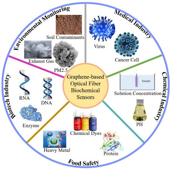
Figure 14.
Diagrams of biochemical sensors for detecting important substances in different fields.
3.1. Graphene Fiber-Grating Sensor
4.1. Graphene Fiber-Grating Sensor
34.1.1. Graphene Long-Period Fiber-Grating Sensor
Long-period fiber gratings are also called transmission fiber gratings, and their period is relatively long, which is generally hundreds of microns. In 2017, Liu Chen’s research team studied biosensors based on graphene oxide nanosheets that functionalized dual-peak long-period grating (dLPG), and proposed a label-free biosensor based on GO-coated dLPG for the real-time detection of the biological affinity between the antibody and the antigen; this was through a new GO deposition technology that combines chemical bonding and physical adsorption, enabling GO to bind to dLPG firmlyand uniformly with good stability and high repeatability.