Your browser does not fully support modern features. Please upgrade for a smoother experience.
Please note this is a comparison between Version 1 by Alanah Sheridan and Version 2 by Vivi Li.
Statins are 3-hydroxy-3-methylglutaryl coenzyme A (HMG-CoA) reductase inhibitors used worldwide to manage dyslipidaemia and thus limit the development of atherosclerotic disease and its complications. These atheroprotective drugs are now known to exert pleiotropic actions outside of their cholesterol-lowering activity, including altering immune cell function. Macrophages are phagocytic leukocytes that play critical functional roles in the pathogenesis of atherosclerosis and are directly targeted by statins. Early studies documented the anti-inflammatory effects of statins on macrophages, but emerging evidence suggests that these drugs can also enhance pro-inflammatory macrophage responses, creating an unresolved paradox.
- statins
- macrophages
- atherosclerosis
- inflammation
- cholesterol
- atorvastatin
- simvastatin
- rosuvastatin
- fluvastatin
- lovastatin
- pitavastatin
- cerivastatin
- metavastatin
- pravastatin
1. Introduction
1.1. Statins Are the Most Widely Prescribed Medications for the Prevention of Cardiovascular Disease
Cardiovascular disease (CVD) is the leading cause of mortality worldwide, accounting for an estimated 17.9 million deaths in 2019 [1], which equates to 32% of all global deaths. Atherosclerosis, a chronic inflammatory disease characterised by a narrowing of the arteries, is the main underlying cause of CVD [2] and is driven by an imbalance in lipid metabolism and a maladaptive immune response [3]. Despite its causal role in deaths globally, CVD-related mortality in the UK and other industrialised countries has declined over the last 40 years [4][5][4,5], and statins, which have revolutionized the prevention of atherosclerotic CVD, have significantly contributed to this change [6]. The efficacy of statins in the preventative treatment of CVD has led to them becoming one of the most prescribed medications worldwide, with over 200 million people taking them [7].
The clinical benefit of statins in CVD prevention is thought to be primarily driven by their lipid-lowering effects [8][9][8,9], as epidemiological studies have revealed high plasma levels of low-density lipoprotein cholesterol (LDL-C) to be a significant risk factor for atherosclerosis [10]. Mechanistically, statins inhibit cellular cholesterol biosynthesis through the inhibition of the mevalonate pathway via the rate-limiting enzyme 3-hydroxy-3-methylglutaryl coenzyme-A (HMG-CoA) reductase. In addition, statins upregulate hepatic low-density lipoprotein receptor transcription, increasing blood LDL-C removal. Together, these factors result in a 20–60% reduction in circulating LDL-C depending on the particular statin type and dose administered (Table 1).
Table 1.
Pharmacokinetic properties of different statins.
| Statin Name | Brand Name | Daily Dose (mg) | Effect on LDL Cholesterol (% Decrease) | Lipophilicity | Marketed Drug Form | Half-Life (h) | Primary Metabolizing Enzyme(s) |
|---|---|---|---|---|---|---|---|
| Atorvastatin | Lipitor | 10–80 [11][20] | 37–55 [12][13][21,22] | Lipophilic [14][15] | Acid [14][15] | 14 [11][14][15,20] | CYP3A4 [14][15] |
| Cerivastatin a | Baycol | 0.02–0.8 [15][23] | 12–42 [16][24] | Lipophilic [14][15] | Acid [14][15] | 2–4 [15][23] | CYP3A4, 2C8 [14][15][15,23] |
| Fluvastatin | Lescol | 20–80 [17][25] | 21–33 [12][13][21,22] | Lipophilic [14][15] | Acid [14][15] | 3 [17][25] | CYP2C9 [14][17][15,25] |
| Lovastatin | Mevacor | 10–80 [18][26] | 21–45 [12][21] | Lipophilic [14][15] | Lactone [14][15 |
. The different statins vary in their lipophilicity, metabolism, elimination half-lives, and potency and evidence suggests that these distinct characteristics may lead to differential effects on their efficacy (Table 1). For example, studies have suggested that the variability in different statins’ solubility affects their ability to enter cells, with lipophilic statins being found to passively diffuse into numerous cell types, whilst hydrophilic statins are hypothesized to be more liver-selective due to their dependence on membrane transporters [29][30][13,14]. These different properties have been suggested to potentially result in varying distributions of the drugs in different tissues, thereby resulting in differential effects on the mevalonate pathway [14][15].
1.2. The Central Role of Macrophages in Inflammation and CVD
Atherosclerosis is recognised as a chronic inflammatory disease characterised by a lipid imbalance and maladaptive inflammation exacerbated by the accumulation of inflammatory cells in the arterial wall. Cholesterol-laden macrophages (known as foam cells) are protagonists in the development and progression of atherosclerosis, making up the main immune cellular constituents of atherosclerotic lesions [31][32][16,17]. Foam cells contribute to the maintenance of the local endothelial inflammatory response by secreting proinflammatory cytokines and chemokines, as well as producing reactive oxygen and nitrogen species. Macrophages also engage in crosstalk with vascular smooth muscle cells, amplifying the inflammatory cycle by producing additional proinflammatory signals, promoting the growth of lipid-rich lesions [33][18]. Over time, these lesions can undergo further remodelling and form a fibrous cap, a layer of connective tissue that shields the lesion from the lumen (together with the lesion, this is known as an atherosclerotic plaque) [34][19]. Plaques can become unstable and rupture unexpectedly, exposing the lipid core to the blood and triggering thrombosis, which can result in partial or complete vessel occlusion and culminate in myocardial infarction, stroke, and other ischemic events.
Macrophages are tissue-resident leukocytes present in virtually all tissues of the body and have diverse roles, acting as both pro and anti-inflammatory mediators and being associated with the resolution of infections, tissue development, homeostasis, repair, and remodelling [35]. Macrophages display remarkable plasticity, which is shaped by their specific microenvironment [36]. Following their differentiation from monocytes, macrophages are often classified into one of two distinct functional polarization states (based on surface expression markers), M1, classically activated, or M2, alternatively activated [37]. These states represent the two extremes of a spectrum of macrophage phenotypes, describing a pro-inflammatory and anti-inflammatory/pro-resolving phenotype, respectively. Additionally, M0 is used to denote resting/non-activated cells.
M1-like activated macrophages are induced by microbial products, such as lipopolysaccharides (LPS) and toll-like receptor (TLR) ligands, or by cytokines secreted from other immune cells, such as interferon (IFN)-gamma (IFN-γ) [38] (Figure 1). These inflammatory signals trigger both transmembrane receptors (e.g., TLRs and IFN-γ receptor (IFN-γR)) and cytoplasmic receptors (e.g., nucleotide-binding and oligomerization domain (NOD)-like receptors (NLRs)). Traditionally, M1-like macrophages are functionally associated with pathogen clearance and antigen presentation to T cells to initiate the adaptive immune response, which they achieve by secreting high levels of pro-inflammatory cytokines, such as tumour necrosis factor-alpha (TNFα), interleukin (IL) 1β (IL-1β), IL-6, and IL-12, and by expressing activation markers including cluster of differentiation (CD)80, CD86, class II transactivator (CIITA), and major histocompatibility complex class II receptor (MHC-II). Pro-inflammatory macrophages also express high levels of inducible nitric oxide synthase, which enables the synthesis of nitric oxide (NO) that can, in turn, form reactive oxygen species (ROS) with microbicidal properties. The expression of these inflammatory mediators is predominantly controlled by the activation and nuclear translocation of transcription factors in response to initial receptor recognition of inflammatory stimuli. NF-κB (nuclear factor kappa-light-chain enhancer of B-cell) [39], together with STAT1 (Signal transducer and activator of transcription) [38], STAT3 [40], IRF (IFN-γ regulatory factor) [41], and AP-1 (activator protein 1) [42] are all associated with the polarization of macrophages to an M1-like phenotype.
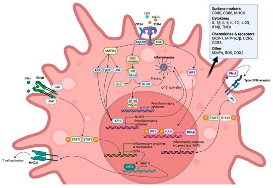
Figure 1. M1-like polarised macrophage signalling pathways (simplified) induced by toll-like receptor (TLR) and IFN-γ receptor (IFN-γR) endogenous and exogenous agonists. Created with BioRender.com, accessed on 7 March 2022.
The switch to M2-like, or alternatively activated, macrophages is mediated by factors such as IL-4 and IL-13 released from innate and adaptive immune cells [38]. M2-like macrophages are considered to be anti-inflammatory as they are noted to resolve inflammation and stimulate tissue repair. They exhibit increased expression of pro-inflammatory cytokine decoy and scavenger receptors, such as IL-1R [43], which act as molecular traps, preventing canonical signalling and thereby regulating inflammation. In addition, they secrete high levels of IL-10, transforming growth factor β, and vascular endothelial growth factor, which ameliorate the excessive activity of both innate and adaptive immune cells, stimulate fibroblast and endothelial cell proliferation, and promote blood-vessel development, allowing wound healing [38][44][38,44]. M2 polarization is also characterised by the expression of the transcription factors STAT6, SOCS1 (suppressor of cytokine signalling), and PPARγ (peroxisome proliferator-activated receptor gamma), along with the markers CD163 and CD36.
Recent evidence suggests that the M1/M2 classification system greatly oversimplifies macrophage heterogeneity. Instead, research indicates that macrophages exist on an activation spectrum with a wide array of phenotypes between these M1 and M2 extremes, dependent on their exposure to biochemical stimuli. ResWearchers refer the reader to recent reviews [45][46][47][45,46,47] for detailed discussion. Despite the evolving views of macrophage polarization, to better compare the findings of the literature referenced in this rentryview, the simplified M1/M2 nomenclature will be used as appropriate.
Atherosclerotic lesions house a heterogeneous population of macrophages, although M1-like cells are the predominant sub-type [48][49][48,49]. M1-like macrophages, expressing pro-inflammatory markers, are known to be associated with unstable and rupture-prone areas, whilst M2-like macrophages are found in stable regions [48]. M2-like macrophages have also been implicated in plaque regression in several different models suggesting that this polarization state’s enrichment may aid the resolution of atherosclerosis [50][51][52][50,51,52]. Therefore, therapeutic agents that encourage this switch from an M1 to an M2-like state, suppressing inflammation, could be a promising treatment strategy to reduce cardiovascular events [53]. Macrophages also play a central role in many other disease states and have therefore emerged as important therapeutic targets in several other pathologies, such as the development and progression of cancerous tumours [54], autoimmune disorders [55] and sepsis [56].
2. In Vitro Evidence Demonstrating the Direct Effects of Statins on Macrophages
An abundance of in vitro studies have reported paradoxical statin-mediated effects on inflammation (Table 2, Figure 23 and Figure 34), resulting from either blunting or enhancing pro-inflammatory signalling cascades. However, a limited number of studies have also reported that statins may alter the differentiation of macrophages rather than simply acting as regulators of inflammatory signalling pathways.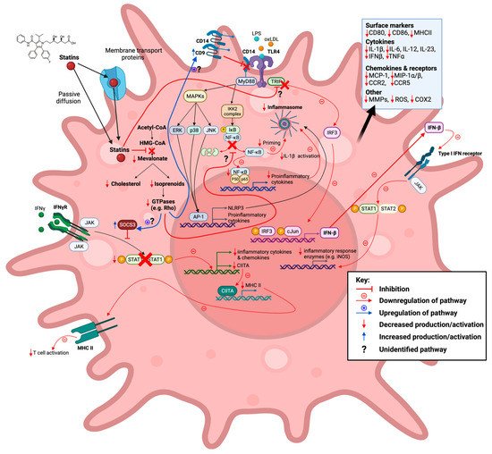
Figure 23. Statins inhibit the mevalonate pathway leading to both reduced cholesterol and isoprenoid biosynthesis, thereby also blocking farnesylation and geranylgeranylation of GTPases. Reduction in these downstream mevalonate intermediates is demonstrated to affect M1-associated macrophage inflammatory signalling pathways in vitro in an anti-inflammatory manner. This action of statins is seen in response to exogenous lipopolysaccharide (LPS), endogenous (interferon gamma (IFN-γ), and oxidized low-density lipoprotein (oxLDL) ligands. Created with BioRender.com, accessed on 7 March 2022.
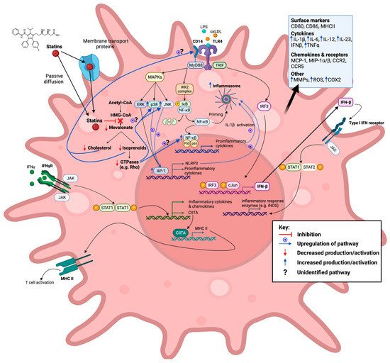
Figure 34. Statins inhibit the mevalonate pathway leading to both reduced cholesterol and isoprenoid biosynthesis, thereby also blocking farnesylation and geranylgeranylation of GTPases. Reduction in these downstream mevalonate intermediates is demonstrated to affect M1-associated macrophage inflammatory signalling pathways in vitro in a pro-inflammatory manner. This action of statins is seen in response to exogenous lipopolysaccharide (LPS) and oxidized low-density lipoprotein (oxLDL) ligands. Created with BioRender.com, accessed on 7 March 2022.
Table 2. The effects of statins on macrophages in vitro. Abbreviations: PBMC, peripheral blood mononuclear cells; BMDMs, bone-marrow derived macrophages; iNOS, inducible nitric oxide synthase; COX-2, cyclooxygenase-2; uPAR, urokinase plasminogen activator receptor; AdipoR, adiponectin receptors; mTOR, mechanistic target of rapamycin; mRNA, messenger ribonucleic acid; MWCNT, multi-walled carbon nanotubes; TF, tissue factor; GILZ, glucocorticoid-induced leucine zipper; TPA, 12-O-tetradecanoyl-phorbol-13-acetate; PMA, phorbol myristate acetate; acLDL, acetylated LDL; M-CSF, macrophage colony-stimulating factor; CC, cholesterol crystal; MSU, monosodium urate; SOD1, superoxide dismutase-1; AGE-RAGE, advanced glycation endproducts-receptor for advanced glycation endproducts; C/EBP, CCAAT/enhancer binding proteins; IP-10, interferon gamma-induced protein-10; agLDL, aggregated LDL; ETS-1, erythroblast transformation specific-1; KLF-2, Krüppel-like factor-2; ICAM-1, intercellular adhesion molecule-1.
| Statin | Model | Summary | Inflammatory Effect |
Ref. | |||||||
|---|---|---|---|---|---|---|---|---|---|---|---|
| Pro | Anti | ||||||||||
| Atorvastatin | Human PBMC derived macrophages | Statins acted as inhibitors of the induction of MHC-II expression by IFN-γ due to suppression of CIITA transcription. Statins repressed MHC-II mediated T-cell activation. | ✓ | [57] | |||||||
| Primary macrophages from B10.PL mice | Atorvastatin prevented IFN-γ induced MHC-II, CD40, CD80, and CD86 expression. | ✓ | [58] | ||||||||
| RAW 264.7 macrophages | Atorvastatin inhibited LPS and IFN-γ-induced NO formation and iNOS induction—thought to be mediated through suppression of NF-κB activation and IFN-γ through STAT1. | ✓ | [59] | ||||||||
| Murine peritoneal macrophages | Atorvastatin pretreatment enhanced TLR2 and TLR4 ligand-stimulated IL-6 and TNF production.] | 3 [14][15] | CYP3A4 [14][18] | ✓[15, | 26] | ||||||
| [ | 60 | ] | Metavastatin b | Lipophilic [19][27] | |||||||
| RAW 264.7 macrophages | Enhanced LPS-mediated MMP-9 gene expression. | ✓ | [61] | Pitavastatin | Livalo | 1–4 [20][28] | 33–44 [21][29] | Lipophilic [ | |||
| 14 | ] | RAW 264.7 macrophages[ | Atorvastatin pretreatment inhibited oxLDL-induced increase in COX-2, TNFα, and MCP-1 secretion.15 | ] | Acid [14][ | ✓15] | [62]12 [20][28] | CYP2C8, 2C9 [14][20][15,28] | |||
| Pravastatin | |||||||||||
| Pravachol | Murine BMDMs10–80 [22][30] | 20–33 [12][21] | Hydrophilic [14][15] | Acid [14][15] | 1.8 [14][15[22],30] | Atorvastatin pretreatment exacerbated LPS-induced upregulation of Il-1b, IL-6, and NLRP3 transcript levels. | ✓ | Non-CYP [14][15] | |||
| [ | 63 | ] | Rosuvastatin | Crestor | 5–40 [23][31] | ||||||
| 38–53 | [12][13][21,22] | Hydrophilic [14][15] | Acid [14][15] | 19 [14][23][15,31] | CYP2C9 [23][ | Human PBMC derived macrophages31] | |||||
| Statin treatment in combination with IL-4 during the macrophage differentiation phase led to increased M2 polarization via PPARγ activation. | ✓ | [64] | Simvastatin | Zocor | 5–80 [24][32] | 23–42 [12][13][21,22] | Lipophilic [14][15] | Lactone [14][15] | 2 [14][15] | CYP3A4 [14][24][15,32] | |
There are several statins clinically available, with atorvastatin, simvastatin, and rosuvastatin being the most popular and most widely prescribed [27][28][11,12]
| RAW 264.7 macrophages | |||||
| Atorvastatin pretreatment inhibited LPS-induced IL-1β and TNFα production in RAW 264.7 macrophages through the enhancement of autophagy. Statin treatment was seen to attenuate NLRP3 inflammasome induction in response to LPS stimulation. Atorvastatin pretreatment inhibited the expression of IL-1β in response to LPS stimulation in peritoneal murine macrophages through autophagy activation, but not that of TNFα. | |||||
| ✓ | [ | 65 | ] | ||
| Human PBMC derived macrophages | Atorvastatin reduced matrix degradation capability via reduced MMP-14 activation and uPAR localization to filipodia in LPS and IFN-γ stimulated macrophages. | ✓ | [66] | ||
| RAW 264.7 macrophages and J774 macrophages | Atorvastatin increased Rac1 GTP-loading in LPS stimulated macrophages, enhancing production of the proinflammatory cytokines IL-1β, TNFα, and IL-6. | ✓ | [67] | ||
| Human monocyte derived macrophages | Statin treatment during macrophage differentiation phase led to enhanced LPS-induced IL-1β and IL-6 secretion. | ✓ | [68] | ||
| THP1 derived macrophages | Statin treatment led to increased pro-inflammatory cytokine (IL-1β, TNFα, and IL-6) and AdipoR expression (also seen in combination with oxLDL stimulation); 24 h statin treatment resulted in increased IL-10 mRNA levels, whilst 72 h treatment resulted in decreased expression. | ✓ | [69] | ||
| Murine BMDMs | Statin-treated macrophages exhibited increased LPS-induced activation of NF-κB and IL-1β protein secretion in response to inflammasome stimulation. | ✓ | ✓ | [70] | |
| Murine BMDMs | Statin pretreatment exacerbated LPS-induced upregulation of IL-1β and NLRP3 transcript levels via p38 and mTOR. | ✓ | [71] | ||
| THP1 derived macrophages | Impaired MWCNT-elicited IL-1β secretion. | ✓ | [72] | ||
| Cerivastatin | Human PBMC derived macrophages | Cerivastatin treatment suppressed growth of macrophages expressing MMPs and TFs. | ✓ | [73] | |
| Rabbit foamy macrophages | Decreased protein expression and activity of MMP-1, MMP-2, and MMP-9. | ✓ | [74] | ||
| RAW-Blue™ cells and Murine BMDMs | Cerivastatin increased NF-κB/AP-1 activation in unstimulated and LPS-activated macrophages. LPS-induced TNF, IL-1β, and IL-6 expression was amplified. Expression of arginase-1 and GILZ was enhanced in unstimulated, LPS- and IL-4-activated macrophages. | ✓ | ✓ | [75] | |
| Fluvastatin | human PBMC derived macrophages | Fluvastatin decreased TF activity in both unstimulated and LPS-, or ac-LDL-stimulated macrophages, but enhanced IL-1β cytokine release. | ✓ | ✓ | [76] |
| Murine peritoneal macrophages and human PBMC derived macrophages | Simvastatin decreased MMP-9 protein secretion and inhibited TPA-induced enhanced MMP-9 release. | ✓ | [77] | ||
| RAW 264.7 macrophages | Fluvastatin inhibited LPS and IFN-γ-induced NO formation and iNOS induction.Thought to be mediated through suppression of NF-κB activation and IFN-γ through STAT1. | ✓ | [59] | ||
| RAW 264.7 macrophages | Fluvastatin upregulated macrophage Socs3 expression, resulting in low responsiveness to inflammatory signals (IFN-γ, IL-6, and M-CSF) due to lower activation of STAT1, STAT3, and STAT5. | ✓ | [78] | ||
| THP1 derived macrophages and THP1 derived acLDL loaded macrophages | Fluvastatin reduced both the expression, secretion, and proportion of active MMP-9 in PMA stimulated and acLDL-loaded THP1 derived macrophages. | ✓ | [79] | ||
| RAW 264.7 macrophages and murine BMDMs | Fluvastatin inhibited LPS-induced suppression of CD9, leading to reduced formation of CD14/TLR4 complexes and TNFα and MMP-9 release. | ✓ | [80] | ||
| Murine BMDMs | Fluvastatin pre-treatment exacerbated LPS-induced upregulation of IL-1b, IL-6, and NLRP3 transcript levels. Statin and LPS treatment of BMDMs harvested from NLRP3−/− mice synergistically enhanced IL-6 but did not affect IL-1β secretion. Statin treatment alone had no effect on the production of inflammatory mediators. | ✓ | [63] | ||
| Human monocyte derived macrophages | Statin treatment during macrophage differentiation phase led to enhanced LPS-induced IL-1β and IL-6 secretion. | ✓ | [68] | ||
| Murine BMDMs | Statin pretreatment exacerbated LPS-induced upregulation of IL-1b and NLRP3 transcript levels via p38 and mTOR. | ✓ | [71] | ||
| THP1 derived macrophages | Impaired MWCNT-elicited IL-1β secretion. | ✓ | [72] | ||
| Human PBMC derived macrophages | Decreased the activity of iNOS in M1 macrophages. | ✓ | [81] | ||
| Lovastatin | Rat peritoneal macrophages and microglia | Inhibited LPS-induced production of NO, TNFα, IL-1β, and IL-6 in rat primary microglia and macrophages. | ✓ | [82] | |
| Human PBMC derived macrophages | Statins acted as inhibitors of the induction of MHC-II expression by IFN-γ due to suppression of CIITA transcription. Statins repressed MHC-II mediated T-cell activation. | ✓ | [57] | ||
| RAW 264.7 macrophages | Lovastatin inhibited LPS and IFN-γ-induced NO formation and iNOS induction—thought to be mediated through suppression of NF-κB activation and IFN-γ through STAT1. | ✓ | [59] | ||
| RAW 264.7 macrophages | Lovastatin upregulated macrophage Socs3 expression, resulting in low responsiveness to inflammatory signals (IFN-γ, IL-6, and M-CSF) due to lower activation of STAT1, STAT3, and STAT5. | ✓ | [78] | ||
| Rabbit foamy macrophages | Decreased protein expression and activity of MMP-1, MMP-2, and MMP-9. | ✓ | [74] | ||
| RAW 264.7 macrophages | Lovastatin increased LPS-induced TNFα production. | ✓ | [83] | ||
| P388D1 macrophages | Statins increased production of MMP-12 in activated macrophage. | ✓ | [84] | ||
| RAW 264.7 macrophages | Lovastatin increased CD14 expression and enhanced LPS-induced membrane levels leading to greater TNFα production, but simultaneously suppressed soluble CD14. | ✓ | [85] | ||
| BMDMs from C57BL/6J mice and RAW 264.7 macrophages | Lovastatin blocked IFN-γ-induced Citta gene expression by inhibiting transcriptional events at Citta pIV, thereby suppressing MHC-II expression. | ✓ | [86] | ||
| RAW 264.7 macrophages | Lovastatin treatment induced NO release but did not affect pro-inflammatory cytokine levels in unstimulated cells. However, with LPS it synergistically enhanced IL-6, IL-12p40, IL-1β, and NO release. | ✓ | [87] | ||
| Murine BMDMs | Lovastatin pretreatment exacerbated LPS-induced upregulation of IL-1b, IL-6, and NLRP3 transcript levels. | ✓ | [63] | ||
| THP1 derived macrophages | Impaired MWCNT-elicited IL-1β secretion. | ✓ | [72] | ||
| Metavastatin | P388D1 cell line | Statins increased production of MMP-12 in activated macrophages. | ✓ | [84] | |
| U937 derived macrophages and RAW 264.7 macrophages | Metavastatin pretreatment significantly increased bacterial clearance, despite reducing oxidative burst and phagocytosis due to increased induction of extracellular traps. | ✓ | ✓ | [88] | |
| J774A.1 mouse macrophages | Increased levels of iNOS and killing of internalized S. pneumoniae. | ✓ | [89] | ||
| Pitavastatin | RAW 264.7 macrophages | Suppressed LPS-induced upregulation of MCP-1, iNOS, and IL-6 gene expression. | ✓ | [90] | |
| THP1 derived macrophages, and murine peritoneal macrophages and BMDMs (BALB/cCrSlc mice) | Pravastatin repressed mature IL-1β release elicited by MWCNT/CC/MSU exposure in THP1-derived macrophages, and LPS + MWCNT induced mature IL-1β release in peritoneal macrophages. Pravastatin pretreatment strongly enhanced mature IL-1β release in LPS + MWCNT exposed BMDMs. | ✓ | ✓ | [72] | |
| Pravastatin | Human PBMC derived macrophages | Statins acted as inhibitors of the induction of MHC-II expression by IFN-γ due to suppression of CIITA transcription. Statins repressed MHC-II mediated T-cell activation. | ✓ | [57] | |
| RAW 264.7 macrophages | Pravastatin inhibited LPS and IFN-γ-induced NO formation and iNOS induction—thought to be mediated through suppression of NF-κB activation and IFN-γ through STAT1. | ✓ | [59] | ||
| RAW 264.7 macrophages | Pravastatin upregulated macrophage Socs3 expression, resulting in low responsiveness to inflammatory signals (IFN-γ, IL-6, and M-CSF) due to lower activation of STAT1, STAT3, and STAT5. | ✓ | [78] | ||
| RAW 264.7 macrophages | Suppressed LPS-induced upregulation of MCP-1, iNOS, and IL-6 gene expression. | ✓ | [90] | ||
| Rosuvastatin | Human monocyte derived macrophages | Rosuvastatin reduced MMP-7 and MMP-9 production. | ✓ | [91] | |
| oxLDL induced THP1 foam cells | Rosuvastatin inhibited ox-LDL-induced reduction of SOD1 expression. | ✓ | [92] | ||
| THP1 derived macrophages | Rosuvastatin inhibited the AGE-RAGE axis and ROS production. | ✓ | [93] | ||
| RAW 264.7 macrophages and J774 macrophages | Rosuvastatin increased Rac1 GTP-loading in LPS-stimulated macrophages, enhancing production of the proinflammatory cytokines IL-1β, TNFα, and IL-6. | ✓ | [67] | ||
| Human monocyte derived macrophages | Statin treatment during macrophage differentiation phase led to enhanced LPS-induced IL-1β and IL-6 secretion | ✓ | [68] | ||
| THP1 derived macrophages | Statin treatment led to increased pro-inflammatory cytokine (IL-1β, TNFα, and IL-6) and AdipoR expression (also seen in combination with oxLDL stimulation); 24 h statin treatment resulted in increased IL-10 mRNA levels, whilst 72 h treatment resulted in decreased expression. | ✓ | [69] | ||
| THP1 derived macrophages | Inhibited foam cell formation and lessened the secretion of inflammatory cytokines (e.g., TNFα, IL-1β, and IL-6) from oxLDL-treated macrophages | ✓ | [94] | ||
| Simvastatin | Human monocyte derived macrophages | Simvastatin decreased superoxide production and therefore LDL oxidation | ✓ | [95] | |
| human PBMC derived macrophages | Simvastatin decreased TF activity in both unstimulated and LPS-stimulated/ac-LDL-stimulated macrophages. The suppression of TF activity induced by statin treatment was accompanied by a diminution in TF mRNA expression. | ✓ | [76] | ||
| Murine peritoneal macrophages | Simvastatin decreased MMP-9 protein secretion and inhibited TPA-induced enhanced MMP-9 release. | ✓ | [77] | ||
| Rabbit foamy macrophages | Decreased protein expression and activity of MMP-1, MMP-2, and MMP-9. | ✓ | [74] | ||
| Peritoneal murine macrophages and RAW 264.7 macrophages | Simvastatin pretreatment enhanced both IL-12p40 and TNFα LPS-induced mRNA expression and protein production by a mechanism involving the AP-1 and C/EBP transcription factors, but IP-10 levels were reduced. | ✓ | ✓ | [96] | |
| PBMC derived human macrophages | Simvastatin inhibited IFN-γ-induced upregulated mRNA expression of the chemokines MCP-1, MIP-1a, and MIP-1b and the chemokine receptors CCR1, CCR2, and CCR5. MCP-1 protein expression was also notably reduced. | ✓ | [97] | ||
| human primary monocyte derived macrophages | Statin administration significantly increased the secretion of IL-1β but had no significant effect on IL-8 or IL-6 and inhibited the secretion of TNFα. In combination with agLDL loading, statin treatment enhanced secretion of IL-1β and IL-8, but had no effect on TNFα or IL-6 secretion. | ✓ | ✓ | [98] | |
| BMDMs from C57BL/6J mice and RAW 264.7 macrophages | Simvastatin blocked IFN-γ-induced Citta gene expression by inhibiting transcriptional events at Citta pIV, thereby suppressing MHC-II expression. | ✓ | [86] | ||
| PBMC derived human macrophages and THP1 derived macrophages | Simvastatin treatment led to the downregulation of inflammatory signalling pathways, marked by a reduction in the gene expression of proinflammatory associated chemokines (MCP-1, MIP-1, and tissue factor) and transcription factors (NF-κB and ETS-1). The anti-inflammatory associated transcription factor KLF-2 had upregulated gene and protein expression. | ✓ | [99] | ||
| Murine peritoneal macrophages | Simvastatin pretreatment enhanced TLR2 and TLR4 ligand-stimulated IL-6 and TNF production. | ✓ | [60] | ||
| RAW 264.7 macrophages | Enhanced LPS-mediated MMP-9 gene expression. | ✓ | [61] | ||
| PBMC derived human macrophages, HL-60 derived macrophages and murine peritoneal macrophages (treated with simvastatin in vivo) | Simvastatin reduced phagocytosis and oxidative burst of IgG opsonized bacteria but enhanced the production of inflammatory mediators (TNFα and COX-2). No effect was seen on inflammatory mediators in response to non-opsonized bacteria, but impairment of phagocytosis remained. | ✓ | ✓ | [100] | |
| RAW 264.7 macrophages | Simvastatin pretreatment reduced basal and S. aureus-stimulated levels of C5aR and dampened macrophage sensitivity to membrane vesicles released from infected cells, decreasing TNFα production. | ✓ | [101] | ||
| RAW 264.7 macrophages and murine BMDMs | Simvastatin inhibited LPS induced suppression of CD9, leading to reduced formation of CD14/TLR4 complexes and TNFα and MMP-9 release. | ✓ | [80] | ||
| RAW 264.7 macrophages and murine BMDMs | Simvastatin pretreatment enhanced IL-12p40 and TNFα production in IFN-γ and L. monocytogenes stimulated macrophages. Statins suppressed MHC-II surface expression on IFN-γ-activated macrophages | ✓ | ✓ | [102] | |
| THP1 derived macrophages | Simvastatin pretreatment inhibited IFN-γ induced expression of MCP-1 and ICAM-1. | ✓ | [103] | ||
| Murine BMDMs and human PBMCs | Simvastatin enhanced LPS-stimulated pro-IL-1β (28 kDa form), which disrupted mature IL-1β inflammatory actions. | ✓ | [104] | ||
| Murine BMDMs | Simvastatin pretreatment exacerbated LPS-induced upregulation of IL-1b, IL-6, and NLRP3 transcript levels. | ✓ | [63] | ||
| Murine BMDMs | Simvastatin reduced parasite burden by enhancing oxidative burst and phagosome maturation. | ✓ | [105] | ||
| Raw 264.7 macrophages | Simvastatin repressed IL-1β secretion in response to H. pylori infection and increased autophagy. | ✓ | [106] | ||
| Human monocyte derived macrophages | Statin treatment during macrophage differentiation phase led to enhanced LPS-induced IL-1β and IL-6 secretion | ✓ | [68] | ||
| RAW-Blue™ cells and Murine BMDMs | Simvastatin increased NF-κB/AP-1 activation in unstimulated and LPS-activated macrophages. LPS-induced TNF, IL-1β, and IL-6 expression was amplified. Expression of arginase-1 and GILZ was enhanced in unstimulated, LPS-, and IL-4-activated macrophages. | ✓ | ✓ | [75] |
2.1. Statins Modulate TLR Inflammatory Signalling Pathways
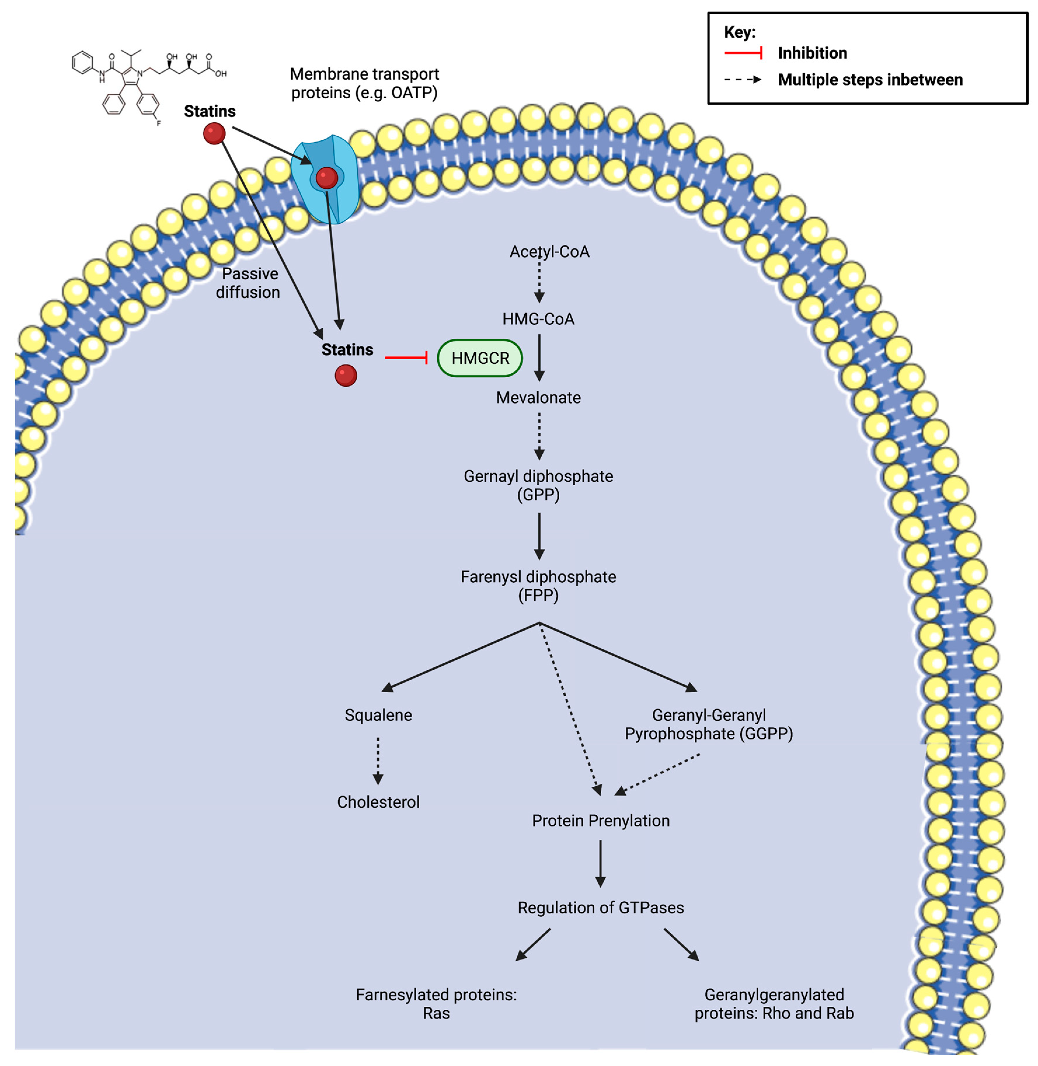
Cell surface TLRs, such as TLR1, TLR2, TLR4, TLR5, and TLR6, are key initiators of innate immune responses. They are predominantly involved in host defence mechanisms through their recognition of a diverse array of stimulatory signals related to microbial membrane components, such as lipids, lipoproteins, proteins, and LPS [107][129]. TLR engagement triggers a range of antimicrobial responses, including the production of reactive nitrogen and oxygen species, inflammatory cytokines, and matrix metalloproteinases (MMPs). However, alongside their responsiveness to exogenous ligands, TLRs also recognise endogenous ligands (e.g., oxLDL) released from damaged tissues or dead cells, thereby regulating sterile inflammatory processes [108][130]. Indeed, prolonged TLR activation has been associated with uncontrolled chronic inflammatory diseases, including atherosclerosis [109][110][111][131,132,133]. TLR4, in particular, is upregulated in atherosclerotic plaques and demonstrates increased expression as a result of ox-LDL exposure [112][113][134,135]. TLR4 signalling is mediated by the adaptor proteins myeloid differentiation primary response 88 (MyD88) and TIR-domain-containing adapter-inducing interferon-β (TRIF), which initiate two separate signal transduction pathways that culminate in the activation of a multitude of transcription factors [114][115][136,137], including members of the NF-κB [116][138] and IRF [117][139] families. MyD88-dependent signalling cascades include the activation of NF-κB and mitogen-activated protein kinase (MAPK) family members, such as extracellular signal-regulated kinase1/2, p38, and c-Jun N-terminal kinase (JNK), which, in turn, mediate the activation of AP-1 family transcription factors or the stabilization of mRNA to regulate inflammatory responses [107][129]. In contrast, TRIF-mediated TLR4 signalling occurs through the activation of IFN3 and STAT1, which induce the expression of IFN genes (e.g., IFN-B) and are also involved in late-phase NF-κB activation [116][118][138,140]. A number of accessory proteins, such as CD14 and CD36, are also suggested to play a role in macrophage inflammation cascades through their association with TLR4 [38].2.1.1. Anti-Inflammatory Modulation of TLR Signalling Pathways
As noted, NF-κB, through its activation in the TLR4 signalling pathways, is a key regulator of both macrophage inflammatory responses to pathogens and their role in sterile inflammatory diseases. Multiple statins (atorvastatin [59][81], fluvastatin [59][76][81,98], lovastatin [59][82][81,104], pravastatin [59][81], and simvastatin [76][99][98,121]) have been shown to inhibit NF-κB activation. The effects of statins on NF-κB activation are suggested to be the result of statins’ inhibition of the mevalonate pathway, specifically the isoprenoid branch, as various studies have reported that the addition of mevalonate, FPP, and GGPP reverses their action on NF-κB [76][99][98,121]. The exact links between statins’ inhibitory action on both protein prenylation and NF-κB activation have yet to be fully elucidated, although it has recently been reported that statins attenuate the degradation of the NF- κB inhibitor protein IκB [119][141]. IκB degradation is reliant on the phosphorylation of the IKK2 complex, which may be regulated by Rac1 in macrophages [120][142]. The upregulated gene and protein expression of Krüppel-like factor 2 [99][121] (a potent regulator of pro-inflammatory activation) and SOD1 [92][114] (associated with increased antioxidant enzyme activity and decreased ROS production [121][143]) have also been reported to occur in statin-treated macrophages and may contribute to the suppression of NF-κB-driven signalling pathways. Statin-mediated inhibition of the IκB/NF-κB pathway has been shown to result in a global anti-inflammatory effect on macrophages, with mRNA and protein analysis revealing the attenuated expression of many pro-inflammatory associated mediators, including cytokines (TNFα, IL-1β, and IL-6) [82][99][104,121], chemokines (MCP-1 and MIP-1α/β) [99][121], and tissue factor (a membrane-bound glycoprotein that plays a prominent role in the extrinsic pathway of blood coagulation and fibrin deposition) [76][98], and NO production [59][81][82][81,103,104]. Importantly, the inhibitory effects of statin treatment on NF-κB-induced cytokine synthesis have also been seen when using the CVD-relevant endogenous ligand oxLDL and are associated with reduced macrophage oxLDL loading and foam cell formation [62][92][94][95][84,114,116,117]. Interestingly, statin-mediated inhibition of the MyD88/NF-κB pathway has also been implicated in reducing inflammatory responses through enhancing autophagy [65][106][122][123][87,128,144,145] via the Akt-mTORC1 axis [65][122][87,144], but there are conflicting thoughts on whether this results from the inhibition of the cholesterol or isoprenoid biosynthesis branch of the mevalonate pathway [106][122][123][128,144,145]. The increased autophagy resulting from statin treatment has been noted to restrict NLRP3 (NOD-, LRR-, and pyrin domain-containing protein 3) inflammasome activation and thus reduce pro-inflammatory cytokine release [65][106][87,128]. In addition to signalling through NF-κB-dependent pathways, which are thought to be induced predominantly by MyD88-signalling, it has been proposed that statins’ inhibitory effects on macrophage inflammatory responses result from a downstream suppression of TRIF-mediated signalling [90][112]. Pravastatin and pitavastatin treatment of TLR4-stimulated RAW264 macrophages have a strong inhibitory effect on the TRIF/IRF3/IFN-β pathway in macrophages. The reduction in IFN-β expression resulting from statin treatment led to decreased STAT1 phosphorylation and the attenuation of pro-inflammatory gene expression in macrophages, evidenced by the reduced secretion of MCP-1, NO, and IL-6. Unlike previous studies, the researchers could not identify whether this action was the result of mevalonate or isoprenoid inhibition by statins, as they noted that mevalonate itself also suppressed LPS-induced expression of IFN-β [90][112]. Statin treatment has also been reported to reduce the matrix degrading capacity of M1-like polarized macrophages through the modulation of matrix metalloproteinase (MMP) expression [66][74][77][79][88,96,99,101]. This is particularly relevant to CVD, as atherosclerotic lesions show enhanced MMP expression, and this is thought to contribute to the weakening of the vascular wall, aiding plaque rupture [124][146]. Atorvastatin co-incubation during the polarization of classically activated macrophages was found to reduce MMP-14 activation [66][88], which is thought to mediate the expression of other MMPs, such as MMP-9. MMP-9 is one of the most widely investigated MMPs and is known to be involved in inflammation (e.g., extracellular processing of IL-1β [125][147]) and fibrosis in CVD [126][148]. In line with this, various studies have reported that statin treatment decreases MMP-9 protein secretion, thereby reducing its activity [77][79][99,101]. Importantly, this effect was also seen in in vitro studies of foamy macrophages [74][96], which are abundant in atherosclerotic plaques. This effect of statins is thought to be dependent on their action as mevalonate inhibitors [66][77][88,99], and there is evidence that the uncoupling of JAK/STAT signalling plays a role [79][101]. However, it should be noted that most of the studies examining statin-mediated effects on MMP expression in macrophages have not investigated the potential underlying mechanisms, and the exact point in the TLR-signalling pathway that is impacted awaits clarification. Macrophage production of MMPs in the absence of statin treatment is regulated via both the NF-κB [127][128][149,150] and MAPK [129][151] pathways. A final means by which statins are thought to blunt TLR4-induced macrophage inflammation is not via inhibition of its signalling cascade but rather via the enhancement of anti-inflammatory response elements. In this respect, it has been reported that fluvastatin and simvastatin upregulate CD9 expression in both RAW264.7 cells and murine bone-marrow derived macrophages (BMDMs) treated with LPS [80][102], consequently leading to reduced TNFα and MMP-9 production. CD9 is a recognised anti-inflammatory marker of macrophages [130][152] and negatively regulates LPS-induced macrophage activation by preventing the formation of CD14/TLR4 complexes [131][153]. Indeed, statin treatment no longer resulted in significant inhibition of TNFα and MMP-9 in BMDMs from CD9 knock-out mice, suggesting that statins’ anti-inflammatory effects are, to a degree, dependent on CD9 [80][102]. The upregulation of CD9 observed following statin treatment appears to be dependent on their inhibitory action on protein prenylation (Figure 42), specifically geranylgeranylation, as GGTI-298 (a geranylgeranyltransferase inhibitor), but not FTI-277 (a farnesyl transferase inhibitor) increased LPS-treated CD9 levels to a comparable degree. However, the precise mechanism by which decreased isoprenoid synthesis confers CD9 upregulation is currently unknown.Figure 4. Statin inhibition of 3-hydroxy-3methylglutaryl coenzyme A (HMG-CoA) reductase (HMGCR) and the subsequent implications on downstream metabolites of the mevalonate pathway, including the synthesis of cholesterol and the isoprenoids farnesyl pyrophosphate (FPP) and geranylgeranyl pyrophosphate (GGPP). Protein prenylation, via isoprenoids, is essential for the activation of small guanosine triphosphate (GTP)-binding proteins (Ras, Rho, Rac). The cellular uptake of the drug depends on its solubility. Lipophilic statins are more likely to enter the cell via passive diffusion, whereas hydrophilic statins require protein transporters, such as organic anion transporting polypeptides (OATPs) in hepatocytes. Created with BioRender.com (accessed on 7 March 2022) and Smart.Servier.com (accessed on 7 March 2022).