Direct Cardiac Reprogramming: Comparison
Please note this is a comparison between Version 2 by Jessie Wu and Version 1 by Emre Bektik.

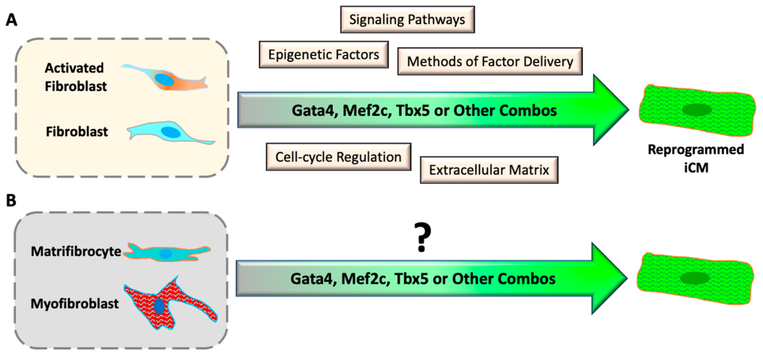
Coronary artery disease is the most common form of cardiovascular diseases, resulting in the loss of cardiomyocytes (CM) at the site of ischemic injury. To compensate for the loss of CMs, cardiac fibroblasts quickly respond to injury and initiate cardiac remodeling in an injured heart. In the remodeling process, cardiac fibroblasts proliferate and differentiate into myofibroblasts, which secrete extracellular matrix to support the intact structure of the heart, and eventually differentiate into matrifibrocytes to form chronic scar tissue. Discovery of direct cardiac reprogramming offers a promising therapeutic strategy to prevent/attenuate this pathologic remodeling and replace the cardiac fibrotic scar with myocardium in situ. Since the first discovery in 2010, many progresses have been made to improve the efficiency and efficacy of reprogramming by understanding the mechanisms and signaling pathways that are activated during direct cardiac reprogramming.

  • cardiac remodeling
  • direct cardiac reprogramming
  • heart disease
  • myocardial infarction
  • heart regeneration

1. Direct Reprogramming of Mouse Fibroblasts into iCMs In Vitro

Since lost CMs in an injured heart are replaced by cardiac fibroblasts, it will be a promising therapy for cardiac regenerative medicine if proliferated cardiac fibroblasts can be transdifferentiated into functional CMs. Transdifferentiation was initially reported in the early 1990s whereby a single transcription factor, MyoD, was sufficient to convert fibroblasts and epithelial cells into skeletal muscle cells [43][1]. After decades of efforts, the discovery of iPSCs [44,45][2][3] demonstrated that, rather than a single transcription factor, a combination of several transcription factors might be required to directly convert a terminally-differentiated cell type into another. Indeed, in 2010, Ieda et al. [17][4] cultured cardiac fibroblasts, isolated from αMHC-GFP transgenic mice, and successfully identified a minimal combination of three transcription factors (GMT: Gata4, Mef2c, and Tbx5) that could directly convert cardiac fibroblasts into induced cardiomyocyte-like cells (iCMs) without undergoing an intermediate pluripotency or progenitor state. Reprogrammed αMHC-GFP+ iCMs expressed a group of cardiac genes, e.g., Myh6, Actc1, Actn2, etc., and showed assembled sarcomere structures. Spontaneous calcium oscillations/transients were observed in many iCMs two weeks after reprogramming induction, but action potential and cell contraction were developed only in a very small population (0.01–0.1%) of reprogrammed cells.
Soon after the first study by Ieda et al. [17][4], Song et al. [21][5] included Hand2 in the GMT cocktail (GHMT), which yielded more αMHC-GFP/cardiac Troponin-T (cTnT) double-positive iCMs in adult murine tail-tip fibroblasts and cardiac fibroblasts than GMT only. Addis et al. [46][6] used a different reporter system in which a genetically encoded calcium indicator GCaMP was driven by cTnT promoter. They found that Nkx2.5 could significantly enhance the efficiency of GHMT to reprogram more functional iCMs with spontaneous calcium oscillations and beating. Protze et al. [47][7] screened triple combinations of a group of candidate transcription factors and found that an optimal combination of Mef2c, Tbx5, and Myocd could activate expression of a broader spectrum of cardiac genes than GMT did. Hirai et al. [48][8] fused MyoD domain to Mef2c and found that MyoD-Mef2c fused together with three other wild-type genes (Gata4, Hand2 and Tbx5) could yield 15-fold more beating iCMs than wild-type GHMT. In contrast to the transcription-factor-based approach, Jayawardena et al. [19][9] investigated various combinations of cardiac enriched microRNAs (miRs) for iCM induction and found that a combination of muscle-specific miRs (miR combo: miR1, miR133, miR208, and miR409) could induce iCMs from mouse cardiac fibroblasts. Moreover, JAK inhibitor improved the reprogramming capability of the miR combo. Consistently, Muraoka et al. [49][10] found that miR133 suppressed Snai1, a master regulator of epithelial-to-mesenchymal transition improved GMT-mediated iCM reprogramming, including a higher yield and shortened induction time of beating iCMs. Importantly, the reprogramming trajectory is acquired within 48 h of GMT virus infection into mouse cardiac fibroblasts [50][11], and a sufficient expression of all reprogramming factors in individual fibroblasts is critical to overcome the initial barrier for a successful iCM reprogramming [50,51][11][12].

2. In Situ Reprogramming of iCMs in The Heart

After the success of in vitro iCM-reprogramming, converting resident cardiac fibroblasts into functional CMs in situ is critical to translating this promising approach into a practical therapeutic paradigm for cardiac regenerative medicine. The first success of in vivo reprogramming was reported by Qian et al. [20][13]. They found that a local injection of GMT retroviruses into the ischemic region of the heart, right after coronary artery ligation, could reprogram new iCMs with improved ejection-fraction of the heart and decreased scar size. They utilized multiple lineage-tracing animals, including periostin-Cre and fibroblasts-specific protein 1 (Fsp1)-Cre mice, and demonstrated that in vivo reprogrammed iCMs predominantly originated from resident cardiac fibroblasts. Importantly, in vivo reprogrammed iCMs were highly similar to native CMs with a well-formed sarcomere and functionally coupled well with endogenous CMs. Meanwhile, Song et al. [21][5] also showed the success of in vivo iCM-reprogramming with GHMT factors. Most importantly, both studies have shown that in vivo generated iCMs improve the ejection-fraction and decrease scar size four weeks after MI. Additional Thymosin-β4 could improve the outcome of GMT-reprogramming and further enhance the functional recovery of the infarcted heart through improved neovascularization [52][14]. The success of in vivo reprogramming has also been reproduced with a miR combo (miR-1, miR-133, miR-208, miR-499), which decreased scar size and improved heart function in the mouse MI model [19,53][9][15]. Inagawa et al. [18][16] found that GMT viruses majorly infected fibroblasts and other non-myocyte cells within the scar, but only 3% of infected cells expressed αMHC-GFP and formed striated sarcomeres. A single polycistronic vector of three factors (GMT) with self-cleaving peptides could increase the in vivo-reprogramming yield of αMHC-GFP+/α-Actinin+ cells, of which 30% showed well-formed sarcomere structures. Consistently, Wang et al. [54][17] studied different orders of three factors in single polycistronic vector and found that a polycistronic vector of MGT, in which Mef2c is expressed at relatively higher levels than Gata4 and Tbx5, could improve the efficiency and the quality of in vitro-reprogrammed iCMs. However, MGT construct only increased the number but not the quality of in vivo-reprogrammed iCMs [55][18], suggesting that the in vivo environment of the heart has a more significant role in iCM quality than MGT itself. Nonetheless, single-cell transcriptome analysis revalidated that Mef2c is expressed relatively higher than Gata4 and Tbx5 in successfully reprogramming iCMs [56][19].

3. Direct Cardiac Reprogramming of Human Fibroblasts

Another critical step in translating direct cardiac reprogramming into a therapeutic approach is to achieve reprogramming of human cells with optimized reprogramming cocktails. Several research teams, including ours, has shown that neither GMT nor GHMT were sufficient to reprogram human fibroblasts. Wada et al. [57][20] found that the addition of Myocd and Mesp1 to GMT (GMTMM) could convert human cardiac and dermal fibroblasts into iCMs, in which the expression of cardiac genes were increased and fibroblast genes were decreased. In our lReseaboratory, we rchers re-performed a screening of 21 cardiac enriched transcription factors and found that Esrrg and Mesp1 together with GMT (5F) was sufficient to induce αMHC and cTnT double-positive iCMs from human fibroblasts [58][21]. Moreover, addition of Myocd and ZFPM2 to 5F combination (7F) further increased the yield and improved the quality of reprogrammed iCMs. Global gene expression in iCMs shifted from fibroblasts to cardiomyocyte-like state. Calcium transients and a depolarized resting membrane potential were observed in both 5F- and 7F-reprogrammed cells; however, spontaneous contraction was not observed. OuResearchers' single cell qPCR studies demonstrated that both Hand2 and miR1 could further improve the quality of 7F-reprogrammed iCMs, indicated by more activated cardiac genes and silenced fibroblast genes [59][22], although the yield of iCMs was not improved. Meanwhile, TGF-β and Wnt signaling inhibitors could improve the 7F-iCM reprogramming and induced more spontaneous calcium oscillations in 7F-iCMs [60][23]. Nam et al. [61][24] started from GHMT and screened additional transcription factors for human iCM reprogramming. They found that Myocd together with GHMT could activate more cTnT expression in human fibroblasts. They further included miR1 and miR133 and identified a reprogramming cocktail of six factors (Gata4, Hand2, Tbx5, Myocd, miR1, and miR133) that could directly convert human fibroblasts into iCMs. Spontaneous calcium oscillation was observed in 8-week reprogrammed iCMs and spontaneous contraction was observed in a very small portion of iCMs 11 weeks after reprogramming.

4. Mechanistical Understanding of Direct Cardiac Reprogramming

4.1. Activation of Signaling Pathways During Reprogramming

It has been found that different signaling pathways are involved in and directly regulate iCM reprogramming (Figure 1A). Inhibition of transforming growth factor beta (TGF-β) by SB431542 compound [64,65][25][26] could enhance GHMT-reprogramming and yield more iCMs from mouse embryonic fibroblasts (MEFs) and adult cardiac fibroblasts. Inhibition of Rho-associated kinase (ROCK) pathway increased the conversion rate of iCM-reprogramming in MEFs, tail tip fibroblasts, and cardiac fibroblasts [65][26], probably by the prevention of cell apoptosis that happens in some newly-reprogrammed iCMs [66][27]. Inhibition of Notch signaling increased the binding of Mef2c to the promoter regions of cardiac structural genes and subsequently enhanced cardiac reprogramming [67][28]. Furthermore, a combination of Wnt-inhibitor and TGF-β inhibitor could significantly augment GMT reprogramming and accelerate the conversion progression of beating iCMs [60][23]. In addition, activation of IGF1/PI3K/Akt1 signal pathway, in which mTORC1 and Foxo3a act as downstream mediators, enhanced GHMT-mediated reprogramming [68][29]. The reprogramming benefit of activation of p38 MAP kinase and PI3K/Akt pathways was consistently observed with a serum-free culture condition of FGF2, FGF10, and VEGF (FFV) [69][30]. On the other hand, suppression of inflammatory signaling has been shown to enhance direct cardiac reprogramming. For example; the zinc finger transcription factor 281 (ZNF281) enhanced cardiac reprogramming in part by repressing the inflammatory markers in adult mouse tail-tip fibroblasts [70][31]. Recently, inhibition of C-C chemokine signaling in MEFs or neonatal cardiac fibroblasts enhanced cardiac reprogramming efficiency, emphasizing a requirement of immune-response suppression for iCM-reprogramming progression [71][32]. Consistently, inhibition of inflammatory immune signaling through suppression of cyclooxygenase-2 highly improved the yield and quality of reprogrammed iCMs in neonatal and adult mouse fibroblasts [72][33], which was not observed in MEFs.
Figure 1. Progresses and challenges of direct cardiac reprogramming in vitro. (A) Understanding the mechanism of iCM reprogramming with cultured inactive and activated fibroblasts. (B) It is unknown if differentiated myofibroblasts and matrifibrocytes can be reprogrammed into iCMs.

4.2. Epigenetic Barriers of Reprogramming

Direct reprogramming of somatic cells into other type of cells, including iPSCs [73[34][35],74], neurons [75,76][36][37] and CMs, requires overcoming epigenetic barriers and the opening of chromatin structures on critical genes related to specific cell-fate identity. Indeed, the enrichment of trimethylated histone H3 of lysine 4 (H3K4me3), a mark of actively-transcribed genes, was increased and trimethylated histone H3 of lysine 27 (H3K27me3), a mark of inactive genes, was decreased in cardiac gene promoter regions as early as Day 3 of cardiac reprogramming [77][38]. Interestingly, the most effective timing of H3K27me3 diminishment to improve iCM-reprogramming was observed from Day 1 to Day 4 after virus infection, while diminishment of H3K9me2, another mark of inactivated genes, was most effective in enhancing the reprogramming from Day 3 to Day 7 [78][39], suggesting that the timing of epigenetic regulation is critical for a successful direct cardiac reprogramming. Inhibition of H3K27 methyltransferases or of polycomb repressive complex 2 (PRC2) could induce cardiac gene expression in fibroblasts; while inhibition of H3K27 demethylases blocked the induction of cardiac gene expression in miR-combo-mediated reprogramming [79][40]. Consistently, suppression of Bmi1, one component of PRC1, enhanced open chromatin states of cardiac genes, especially of Gata4, which improved cardiac reprogramming and eliminated the need for exogenous Gata4 [80][41]. Nonetheless, ZNF281, a transcriptional regulator, co-occupied cardiac gene enhancers with Gata4 and promoted GHMT(+Akt1)-mediated cardiac reprogramming in adult mouse fibroblasts through synergistic activation of diverse set of cardiac genes [70][31]. Very recently, it was found that Mef2c orchestrates chromatin accessibility of Gata4 and Tbx5 factors by recruiting them to Mef2 binding sites [81][42]. On the other hand, ectopic Gata4 was shown to be enriched at low levels on additional target gene loci and co-expression with another pioneer factor, Foxa2, increased enrichment on sampling genomic sites [82][43].

4.3. Cell-Cycle Regulation During Direct Cardiac Reprogramming

Although cardiac fibroblasts are quiescent in the healthy heart [31][44]; fibroblasts in an injured heart or in culture are quickly activated to re-enter the cell cycle. It is known that each cell-cycle phase constitutes a chain of interconnected events with a dynamic fluctuation of epigenetic chromatin modifications, including genomic DNA methylation and histone modifications [83,84][45][46]; therefore, it is important to understand how cell cycle is regulated in reprogrammed cells during direct cardiac reprogramming. Time-lapse imaging from Day 2 to Day 4 post-GMT-infection showed directly that nearly 40% of αMHC-GFP+ iCMs reprogrammed from MEFs were still active in the cell cycle at early stages of reprogramming [66][27], but αMHC-GFP+ iCMs gradually exited the cell cycle along with the progression of reprogramming. Interestingly, time-lapse imaging of early-stage reprogrammed cells suggested that iCM-reprogramming was mostly initiated at late-G1- or S-phase [66][27], indicating that a particular phase of the cell cycle might offer a more optimal epigenetic status for reprogramming induction. Indeed, depletion of Foxa2 in human foreskin fibroblasts resulted in a decreased demethylation on Foxa2 target genes in S-phase, but not in G1-arrested cells [82][43], suggesting that a loosened structure of S-phase chromosomes assists transcription factors access to their targeted genes. In our study, cCell-cycle synchronization at S-phase facilitated cell-cycle exit and accelerated iCM reprogramming with enhanced expression of cardiac genes [66][27]. Other studies have also demonstrated that cell-cycle exit is critical for the maturation of reprogrammed iCMs from MEFs and mouse cardiac fibroblasts [66[27][47],85], while overexpression of large T antigen, which endows cardiac fibroblasts a constitutive proliferation ability, inhibited reprogramming induction [56][19]. Recently, by single-cell analysis techniques, critical function of cell-cycle exit is validated in iCM reprogramming of human cardiac fibroblasts [86][48]. In summary, cell-cycle exit, which is a developmentally required process of CM maturation in mammalian hearts [87][49], is an important and necessary event for direct cardiac reprogramming.

4.4. Modification of Extracellular Matrix

Extracellular matrix (ECM) has a significant impact on the function of cardiomyocytes. Matrices that mimic mechanical stiffness of the developing heart is optimal for promoting actomyosin striation and for transmitting contraction of cardiomyocytes to the matrix; neither harder matrices, which mimic the ECM of infarcted heart, nor softer matrices are good for developing well organized sarcomeres in cardiomyocytes [88,89][50][51]. Manipulating the stiffness of culture matrix could improve the functional maturation of human iPSC-derived cardiomyocytes [90][52]. Indeed, ECM also plays an important role in cardiac reprogramming. Although adjusting the substrate stiffness alone didn’t improve reprogramming yield, a micro-grooved substrate could enhance reprogramming efficiency and yielded more mature iCMs with improved quality, including the higher expression of cardiac genes and more beating cells, through an increased nuclear localization of Mkl1, a mechanosensitive transcription factor [91][53]. Li et al. [92][54] encapsulated reprogrammed fibroblasts in three-dimensional (3D) hydrogels and found that the 3D tissue bundle environment increased the expression of matrix metalloproteinases (MMPs) and enhanced direct cardiac reprogramming with increased yield and improved quality of iCMs. These findings demonstrate the importance of a proper extracellular-matrix for reprogramming, which is one of the mysterious in vivo environmental factors in the native heart. Although these studies partly mimic the in vivo environment of a scar, an actual scar exhibits different levels of stiffness [93][55] with different amounts of collagen deposition [94][56] at various time points after infarction, which may affect reprogramming quality differently. Therefore, a specific period of post-MI time window may have an optimal stiffness environment to maximize the reprogramming efficiency in vivo. Indeed, infarcted myocardium progressively increased stiffness within a month and the highest level of regeneration was achieved at Day 7–14 post-MI, which provided an optimal stiffness for the endothelial progenitor lineage commitment of transplanted bone-marrow mononuclear cells [93][55]. Therefore, future reprogramming studies should pay greater attention to the mechanical microenvironments in cell cultures and in vivo studies.

References

  1. Choi, J.; Costa, M.L.; Mermelstein, C.S.; Chagas, C.; Holtzer, S.; Holtzer, H. MyoD converts primary dermal fibroblasts, chondroblasts, smooth muscle, and retinal pigmented epithelial cells into striated mononucleated myoblasts and multinucleated myotubes. Proc. Natl. Acad. Sci. USA 1990, 87, 7988–7992.
  2. Takahashi, K.; Yamanaka, S. Induction of pluripotent stem cells from mouse embryonic and adult fibroblast cultures by defined factors. Cell 2006, 126, 663–676.
  3. Takahashi, K.; Tanabe, K.; Ohnuki, M.; Narita, M.; Ichisaka, T.; Tomoda, K.; Yamanaka, S. Induction of pluripotent stem cells from adult human fibroblasts by defined factors. Cell 2007, 131, 861–872.
  4. Ieda, M.; Fu, J.D.; Delgado-Olguin, P.; Vedantham, V.; Hayashi, Y.; Bruneau, B.G.; Srivastava, D. Direct Reprogramming of Fibroblasts into Functional Cardiomyocytes by Defined Factors. Cell 2010, 142, 375–386.
  5. Song, K.; Nam, Y.J.; Luo, X.; Qi, X.; Tan, W.; Huang, G.N.; Acharya, A.; Smith, C.L.; Tallquist, M.D.; Neilson, E.G.; et al. Heart repair by reprogramming non-myocytes with cardiac transcription factors. Nature 2012, 485, 599–604.
  6. Addis, R.C.; Ifkovits, J.L.; Pinto, F.; Kellam, L.D.; Esteso, P.; Rentschler, S.; Christoforou, N.; Epstein, J.A.; Gearhart, J.D. Optimization of direct fibroblast reprogramming to cardiomyocytes using calcium activity as a functional measure of success. J. Mol. Cell. Cardiol. 2013, 60, 97–106.
  7. Protze, S.; Khattak, S.; Poulet, C.; Lindemann, D.; Tanaka, E.M.; Ravens, U. A new approach to transcription factor screening for reprogramming of fibroblasts to cardiomyocyte-like cells. J. Mol. Cell. Cardiol. 2012, 53, 323–332.
  8. Hirai, H.; Katoku-Kikyo, N.; Keirstead, S.A.; Kikyo, N. Accelerated direct reprogramming of fibroblasts into cardiomyocyte-like cells with the MyoD transactivation domain. Cardiovasc. Res. 2013, 100, 105–113.
  9. Jayawardena, T.M.; Egemnazarov, B.; Finch, E.A.; Zhang, L.; Payne, J.A.; Pandya, K.; Zhang, Z.; Rosenberg, P.; Mirotsou, M.; Dzau, V.J. MicroRNA-mediated in vitro and in vivo direct reprogramming of cardiac fibroblasts to cardiomyocytes. Circ. Res. 2012, 110, 1465–1473.
  10. Muraoka, N.; Yamakawa, H.; Miyamoto, K.; Sadahiro, T.; Umei, T.; Isomi, M.; Nakashima, H.; Akiyama, M.; Wada, R.; Inagawa, K.; et al. MiR-133 promotes cardiac reprogramming by directly repressing Snai1 and silencing fibroblast signatures. EMBO J. 2014, 33, 1565–1581.
  11. Stone, N.R.; Gifford, C.A.; Thomas, R.; Pratt, K.J.B.; Samse-Knapp, K.; Mohamed, T.M.A.; Radzinsky, E.M.; Schricker, A.; Yu, P.; Ivey, K.N.; et al. Unique Transcription Factor Functions Regulate Epigenetic and Transcriptional Dynamics During Cardiac Reprogramming. bioRxiv 2019.
  12. Zhang, Z.; Zhang, A.D.; Kim, L.J.; Nam, Y.J. Ensuring expression of four core cardiogenic transcription factors enhances cardiac reprogramming. Sci. Rep. 2019, 9, 6362.
  13. Qian, L.; Huang, Y.; Spencer, C.I.; Foley, A.; Vedantham, V.; Liu, L.; Conway, S.J.; Fu, J.-d.D.; Srivastava, D. In vivo reprogramming of murine cardiac fibroblasts into induced cardiomyocytes. Nature 2012, 485, 593–598.
  14. Srivastava, D.; Ieda, M.; Fu, J.; Qian, L. Cardiac repair with thymosin β4 and cardiac reprogramming factors. Ann. N. Y. Acad. Sci. 2012, 1270, 66–72.
  15. Jayawardena, T.M.; Finch, E.A.; Zhang, L.; Zhang, H.; Hodgkinson, C.P.; Pratt, R.E.; Rosenberg, P.B.; Mirotsou, M.; Dzau, V.J. MicroRNA induced cardiac reprogramming in vivo: Evidence for mature cardiac myocytes and improved cardiac function. Circ. Res. 2015, 116, 418–424.
  16. Inagawa, K.; Miyamoto, K.; Yamakawa, H.; Muraoka, N.; Sadahiro, T.; Umei, T.; Wada, R.; Katsumata, Y.; Kaneda, R.; Nakade, K.; et al. Induction of cardiomyocyte-like cells in infarct hearts by gene transfer of Gata4, Mef2c, and Tbx5. Circ. Res. 2012, 111, 1147–1156.
  17. Wang, L.; Liu, Z.; Yin, C.; Asfour, H.; Chen, O.; Li, Y.; Bursac, N.; Liu, J.; Qian, L. Stoichiometry of Gata4, Mef2c, and Tbx5 Influences the Efficiency and Quality of Induced Cardiac Myocyte Reprogramming. Circ. Res. 2015, 116, 237–244.
  18. Ma, H.; Wang, L.; Yin, C.; Liu, J.; Qian, L. In vivo cardiac reprogramming using an optimal single polycistronic construct. Cardiovasc. Res. 2015, 108, 217–219.
  19. Liu, Z.; Wang, L.; Welch, J.D.; Ma, H.; Zhou, Y.; Vaseghi, H.R.; Yu, S.; Wall, J.B.; Alimohamadi, S.; Zheng, M.; et al. Single-cell transcriptomics reconstructs fate conversion from fibroblast to cardiomyocyte. Nature 2017.
  20. Wada, R.; Muraoka, N.; Inagawa, K.; Yamakawa, H.; Miyamoto, K.; Sadahiro, T.; Umei, T.; Kaneda, R.; Suzuki, T.; Kamiya, K.; et al. Induction of human cardiomyocyte-like cells from fibroblasts by defined factors. Proc. Natl. Acad. Sci. USA 2013, 110, 12667–12672.
  21. Fu, J.-D.D.; Stone, N.R.; Liu, L.; Spencer, C.I.; Qian, L.; Hayashi, Y.; Delgado-Olguin, P.; Ding, S.; Bruneau, B.G.; Srivastava, D. Direct reprogramming of human fibroblasts toward a cardiomyocyte-like state. Stem Cell Rep. 2013, 1, 235–247.
  22. Bektik, E.; Dennis, A.; Prasanna, P.; Madabhushi, A.; Fu, J.-D.D. Single cell qPCR reveals that additional HAND2 and microRNA-1 facilitate the early reprogramming progress of seven-factor-induced human myocytes. PLoS ONE 2017, 12, e0183000.
  23. Mohamed, T.M.A.; Stone, N.R.; Berry, E.C.; Radzinsky, E.; Huang, Y.; Pratt, K.; Ang, Y.-S.; Yu, P.; Wang, H.; Tang, S.; et al. Chemical Enhancement of In Vitro and In Vivo Direct Cardiac Reprogramming Clinical Perspective. Circulation 2016, 135, 978–995.
  24. Nam, Y.J.; Song, K.; Luo, X.; Daniel, E.; Lambeth, K.; West, K.; Hill, J.A.; DiMaio, J.M.; Baker, L.A.; Bassel-Duby, R.; et al. Reprogramming of human fibroblasts toward a cardiac fate. Proc. Natl. Acad. Sci. USA 2013, 110, 5588–5593.
  25. Ifkovits, J.L.; Addis, R.C.; Epstein, J.A.; Gearhart, J.D. Inhibition of TGFbeta signaling increases direct conversion of fibroblasts to induced cardiomyocytes. PLoS ONE 2014, 9, e89678.
  26. Zhao, Y.; Londono, P.; Cao, Y.; Sharpe, E.J.; Proenza, C.; O’Rourke, R.; Jones, K.L.; Jeong, M.Y.; Walker, L.A.; Buttrick, P.M.; et al. High-efficiency reprogramming of fibroblasts into cardiomyocytes requires suppression of pro-fibrotic signalling. Nat. Commun. 2015, 6, 8243.
  27. Bektik, E.; Dennis, A.; Pawlowski, G.; Zhou, C.; Maleski, D.; Takahashi, S.; Laurita, K.R.; Deschênes, I.; Fu, J.-D.D. S-phase Synchronization Facilitates the Early Progression of Induced-Cardiomyocyte Reprogramming through Enhanced Cell-Cycle Exit. Int. J. Mol. Sci. 2018, 19, 1364.
  28. Abad, M.; Hashimoto, H.; Zhou, H.; Morales, M.G.; Chen, B.; Bassel-Duby, R.; Olson, E.N. Notch Inhibition Enhances Cardiac Reprogramming by Increasing MEF2C Transcriptional Activity. Stem Cell Rep. 2017, 8, 548–560.
  29. Zhou, H.; Dickson, M.E.; Kim, M.S.; Bassel-Duby, R.; Olson, E.N. Akt1/protein kinase B enhances transcriptional reprogramming of fibroblasts to functional cardiomyocytes. Proc. Natl. Acad. Sci. USA 2015, 112, 11864–11869.
  30. Yamakawa, H.; Muraoka, N.; Miyamoto, K.; Sadahiro, T.; Isomi, M.; Haginiwa, S.; Kojima, H.; Umei, T.; Akiyama, M.; Kuishi, Y.; et al. Fibroblast Growth Factors and Vascular Endothelial Growth Factor Promote Cardiac Reprogramming under Defined Conditions. Stem Cell Rep. 2015, 5, 1128–1142.
  31. Zhou, H.; Morales, M.G.; Hashimoto, H.; Dickson, M.E.; Song, K.; Ye, W.; Kim, M.S.; Niederstrasser, H.; Wang, Z.; Chen, B.; et al. ZNF281 enhances cardiac reprogramming by modulating cardiac and inflammatory gene expression. Genes Dev. 2017, 31, 1770–1783.
  32. Yijing, G.; Ienglam, L.; Shuo, T.; Wenbin, G.; Karatas, H.; Yangbing, L.; Shaomeng, W.; Liu, L.; Zhong, W. Enhancing Cardiac Reprogramming by Suppressing Specific C-C Chemokine Signaling Pathways. bioRxiv 2019.
  33. Muraoka, N.; Nara, K.; Tamura, F.; Kojima, H.; Yamakawa, H.; Sadahiro, T.; Miyamoto, K.; Isomi, M.; Haginiwa, S.; Tani, H.; et al. Role of cyclooxygenase-2-mediated prostaglandin E2-prostaglandin E receptor 4 signaling in cardiac reprogramming. Nat. Commun. 2019, 10, 674.
  34. Soufi, A.; Donahue, G.; Zaret, K.S. Facilitators and impediments of the pluripotency reprogramming factors’ initial engagement with the genome. Cell 2012, 151, 994–1004.
  35. Chronis, C.; Fiziev, P.; Papp, B.; Butz, S.; Bonora, G.; Sabri, S.; Ernst, J.; Plath, K. Cooperative Binding of Transcription Factors Orchestrates Reprogramming. Cell 2017, 168, 442–459.
  36. Wapinski, O.L.; Vierbuchen, T.; Qu, K.; Lee, Q.Y.; Chanda, S.; Fuentes, D.R.; Giresi, P.G.; Ng, Y.H.; Marro, S.; Neff, N.F.; et al. Hierarchical mechanisms for direct reprogramming of fibroblasts to neurons. Cell 2013, 155, 621–635.
  37. Chanda, S.; Ang, C.E.; Davila, J.; Pak, C.; Mall, M.; Lee, Q.Y.; Ahlenius, H.; Jung, S.W.; Sudhof, T.C.; Wernig, M. Generation of induced neuronal cells by the single reprogramming factor ASCL1. Stem Cell Rep. 2014, 3, 282–296.
  38. Liu, Z.; Chen, O.; Zheng, M.; Wang, L.; Zhou, Y.; Yin, C.; Liu, J.; Qian, L. Re-patterning of H3K27me3, H3K4me3 and DNA methylation during fibroblast conversion into induced cardiomyocytes. Stem Cell Res. 2016, 16, 507–518.
  39. Hirai, H.; Kikyo, N. Inhibitors of suppressive histone modification promote direct reprogramming of fibroblasts to cardiomyocyte-like cells. Cardiovasc. Res. 2014, 102, 188–190.
  40. Dal-Pra, S.; Hodgkinson, C.P.; Mirotsou, M.; Kirste, I.; Dzau, V.J. Demethylation of H3K27 Is Essential for the Induction of Direct Cardiac Reprogramming by miR Combo. Circ. Res. 2017, 120, 1403–1413.
  41. Zhou, Y.; Wang, L.; Vaseghi, H.R.; Liu, Z.; Lu, R.; Alimohamadi, S.; Yin, C.; Fu, J.-D.D.; Wang, G.G.; Liu, J.; et al. Bmi1 Is a Key Epigenetic Barrier to Direct Cardiac Reprogramming. Cell Stem Cell 2016, 18, 382–395.
  42. Hashimoto, H.; Wang, Z.; Garry, G.A.; Malladi, V.S.; Botten, G.A.; Ye, W.; Zhou, H.; Osterwalder, M.; Dickel, D.E.; Visel, A.; et al. Cardiac Reprogramming Factors Synergistically Activate Genome-wide Cardiogenic Stage-Specific Enhancers. Cell Stem Cell 2019.
  43. Donaghey, J.; Thakurela, S.; Charlton, J.; Chen, J.S.; Smith, Z.D.; Gu, H.; Pop, R.; Clement, K.; Stamenova, E.K.; Karnik, R.; et al. Genetic determinants and epigenetic effects of pioneer-factor occupancy. Nat. Genet. 2018, 50, 250–258.
  44. Souders, C.A.; Bowers, S.L.; Baudino, T.A. Cardiac fibroblast: The renaissance cell. Circ. Res. 2009, 105, 1164–1176.
  45. Brown, S.E.; Fraga, M.F.; Weaver, I.C.; Berdasco, M.; Szyf, M. Variations in DNA methylation patterns during the cell cycle of HeLa cells. Epigenetics 2007, 2, 54–65.
  46. Bou Kheir, T.; Lund, A.H. Epigenetic dynamics across the cell cycle. Essays Biochem. 2010, 48, 107–120.
  47. Zhou, Y.; Wang, L.; Liu, Z.; Alimohamadi, S.; Yin, C.; Liu, J.; Qian, L. Comparative Gene Expression Analyses Reveal Distinct Molecular Signatures between Differentially Reprogrammed Cardiomyocytes. Cell Rep. 2017, 20, 3014–3024.
  48. Zhou, Y.; Liu, Z.; Welch, J.D.; Gao, X.; Wang, L.; Garbutt, T.; Keepers, B.; Ma, H.; Prins, J.F.; Shen, W.; et al. Single-Cell Transcriptomic Analyses of Cell Fate Transitions during Human Cardiac Reprogramming. Cell Stem Cell 2019.
  49. Takeuchi, T. Regulation of cardiomyocyte proliferation during development and regeneration. Dev. Growth Differ. 2014, 56, 402–409.
  50. Chopra, A.; Lin, V.; McCollough, A.; Atzet, S.; Prestwich, G.D.; Wechsler, A.S.; Murray, M.E.; Oake, S.A.; Kresh, J.Y.; Janmey, P.A. Reprogramming cardiomyocyte mechanosensing by crosstalk between integrins and hyaluronic acid receptors. J. Biomech. 2012, 45, 824–831.
  51. Engler, A.J.; Carag-Krieger, C.; Johnson, C.P.; Raab, M.; Tang, H.Y.; Speicher, D.W.; Sanger, J.W.; Sanger, J.M.; Discher, D.E. Embryonic cardiomyocytes beat best on a matrix with heart-like elasticity: Scar-like rigidity inhibits beating. J. Cell Sci. 2008, 121, 3794–3802.
  52. Ribeiro, A.J.; Ang, Y.S.; Fu, J.D.; Rivas, R.N.; Mohamed, T.M.; Higgs, G.C.; Srivastava, D.; Pruitt, B.L. Contractility of single cardiomyocytes differentiated from pluripotent stem cells depends on physiological shape and substrate stiffness. Proc. Natl. Acad. Sci. USA 2015, 112, 12705–12710.
  53. Sia, J.; Yu, P.; Srivastava, D.; Li, S. Effect of biophysical cues on reprogramming to cardiomyocytes. Biomaterials 2016, 103, 1–11.
  54. Li, Y.Z.; Dal-Pra, S.; Mirotsou, M.; Jayawardena, T.M.; Hodgkinson, C.P.; Bursac, N.; Dzau, V.J. Tissue-engineered 3-dimensional (3D) microenvironment enhances the direct reprogramming of fibroblasts into cardiomyocytes by microRNAs. Sci. Rep. 2016, 6.
  55. Zhang, S.; Sun, A.; Ma, H.; Yao, K.; Zhou, N.; Shen, L.; Zhang, C.; Zou, Y.; Ge, J. Infarcted myocardium-like stiffness contributes to endothelial progenitor lineage commitment of bone marrow mononuclear cells. J. Cell. Mol. Med. 2011, 15, 2245–2261.
  56. Voorhees, A.P.; DeLeon-Pennell, K.Y.; Ma, Y.; Halade, G.V.; Yabluchanskiy, A.; Iyer, R.P.; Flynn, E.; Cates, C.A.; Lindsey, M.L.; Han, H.C. Building a better infarct: Modulation of collagen cross-linking to increase infarct stiffness and reduce left ventricular dilation post-myocardial infarction. J. Mol. Cell. Cardiol. 2015, 85, 229–239.
More
Video Production Service