Metabolic syndrome (MetS) constitutes a cluster of at least three out of five of the conditions including central obesity, high blood pressure, high blood sugar, high serum triglycerides, and low serum high-density lipoprotein (HDL). Patients diagnosed with MetS exhibit hallmarks of redox imbalance while oxidative stress is now perceived as both the cause and the consequence of MetS.
- oxidative stress
- metabolic syndrome
- reactive oxygen species
1. 1. Introduction
Metabolic syndrome (MetS) constitutes a cluster of at least three out of five of the conditions including central obesity, high blood pressure, high blood sugar, high serum triglycerides, and low serum high-density lipoprotein (HDL). It is estimated that at least one third of European and American populations and 27% of Chinese population suffer from MetS [1][2]. The most popular definition used for surveys and health care plan is by IDF (International Diabetes Federation) 2006 [3][4]:
Waist > 94 cm (men) or > 80 cm (women) in Europe, > 102 cm (men) or > 88cm (women) in USA, > 90 cm (men) or > 80 cm (women) in Asia, along with the presence of 2 or more of the following:
Blood glucose greater than 5.6 mmol/L (100 mg/dl) or diagnosed diabetes
HDL cholesterol < 1.0 mmol/L (40 mg/dl) in men, < 1.3 mmol/L (50 mg/dl) in women or drug treatment for low HDL-C
Blood triglycerides (TG) > 1.7 mmol/L (150 mg/dl) or drug treatment for elevated triglycerides
Blood pressure > 130/85 mmHg or drug treatment for hypertension (HT)
The incidence of MetS is rising worldwide contributing not only to the increased morbidity and mortality but also to the dramatic increase of treatment costs. Except for pharmacological management, current therapeutic methods include primarily lifestyle changes and diet. Unfortunately, most patients do not follow these recommendations. Bariatric surgery, reserved for patients with morbid obesity, made significant progress in the treatment of obesity and related metabolic disorders, however, it is associated with serious risk and side effects. OxS is a vital phenomenon occurring in plethora of metabolic disorders including type 2 diabetes (T2DM), obesity and cancer [5].
2. Overview of Oxidative Str
2. Overview of Oxidative Stress
ess
OxS, as a prolonged state of a disbalance between the oxidative and antioxidative systems of the cells, results in the overproduction of free radicals and reactive oxygen species (ROS), e.g., superoxide anion (O−•2−•), hydroxyl radical (•OH), singlet oxygen1 (21O2), hydrogen peroxide (H2O2), hypochlorous radical (C−lO−), peroxinitrite radical (ON−OO−), and nitric oxide (NO)). Nevertheless, O−•2−• constitutes the precursor for H2O2, •OH, and ON−OO− [6]. ROS could attack virtually all types of biological molecules, leading to further cellular and tissue damage. Lipid peroxidation (LPO) is an autocatalytic process generating reactive aldehydes, e.g., malondialdehyde (MDA), trans-4-oxo-2-nonenal (4-ONE) and trans-4-hydroxy-2-nonenal (4-HNE). Protein carbonylation appears due to protein oxidation via ROS (“direct protein carbonylation” of prolines, lysines, threonines, and arginines). The “secondary protein carbonylation” occurs non-oxidatively within side chains of arginines, lysines, and cysteines with adduction of reactive carbonyl species originating from products of LPO as well as autooxidation of carbohydrates (methylglyoxal, glyoxal) [7]. Genotoxic stress is elicited upon oxidative damage to nitrogenous bases within DNA. The most well-known one, 8-oxo-7,8-dihydroguanine (8-oxoG), is perceived as an indicator of whole-body marker of OxS in urine [8].
In general, elevated level of ROS is capable of posing tremendous threat to each cell due to altering its function, metabolism, cell cycle as well as introducing genetic mutations and triggering apoptosis. Importantly, ROS modulate function of cells through modifying proteins at the posttranslational level via phosphorylation and sulfonylation, nitrosylation, carbonylation, and glutathionylation [9][10]. OxS is generated by incomplete reduction of oxygen as electrons flow from one complex to the next in various dynamic intracellular organelles such as the endoplasmic reticulum (ER), lysosomes and mitochondria as by-products of oxidative protein folding, dysfunctional autophagy, mitochondrial respiration and detoxification [5][11]. The essential sources of ROS are the following enzymes: the nicotinamide adenine dinucleotide phosphate (NADPH) oxidase family of enzymes (NOX), as well as cyclooxygenases, xanthine oxidase (XO), myeloperoxidase (MPO), lipooxygenases and nitric oxide synthase (NOS) [12]. Enzymes critical for the strategy of preventing ROS formation are: superoxide dismutases (SODs), glutathione peroxidases (GPx), catalase (CAT) and glutathione reductase (GR) [1 3
Secondly, carbonyl groups are indirectly introduced into proteins by non-oxidative covalent adduction of reactive car-bonyl species (RCS) to side chains of the nucleophilic amino acids arginine, lysine, and cysteine. Secondly, carbonyl groups are indirectly introduced into proteins by non-oxidative covalent adduction of reactive car-bonyl species (RCS) to side chains of the nucleophilic amino acids arginine, lysine, and cysteine. In general, ROS are capable of posing tremendous threat to each cell due to altering its function, metabolism, cell cycle as well as introducing genetic mutations and triggering apoptosis. OxS is generated by incomplete reduction of oxygen as electrons flow from one complex to the next in various dynamic intracellular organelles such as the endoplasmic reticulum, lysosomes and mitochondria as by-products of oxidative protein folding, dysfunctional autophagy, mitochondrial respiration and detoxification [10][36]. The essential sources of ROS generated also within the vascular system are the following enzymes : the nicotinamide adenine dinucleotide phosphate (NADPH) oxidase family of enzymes (Nox), as well as cyclooxygenases, xanthine oxidase (XO), myeloperoxidase (MPO), lipooxygenases and nitric oxide synthase (NOS) [37]. The reactive molecules are formed by reduction–oxidation (redox) reactions in response to the activation of several types of oxidases, that catalyze reactions to neutralize free radicals, either directly or via reactions catalyzed by free metals (e.g., Cu, Fe, Cd and Pb) or metals-containing components. These enzymes include: superoxide dismutases (SOD), glutathione peroxidases (GPx), glutathione reductase (GSR), catalase (CAT). SOD is an enzyme that catalyzes the dismutation (or partitioning) of the O2 −• into ordinary molecular oxygen (O2) and H2O2. O2 −• is produced as a by-product of oxygen metabolism and, if not regulated, causes many types of cell damage [43]. Three forms of SOD are present in humans, all other mammals, and most chordates. SOD1 is located in the cytoplasm, SOD2 in the mitochondria, and SOD3 is extracellular. GPxs are enzymes with peroxidase activity with 8 isozymes, which are encoded by different genes and the biochemical function of GPx is to reduce lipid hydroperoxides to their corresponding alcohols and to reduce free hydrogen peroxide to water. Mammalian GPx1, GPx2, GPx3, and GPx4 have been shown to be selenium-containing enzymes, whereas GPx6 is a selenoprotein in humans with cysteine-containing homologues in rodents [44]. Glutathione reductase (GSR) catalyzes the reduction of glutathione disulfide (GSSG) to the sulfhydryl form of glutathione (GSH), which is a critical molecule in resisting OxS, maintaining the reducing environment of the cell and its proper function. Glutathione can act as a scavenger for hydroxyl radicals, singlet oxygen, and various electrophiles. Reduced glutathione reduces the oxidized form of the enzyme glutathione peroxidase, which in turn reduces H2O2. In addition, it plays a key role in the metabolism and clearance of xenobiotics, acts as a cofactor in certain detoxifying enzymes, participates in transport, and regenerates antioxidants such as vitamins E and C to their reactive forms [41]. CAT catalyzes the decomposition of H2O2 to water and oxygen, it contains four iron-containing heme groups that allow the enzyme to react with the H2O2 and is usually located in a cellular organelle called the peroxisome, whose major function is the breakdown of very long chain fatty acids through β-oxidation [39][40].
].
2.1. The Interplay between Oxidative Stress and Metabolic Syndrome
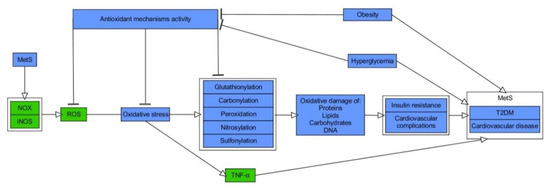
While it is broadly accepted that OxS has a role in the pathogenesis of MetS, it is the matter of debate about its causal impact [14]. From the less controversial point of view, OxS constitutes both the consequence and the trigger for MetS, forming a pathogenic vicious cycle initiated in hypertrophic WAT [14][15][16]. Patients diagnosed with MetS exhibit serum hallmarks of redox imbalance in the form of, e.g., increased levels of protein oxidation products, MDA, elevated XO activity, hyperglycemia (HG), elevated TG as well reduced concentrations of HDL-C, vitamin E and C along with declined levels of heat shock response proteins (HSP70) and SOD as compared to healthy probands [17][18][19]. Other studies indicated raised activity of erythrocyte-specific SOD and MPO with elevation of plasma concentrations of H2O2 and MDA in MetS patients in comparison to controls [20][21]. Moreover, a cross-sectional study conducted on the limited number of Japanese MetS patients and healthy subjects indicated an increase of systemic OxS, as determined by urinary 8-epiprostaglandin F2α (8-epi-PGF2α) in single urine samples, being correlated with visceral AT (VAT) accumulation [22]. MetS patients, including those with T2DM, also exhibited elevation of plasma thiobarbituric acid reactive substances (TBARS), protein carbonylation products, and NOx, where the latter indicated the phenomenon of nitrosative stress (NS) [23][24][25]. Additionally, study on MetS and healthy subjects reported raised values for advanced oxidation protein products (AOPP) and pro-oxidant-antioxidant balance (PAB), in plasma and serum, respectively [26]. Importantly, regression analysis indicated positive and independent association between MetS and higher PAB values 56[27]. Another report showed that levels of ischemia modified albumin (IMA), a protein oxidation marker typical of hypoxia and acidosis, and AOPP were increased with the number of risk factors for MetS, yet it was more significant for AOPP. The latter was also revealed as an independent determinant for occurrence of MetS in studied population of Poles 27[28]. Interestingly, according to data obtained by Venturini et al., AOPP are more related to components of MetS than markers of LPO 28[29]. The elevated release of O2−• from the monocytes of MetS patients, plasma levels of ox-LDL, and nitrotyrosine as compared with healthy probands was also found 29[30]. Appealingly, Yubero-Serrano et al. investigated the relationship between OxS degree and the number of components of MetS in patients. They indicated that activity of SOD and GPx was substantially declined in patients suffering from 2 MetS components than probands with 4/5 MetS components 30[31]. The general relationship between OxS and MetS is presented in Figure 1
.
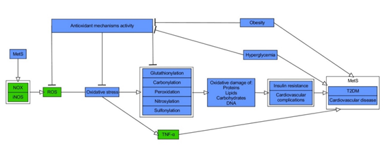
Figure 1. Molecular relationship between oxidative stress and metabolic syndrome. Stimulatory interactions are indicated by arrows and inhibition by T-bars. Actions related to boxes refer to all items inside the box. The figure is a part of paper by Włodarski et al. 31[32]. MetS–metabolic syndrome, NOX–NADPH oxidase, iNOS–inducible nitric oxide synthase, ROS–reactive oxygen species, TNF-α–tumour necrosis factor-α, T2DM–type 2 diabete
s.
2.2. Hypertrophic, Hypoxic and Inflamed White Adipose Tissue—The Initial Fire for Pathogenic Vicious Cycle of Oxidative Stress in Metabolic Syndrome
The pathogenic mechanisms of MetS are complex, thus remaining to be fully elucidated. WAT is a structure comprised mainly of adipose-derived stem cells (ASCs), preadipocytes, adipocytes and immune cells, which is responsible for fat storage. It deposits an excess of energy in triglycerides or mobilizes fatty acids (FA) according to current metabolic needs. It is perceived as an immunological organ and it releases polypeptides (adipo-/cytokines) as well as metabolites capable of exerting systemic actions, including body weight/energy balance, appetite regulation, glucose homeostasis, insulin signaling, and blood pressure contr[32]ol [33][34]. The first known adipocyte hormone, leptin, whose genetic absence causes massive obesity, suppresses appetite, while other hormones, like adiponectin, have just the opposite effe[34]ct [35][36]. Adiponectin increases sensitivity of cells to insulin as well as pancreatic β-cells survival and functionality. Furthermore, it exerts cardio- and vasculoprotective impact along with regulating the function of macrophages 36[37]. Interestingly, Benrick A et al. determined that overexpression of adiponectin has a positive influence on WAT. It decreases the size of adipocytes, increases mitochondrial density, and mediates transcriptional upregulation of factors related to efficient esterification of free fatty acids (FFA) 37[38]. In overall, due to its insulin-sensitizing, antioxidative, anti-inflammatory, and anti-atherogenic impact, adiponectin protects against the MetS 38[39].
However, the primary trigger for most of the pathways investigated in MetS is adiposity, especially visceral one, thus stressing the importance of a high caloric intake as a major causative factor 39[40]. Indeed, prolonged and excessive intake of calories, which exceeds white adipocytes’ storage capacity, induces their hypertrophy and hyperplasia, leading to WAT’s hypoxia, along with consequent necrosis and apoptosis of fat cells. These events elicit burst of OxS and M1 type macrophages’ infiltration 40[41]. Both macrophages and adipocytes produce and secrete proinflammatory adipo-/cytokines and chemokines (e.g., resistin, visfatin, tumour necrosis factor α (TNF-α), monocyte chemoattractant protein-1 (MCP-1, also known as CCL2), interleukin 1β (IL-1β), plasminogen activator inhibitor-1 (PAI-1), interleukin 6 (IL-6), retinol-binding protein 4 (RBP4), and C-reactive protein (CRP)). Hypertrophic WAT is characterized by upregulation of CC chemokines (CCL1/2/3/5,7,8) and their respective receptors (CCR1/2/3/5), the molecules responsible for trafficking leukocytes for the site of inflammation, while CCL2/CCR2 axis is a major one for recruitment of macrophages into WA41][T [42][43]. Proinflammatory cytokines induce signaling pathways of c-Jun N-terminal kinase (JNK) and IκB kinase-β (IKK-β), while the latter activates a transcription factor involved in production of cytokines and proinflammatory factors, called nuclear factor kappa-light-chain-enhancer of activated B cells (NF-κB43][) [44][45]. NF-κB is an inducible transcription factor and master regulator of inflammation due to inducing macrophages polarization as well as expression of numerous cytokines (e.g., IL-1, IL-6, TNF-α) and chemokines (e.g., MCP-1, IL-18) 45[46]. Once secreted, these cytokines activate their extracellular receptors. Simultaneously, the levels of anti-inflammatory adipokines, e.g., adiponectin, omentin decrease. The above biological events trigger a redox imbalance between ROS production and their scavenging, leading to induction of low grade chronic inflammation and intensification of OxS–paving the way for MetS 39][43[40]46[44][47][48]. These phenomena vastly affect WAT, which becomes insulin-resistant, and initiates hyperinsulinemia, enhances lipolysis, increases levels of circulating FFA and their deposition in muscles, liver and pancreas, followed by lipotoxicity, elevated production of glucose due to increased gluconeogenesis and glycogenolysis, and finally, systemic IR and HG 39[40]48[49]. Furthermore, atherogenic dyslipidemia, endothelial dysfunction, introduction of hypercoagulable state and HT are observed, thus probably presenting almost the entire spectrum of MetS components [3].
Moreover, the sources of adipose ROS are diversified, being under control of both hormonal and metabolic determinants 49[50]. Aside from inflammatory cells, mitochondria, as mi ni factories for ATP production due to oxidative phosphorylation, constitute a major producer of superoxide anions which are converted into H2O2 via SOD2 50[51]. It was reported that mitochondrial dysfunction and consequent increased levels of ROS repress insulin signaling as well as production of adiponectin promoting IR in fat cells 51[52Moreover, WAT is the source of ]. ROS-generating enzymes such as NOX, xanthine dehydrogenase/oxidoreductase system (XOR), endoplasmic reticular oxidoreductin 1 (ERO1), pyruvate dehydrogenase (PDH), nicotinamide nucleotide transhydrogenase (NNT) 49[50]. Interestingly, diet-induced obesity (DIO) in mice supported the elevation of mitochondrial ROS generated by fat cells, thus accelerating mitochondrial uncoupling, biogenesis, and fatty acid oxidation so as to prevent from weight gain and serving as an adaptive mechanism. In other words, the deficiency of SOD2 in adipocytes resulted in increased superoxide levels but simultaneously the lack of IR and increased body mass in spite of obesogenic conditions 52[53]. Currently, ROS are perceived as second messengers which may facilitate resistance to stress in WAT. However, biological outcomes of short-term and long term-ROS are fully different. For instance, while the first one can be produced upon insulin stimulation, the latter deteriorates insulin response leading to WAT dysfunction 50[51].
2.3. Insulin Resistance, Hyperglycemia and Oxidative Stress
IR is an impaired response of the body to insulin, resulting in elevated levels of glucose in the blood (a key component of T2DM and MetS) 53[54]. OxS has been recognized as especially important mechanism in IR 54[55]. The hormone insulin features a pivotal role in maintaining physiological levels of blood glucose through various effects on insulin target cells 55[56]. For instance, it elicits vasodilatory as well as vasoconstrictive effects due to the stimulation of endothelial cells for the release of endothelin and nitric oxide, thus increasing the distribution of glucose from blood to organs 56[27]. Insulin is also critical for highly insulin-sensitive cells, such as muscle, hepatic, and fat ones. Transduction of insulin signal takes place via transmembrane insulin receptors (INSR), whose activation involves dimerization and autophosphorylation of tyrosines located on the intracellular part of receptors due to their kinase activity 55[56]. Phosphorylated tyrosines are used by adaptor proteins, such as widely known insulin receptor substrates (IRS), as docking sites. These molecules also undergo phosphorylation and mediate the signal via two major pathways: phosphatidyl inositol 3-kinase (PI3K) / protein kinase B (AKT), which activation results in plethora of metabolism-oriented actions, and mitogen-activated kinases (MAPK), which are mainly responsible for growth and differentiation of cells 55[56]. PI3K conducts phosphatidyl inositol 4,5-biphosphate (PIP2) to phosphatidyl inositol 3,4,5-triphosphate (PIP3) conversion. This is indispensable for plasma membrane recruitment of AKT, followed by phosphorylation of its two specific serine sites by 3-phosphoinositide-dependent kinase-1 (PDK) and mammalian target of rapamycin complex 2 (mTORC 2) 55[56]. Activated AKT is capable of phosphorylating numerous downstream proteins so as to exert metabolic functions of insulin such as induction of glycogenesis (glycogen synthase (GS)), repression of gluconeogenesis (forkhead box O1 (FOXO1)) or lipolysis (phosphodieterase-3B (PDE-3B)) 44[45]. Moreover, it initiates intracellular glucose transport due to phosphorylation of Akt substrate of 160 kDa (A160), which is responsible for translocation of glucose transporter 4 (GLUT-4) to cellular membrane in fat and muscle cells [57][58]. IR always involves disturbances in intracellular insulin signaling [59]. It commonly concerns decreased activity or expression of molecules involved in signal transduction (INSR, IRS-1, GLUT-4), decreased expression / translocation of GLUT-4, or increased expression/activity of antagonists of PI3K/AKT pathway, e.g., phosphatase and tensin homolog (PTEN), and polypyrimidine tract binding protein-1 (PTP1B) [60][61][62][63]. The impaired signaling of insulin demands increased concentrations of insulin (hyperinsulinemia). However, an extensive work of pancreatic β-cells to secrete insulin into bloodstream is ultimately pointless, as the vicious cycle of IR is starting to develop, finally leading to a further decrease of available insulin-stimulated GLUT-4 in the cellular membrane and hyperglycemia [64]. Connections between OxS and insulin signaling are illustrated in Figure 2.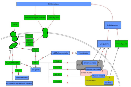
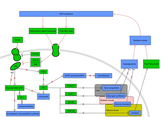
Figure 2. A vicious cycle between oxidative stress and insulin signaling. Stimulatory interactions are indicated by arrows and inhibition by T-bars. Actions related to boxes refer to all items inside the box. Pathological interactions are highlighted in red as well as red crosses denoting withdrawal of physiological insulin signalinThe figure is a part of paper by Włodarski et al. [31]. g. Processes/phenomena are highlighted in blue and proteins/compounds are highlighted in green. The figure is a part of paper by Włodarski et al. [31]. INSR—insulin receptor, IRS—insulin receptor substrate, PTP1B—protein tyrosine phosphatase 1B, SOCS—suppressor of cytokine signaling, JNK–c-Jun N-terminal kinase, PI3K—phosphoinositide 3-kinase, AKT—protein kinase B, FOXO1—forkhead box protein O1, GSK3—glycogen synthase kinase 3, AS160—Akt substrate of 160 kDa, PDE-3B—phosphodiesterase 3 B, eNOS—endothelial nitric oxide synthase.
Lipotoxicity, which is associated with increased plasma level of FFA and intracellular lipid efflux, is vastly involved in muscle and hepatic IR and dysfunction of β-cells [65]. Moreover, FFA stimulate signaling via protein kinase C (PKC) so as to induce NADPH oxidase-mediated OxS and inflammatory signaling via IKK-β and JNK pathways, which perform direct phosphorylation of IRS [66]. The most well-known indicators of lipotoxicity, intracellular lipid intermediates such as diacylglicerol (DAG) and ceramides, act via either different forms of PKC or via protein phosphatase 2A (PPA 2) so as to sequester AKT2 or directly affect IRS protei ns [65][67]. Accumulating amount of data suggests that ROS impair insulin synthesis and secretion and induce IR [68]. Furthermore, in the course of IR, hyperinsulinemia makes PI3K phosphorylate Rac Family Small GTPase 1 (Rac1) protein instead of PIP2, thus potentiating activity of NOX4 and elevating ROS production [69]. OxS impairs intracellular signaling of insulin due to potentiating activity of SH2-containing tyrosine-protein phosphatase (SHO2), PTP1B and glycogen synthase kinase-3 (GSK-3β) [70][71]. One of the major products of LPO, 4-HNE, potentiates activity of GSK-3β, affects IRS, decreases secretion of adiponectin, and induces lipolysis, protein carbonylation and, finally, IR in muscles 50[51][72]. Reactive aldehydes make adducts with numerous cellular proteins in various cellular compartments. They mediate protein carbonylation which results in either accumulation or accelerated degradation of affected proteins, enzymes inactivation, changes in gene expression and mitochondrial dysfunction [7]. For instance, several proteins associated with insulin signaling, lipotoxicity, and response to cellular stresses were reported to be carbonylated in WAT of obese insulin-resistant mice, while fatty acid-binding protein was proved to be carbonylated by 4-HNE in vivo [73]. Indeed, protein carbonylation is becoming a more and more studied component of IR and T2DM pathogenesis [7]. Finally, it is worth highlighting that IR is connected with the coordinated interaction among oxidative, nitrosative, genotoxic, carbonyl, and ER stress 53[54].
HG is a trigger for generation of ROS at the amount which could not be managed by antioxidative system [71]. The central role for HG-mediated OxS is thought to be dependent on the inhibition of glyceraldehyde-3 phosphate dehydrogenase (GAPDH) and accumulation of GAP that upregulates some pathways branching of glycolysis. Namely, HG enhances production of ROS via increasing flux into the polyol, hexosamine and the glyceraldehyde autoxidation pathways as well as activating DAG/PKC signaling pathway, and stimulating formation of advanced glycation end (AGE) products 50[51][71]. Thus, HG is capable of activating numerous pathways associated with inflammation and OxS, e.g., an activation of PKC stimulates NOX enzymes and lipoxygenases [74]. Excessively generated sorbitol (polyol pathway) mediates activation of p38 MAPK and JNK – core proteins in inflammatory response [75]. The formation of glyoxal (product of glucose autooxidation) and methylglyoxal (product of GAP dephosphorylation) take part in the formation of AGE products. Both precursors bind to specific AGE receptors (RAGE) or interact with various biomolecules thus accelerating OxS via PKC-dependent or independent pathways [71]. Interestingly, methylglyoxal itself affects interaction of insulin with its receptor [76][77]. AGE/RAGE pathway promotes vascular endothelium’s expression of MCP-1, known to indicate vascular endothelial dysfunction and prothrombotic impact [77]. Furthermore, it is also involved in promoting expression of NF-κB via toll-like receptor 4 (TLR4) pathway [54]. It is to be noted that the early glycation of proteins can be also reversible, as in Schiff bases or Amadori adducts, including a marker of diabetes, glycated hemoglobin (HbA1C) [78]. For thoroughly depicted net of interactions among ROS, IR, HG, and inflammation, see the review by Luc et al. [79
].
2.4. Dyslipidemia and Oxidative Stress
Dyslipidemia in MetS is a state with elevated level of plasma TG associated with increased level of very low-density lipoprotein (VLDL), small, dense LDL (sdLDL-C), FFAs and low HDL cholesterol level that promotes the development of atherosclerosis [80]. Indeed, oxidatively modified LDL (ox-LDL) is an important player in inducing the process of atherosclerosis as it affects expression of adhesive molecules, cytokines and growth factors and changes function of important vasoactive molecules such as NO, angiotensin II (Ang II) or endothelin 1 (ET 1) [81][82]. Interestingly, treatment of MetS patients with rosuvastatin causes beneficial effect not only on levels of ox-LDL, HDL, and inflammatory markers, but also ameliorates total antioxidant capability [83][84]. Furthermore, high cholesterol level is a promoter of OxS in endothelial cells 84[85]. In an extensive review, Spahis et al. enumerated several types of connection between dyslipidemia and OxS in MetS. For instance, elevation of O−•2−• by NADPH occurring upon obesity/HT/hypertriglyceridemia, as well as lowered level of bilirubin in MetS (a protective agent against LDL oxidation), magnitude of LDL oxidation being dependent on waist circumference (visceral adiposity) or ox-LDL affecting mitochondrial functionality [14]. Hyperlipidemia triggers elevation of ROS and proinflammatory cytokines which may be causative factors for lipotoxicity, being predominantly known to be caused by increased rate of lipolysis and repressed synthesis of TG in obesity-affected, insulin-resistant WAT, triggering increased levels of circulating FFA and accumulation of lipids in non-adipose organs (e.g., liver, pancreas, muscles) [85]. For instance, in the recent study by Feillet-Coudray et al., a high-fat high-fructose diet in Wistar rats led to excessive weight gain along with glucose intolerance and hepatic steatosis with elevation of ceramides and DAG (lipotoxicity indicators). Moreover, there was an increase in hepatic NOX activity and protein level of IL-6 along with decrease of total GSSG and GSH as well as activity of SOD and GPx. As these phenomena were associated with moderately marked OxS and inflammation, metabolic alterations were rather suggested to be the trigger, yet, not the cause of OxS [86]. Interestingly, FFAs were reported to be capable of activating renin-angiotensin system (RAS) in mice adipocytes (3T3L1) by TLR4/NF-κB pathway [87]. In fat cells, RAS is connected with the impairment of preadipocytes differentiation, promotion of lipolysis, along with OxS and inflammatio
n.
2.5. Hypertension and Oxidative Stress
There are numerous studies which reported persistently increased ROS levels in HT, along with vast improvement upon antioxidative treatment [88][89][90][91]][93][94][92. HT is connected with vascular remodeling, increased vasoconstriction and arterial stiffness, activation of immune cells, renal dysfunction, and excitation of sympathetic nervous system. Above phenomena are inseparably connected with endothelial dysfunction, LPO, inflammation, fibrosis and more, thus favoring the notion that OxS constitutes a common molecular phenomenon in multifactorial pathogenesis of HT 95[93]. Cardiovascular cells generate ROS due to action of several major enzymes: NOX, XOR, ERO, and uncoupled NOS. While NOX-mediated ROS production is a prevailing one in HT, more and more pieces of evidence suggest its crosstalk with mitochondrial and ER-specific ROS due to phenomenon of ROS-induced ROS release (RIRR) [89][93][94][95][96][97]. Specifically, in the recent review by Touyz et al., OxS was shown to be connected with HT via several ways 89[90]. Firstly, prohypertensive factors such as salt, aldosterone, Ang II, and ET-1 activate NOX enzymes and ROS production, being a trigger for mitochondrial and ER-located ROS formation, all of which are interconnected with inflammation and immune activation, and consequently, HT. Here, it is worthwhile to enumerate the increased expression of proinflammatory factors (e.g., TNF-α, IL-1/6), adhesion molecules, and activation of signaling via proinflammatory pathways (e.g., NF-κB, JNK). Secondly, elevation of vascular ROS elicits activation of C2+a2+ channels and further activation of C2+a2+-sensitive NOX enzymes. Thirdly, Ang II- and ET-1-dependent signaling via their G-coupled receptors promotes transactivation of growth factor receptors (e.g., insulin-like growth factor 1 (IGF-1R), platelet-derived growth factor receptor (PDGFR)) through various mechanisms, triggering activation of signaling via PI3K/AKT and MAPK pathways 89[90]. Moreover, under physiological conditions, eNOS produces NO, a molecule of critical importance for vasorelaxation. Under NOX-initiated OxS, eNOS produces superoxide, rather than NO, being called uncoupled eNOS and contributing to a sustained increase of ROS levels 97[98]. Finally, as thoroughly described by Spahis et al., the accumulation of ox-LDL in vascular endothelium is a source of mitochondria-derived OxS in HT [1
References
- International Diabetes Federation. IDF Diabetes Atlas, 9th edition; International Diabetes Federation: Brussels, Belgium, 2019
- Saklayen, M.G. The Global Epidemic of the Metabolic Syndrome. Curr. Hypertens. Rep. 2018, 20, 12, doi:10.1007/s11906-018-0812-z.
- Alberti, K.G.M.M.; Zimmet, P.; Shaw, J. The metabolic syndrome--a new worldwide definition. Lancet 2005, 366, 1059–1062, doi:10.1016/S0140-6736(05)67402-8.
- Alberti, K.G.M.M.; Eckel, R.H.; Grundy, S.M.; Zimmet, P.Z.; Cleeman, J.I.; Donato, K.A.; Fruchart, J.-C.; James, W.P.T.; Loria, C.M.; Smith, S.C.J. Harmonizing the metabolic syndrome: A joint interim statement of the International Diabetes Federation Task Force on Epidemiology and Prevention; National Heart, Lung, and Blood Institute; American Heart Association; World Heart Federation; International. Circulation 2009, 120, 1640–1645, doi:10.1161/CIRCULATIONAHA.109.192644.
- Price, N.L.; Ramírez, C.M.; Fernández-Hernando, C. Relevance of microRNA in metabolic diseases. Crit. Rev. Clin. Lab. Sci. 2014, 51, 305–320, doi:10.3109/10408363.2014.937522.Vona, R.; Gambardella, L.; Cittadini, C.; Straface, E.; Pietraforte, D. Biomarkers of Oxidative Stress in Metabolic Syndrome and Associated Diseases. Oxid. Med. Cell. Longev. 2019, 2019, 8267234, doi:10.1155/2019/8267234.
- Rani, V.; Deep, G.; Singh, R.K.; Palle, K.; Yadav, U.C.S. Oxidative stress and metabolic disorders: Pathogenesis and therapeutic strategies. Life Sci. 2016, 148, 183–193, doi:10.1016/j.lfs.2016.02.002.Yan, L.-J. Pathogenesis of chronic hyperglycemia: From reductive stress to oxidative stress. J. Diabetes Res. 2014, 2014, 137919, doi:10.1155/2014/137919.
- Pu, M.; Chen, J.; Tao, Z.; Miao, L.; Qi, X.; Wang, Y.; Ren, J. Regulatory network of miRNA on its target: Coordination between transcriptional and post-transcriptional regulation of gene expression. Cell. Mol. Life Sci. 2019, 76, 441–451, doi:10.1007/s00018-018-2940-7.Hecker, M.; Wagner, A.H. Role of protein carbonylation in diabetes. J. Inherit. Metab. Dis. 2018, 41, 29–38, doi:10.1007/s10545-017-0104-9.
- Krützfeldt, J.; Stoffel, M. MicroRNAs: A new class of regulatory genes affecting metabolism. Cell Metab. 2006, 4, 9–12, doi:10.1016/j.cmet.2006.05.009.Valavanidis, A.; Vlachogianni, T.; Fiotakis, C. 8-hydroxy-2’ -deoxyguanosine (8-OHdG): A critical biomarker of oxidative stress and carcinogenesis. J. Environ. Sci. Heal. Part C Environ. Carcinog. Ecotoxicol. Rev. 2009, 27, 120–139, doi:10.1080/10590500902885684.
- Lin, Y.-H. MicroRNA Networks Modulate Oxidative Stress in Cancer. Int. J. Mol. Sci. 2019, 20, doi:10.3390/ijms20184497.Baba, S.P.; Bhatnagar, A. Role of thiols in oxidative stress. Curr. Opin. Toxicol. 2018, 7, 133–139, doi:10.1016/j.cotox.2018.03.005.
- Vona, R.; Gambardella, L.; Cittadini, C.; Straface, E.; Pietraforte, D. Biomarkers of Oxidative Stress in Metabolic Syndrome and Associated Diseases. Oxid. Med. Cell. Longev. 2019, 2019, 8267234, doi:10.1155/2019/8267234.Griendling, K.K.; Touyz, R.M.; Zweier, J.L.; Dikalov, S.; Chilian, W.; Chen, Y.-R.; Harrison, D.G.; Bhatnagar, A. Measurement of Reactive Oxygen Species, Reactive Nitrogen Species, and Redox-Dependent Signaling in the Cardiovascular System: A Scientific Statement From the American Heart Association. Circ. Res. 2016, 119, e39-75, doi:10.1161/RES.0000000000000110.
- O’Brien, J.; Hayder, H.; Zayed, Y.; Peng, C. Overview of MicroRNA Biogenesis, Mechanisms of Actions, and Circulation. Front. Endocrinol. 2018, 9, 402, doi:10.3389/fendo.2018.00402.Matsuzawa, Y.; Funahashi, T.; Nakamura, T. The concept of metabolic syndrome: Contribution of visceral fat accumulation and its molecular mechanism. J. Atheroscler. Thromb. 2011, 18, 629–639, doi:10.5551/jat.7922.
- Nigi, L.; Grieco, G.E.; Ventriglia, G.; Brusco, N.; Mancarella, F.; Formichi, C.; Dotta, F.; Sebastiani, G. MicroRNAs as Regulators of Insulin Signaling: Research Updates and Potential Therapeutic Perspectives in Type 2 Diabetes. Int. J. Mol. Sci. 2018, 19, doi:10.3390/ijms19123705.Burtenshaw, D.; Hakimjavadi, R.; Redmond, E.M.; Cahill, P.A. Nox, Reactive Oxygen Species and Regulation of Vascular Cell Fate. Antioxidants 2017, 6, doi:10.3390/antiox6040090.
- Sebastiani, G.; Po, A.; Miele, E.; Ventriglia, G.; Ceccarelli, E.; Bugliani, M.; Marselli, L.; Marchetti, P.; Gulino, A.; Ferretti, E.; et al. MicroRNA-124a is hyperexpressed in type 2 diabetic human pancreatic islets and negatively regulates insulin secretion. Acta Diabetol. 2015, 52, 523–530, doi:10.1007/s00592-014-0675-y.Ighodaro, O.M.; Akinloye, O.A. First line defence antioxidants-superoxide dismutase (SOD), catalase (CAT) and glutathione peroxidase (GPX): Their fundamental role in the entire antioxidant defence grid. Alexandria J. Med. 2018, 54, 287–293, doi:10.1016/j.ajme.2017.09.001.
- Qi, R.; Wang, J.; Wang, Q.; Qiu, X.; Yang, F.; Liu, Z.; Huang, J. MicroRNA-425 controls lipogenesis and lipolysis in adipocytes. Biochim. Biophys. Acta Mol. Cell Biol. Lipids 2019, 1864, 744–755, doi:10.1016/j.bbalip.2019.02.007.Spahis, S.; Borys, J.-M.; Levy, E. Metabolic Syndrome as a Multifaceted Risk Factor for Oxidative Stress. Antioxid. Redox Signal. 2017, 26, 445–461, doi:10.1089/ars.2016.6756.
- Tahamtan, A.; Teymoori-Rad, M.; Nakstad, B.; Salimi, V. Anti-Inflammatory MicroRNAs and Their Potential for Inflammatory Diseases Treatment. Front. Immunol. 2018, 9, 1377, doi:10.3389/fimmu.2018.01377.Furukawa, S.; Fujita, T.; Shimabukuro, M.; Iwaki, M.; Yamada, Y.; Nakajima, Y.; Nakayama, O.; Makishima, M.; Matsuda, M.; Shimomura, I. Increased oxidative stress in obesity and its impact on metabolic syndrome. J. Clin. Invest. 2004, 114, 1752–1761, doi:10.1172/JCI21625.
- Haque, R.; Chun, E.; Howell, J.C.; Sengupta, T.; Chen, D.; Kim, H. MicroRNA-30b-mediated regulation of catalase expression in human ARPE-19 cells. PLoS ONE 2012, 7, e42542, doi:10.1371/journal.pone.0042542.Carrier, A. Metabolic Syndrome and Oxidative Stress: A Complex Relationship. Antioxid. Redox Signal. 2017, 26, 429–431.
- Bai, X.-Y.; Ma, Y.; Ding, R.; Fu, B.; Shi, S.; Chen, X.-M. miR-335 and miR-34a Promote renal senescence by suppressing mitochondrial antioxidative enzymes. J. Am. Soc. Nephrol. 2011, 22, 1252–1261, doi:10.1681/ASN.2010040367.Le, N.-A. Postprandial Triglycerides, Oxidative Stress, and Inflammation. In Apolipoproteins, Triglycerides and Cholesterol; Waisundara, V.Y., Jovandaric, M.Z., Eds.; IntechOpen: Rijeka, Croatia, 2020.
- Cheng, Y.; Zhou, M.; Zhou, W. MicroRNA-30e regulates TGF-β-mediated NADPH oxidase 4-dependent oxidative stress by Snai1 in atherosclerosis. Int. J. Mol. Med. 2019, 43, 1806–1816, doi:10.3892/ijmm.2019.4102.Armutcu, F.; Ataymen, M.; Atmaca, H.; Gurel, A. Oxidative stress markers, C-reactive protein and heat shock protein 70 levels in subjects with metabolic syndrome. Clin. Chem. Lab. Med. 2008, 46, 785–790, doi:10.1515/CCLM.2008.166.
- Li, Z.-N.; Ge, M.-X.; Yuan, Z.-F. MicroRNA-182-5p protects human lens epithelial cells against oxidative stress-induced apoptosis by inhibiting NOX4 and p38 MAPK signalling. BMC Ophthalmol. 2020, 20, 233, doi:10.1186/s12886-020-01489-8.Zelzer, S.; Fuchs, N.; Almer, G.; Raggam, R.B.; Prüller, F.; Truschnig-Wilders, M.; Schnedl, W.; Horejsi, R.; Möller, R.; Weghuber, D.; et al. High density lipoprotein cholesterol level is a robust predictor of lipid peroxidation irrespective of gender, age, obesity, and inflammatory or metabolic biomarkers. Clin. Chim. Acta. 2011, 412, 1345–1349, doi:10.1016/j.cca.2011.03.031.
- Wu, Y.; Yao, J.; Feng, K. miR-124-5p/NOX2 Axis Modulates the ROS Production and the Inflammatory Microenvironment to Protect Against the Cerebral I/R Injury. Neurochem. Res. 2020, 45, 404–417, doi:10.1007/s11064-019-02931-0.Simão, A.N.C.; Lozovoy, M.A.B.; Simão, T.N.C.; Venturini, D.; Barbosa, D.S.; Dichi, J.B.; Matsuo, T.; Cecchini, R.; Dichi, I. Immunological and biochemical parameters of patients with metabolic syndrome and the participation of oxidative and nitroactive stress. Brazilian J. Med. Biol. Res. Rev. Bras. Pesqui. Med. Biol. 2011, 44, 707–712, doi:10.1590/s0100-879x2011007500069.
- Varga, Z. V.; Kupai, K.; Szűcs, G.; Gáspár, R.; Pálóczi, J.; Faragó, N.; Zvara, A.; Puskás, L.G.; Rázga, Z.; Tiszlavicz, L.; et al. MicroRNA-25-dependent up-regulation of NADPH oxidase 4 (NOX4) mediates hypercholesterolemia-induced oxidative/nitrative stress and subsequent dysfunction in the heart. J. Mol. Cell. Cardiol. 2013, 62, 111–121, doi:10.1016/j.yjmcc.2013.05.009.Da Fonseca, L.J.S.; Nunes-Souza, V.; Guedes, G. da S.; Schettino-Silva, G.; Mota-Gomes, M.A.; Rabelo, L.A. Oxidative status imbalance in patients with metabolic syndrome: Role of the myeloperoxidase/hydrogen peroxide axis. Oxid. Med. Cell. Longev. 2014, 2014, 898501, doi:10.1155/2014/898501.
- Wang, L.; Huang, H.; Fan, Y.; Kong, B.; Hu, H.; Hu, K.; Guo, J.; Mei, Y.; Liu, W.-L. Effects of downregulation of microRNA-181a on H2O2-induced H9c2 cell apoptosis via the mitochondrial apoptotic pathway. Oxid. Med. Cell. Longev. 2014, 2014, 960362, doi:10.1155/2014/960362.Fujita, K.; Nishizawa, H.; Funahashi, T.; Shimomura, I.; Shimabukuro, M. Systemic oxidative stress is associated with visceral fat accumulation and the metabolic syndrome. Circ. J. 2006, 70, 1437–1442, doi:10.1253/circj.70.1437.
- Wang, P.; Zhu, C.; Ma, M.; Chen, G.; Song, M.; Zeng, Z.; Lu, W.; Yang, J.; Wen, S.; Chiao, P.J.; et al. Micro-RNA-155 is induced by K-Ras oncogenic signal and promotes ROS stress in pancreatic cancer. Oncotarget 2015, 6, 21148–21158, doi:10.18632/oncotarget.4125.Caimi, G.; Hopps, E.; Montana, M.; Noto, D.; Canino, B.; Lo Presti, R.; Averna, M.R. Evaluation of nitric oxide metabolites in a group of subjects with metabolic syndrome. Diabetes Metab. Syndr. 2012, 6, 132–135, doi:10.1016/j.dsx.2012.09.012.
- Zhang, X.; Wang, C.; Shan, S.; Liu, X.; Jiang, Z.; Ren, T. TLR4/ROS/miRNA-21 pathway underlies lipopolysaccharide instructed primary tumor outgrowth in lung cancer patients. Oncotarget 2016, 7, 42172–42182, doi:10.18632/oncotarget.9902.Caimi, G.; Hopps, E.; Noto, D.; Canino, B.; Montana, M.; Lucido, D.; Lo Presti, R.; Averna, M.R. Protein oxidation in a group of subjects with metabolic syndrome. Diabetes Metab. Syndr. 2013, 7, 38–41, doi:10.1016/j.dsx.2013.02.013.
- Simone, N.L.; Soule, B.P.; Ly, D.; Saleh, A.D.; Savage, J.E.; Degraff, W.; Cook, J.; Harris, C.C.; Gius, D.; Mitchell, J.B. Ionizing radiation-induced oxidative stress alters miRNA expression. PLoS ONE 2009, 4, e6377, doi:10.1371/journal.pone.0006377.Caimi, G.; Lo Presti, R.; Montana, M.; Noto, D.; Canino, B.; Averna, M.R.; Hopps, E. Lipid peroxidation, nitric oxide metabolites, and their ratio in a group of subjects with metabolic syndrome. Oxid. Med. Cell. Longev. 2014, 2014, 824756, doi:10.1155/2014/824756.
- Shi, Q.; Gibson, G.E. Up-regulation of the mitochondrial malate dehydrogenase by oxidative stress is mediated by miR-743a. J. Neurochem. 2011, 118, 440–448, doi:10.1111/j.1471-4159.2011.07333.x.Korkmaz, G.G.; Altınoglu, E.; Civelek, S.; Sozer, V.; Erdenen, F.; Tabak, O.; Uzun, H. The association of oxidative stress markers with conventional risk factors in the metabolic syndrome. Metabolism 2013, 62, 828–835, doi:10.1016/j.metabol.2013.01.002.
- Caimi, G.; Hopps, E.; Montana, M.; Noto, D.; Canino, B.; Lo Presti, R.; Averna, M.R. Evaluation of nitric oxide metabolites in a group of subjects with metabolic syndrome. Diabetes Metab. Syndr. 2012, 6, 132–135, doi:10.1016/j.dsx.2012.09.012.Zurawska-Płaksej, E.; Grzebyk, E.; Marciniak, D.; Szymańska-Chabowska, A.; Piwowar, A. Oxidatively modified forms of albumin in patients with risk factors of metabolic syndrome. J. Endocrinol. Invest. 2014, 37, 819–827, doi:10.1007/s40618-014-0111-8.
- Thulasingam, S.; Massilamany, C.; Gangaplara, A.; Dai, H.; Yarbaeva, S.; Subramaniam, S.; Riethoven, J.-J.; Eudy, J.; Lou, M.; Reddy, J. miR-27b*, an oxidative stress-responsive microRNA modulates nuclear factor-kB pathway in RAW 264.7 cells. Mol. Cell. Biochem. 2011, 352, 181–188, doi:10.1007/s11010-011-0752-2.Venturini, D.; Simão, A.N.C.; Dichi, I. Advanced oxidation protein products are more related to metabolic syndrome components than biomarkers of lipid peroxidation. Nutr. Res. 2015, 35, 759–765, doi:10.1016/j.nutres.2015.06.013.
- Hong, J.; Wang, Y.; Hu, B.-C.; Xu, L.; Liu, J.-Q.; Chen, M.-H.; Wang, J.-Z.; Han, F.; Zheng, Y.; Chen, X.; et al. Transcriptional downregulation of microRNA-19a by ROS production and NF-κB deactivation governs resistance to oxidative stress-initiated apoptosis. Oncotarget 2017, 8, 70967–70981, doi:10.18632/oncotarget.20235.Jialal, I.; Devaraj, S.; Adams-Huet, B.; Chen, X.; Kaur, H. Increased cellular and circulating biomarkers of oxidative stress in nascent metabolic syndrome. J. Clin. Endocrinol. Metab. 2012, 97, E1844–E1850, doi:10.1210/jc.2012-2498.
- Al-Rawaf, H.A. Circulating microRNAs and adipokines as markers of metabolic syndrome in adolescents with obesity. Clin. Nutr. 2018, doi:10.1016/j.clnu.2018.09.024.Yubero-Serrano, E.M.; Delgado-Lista, J.; Peña-Orihuela, P.; Perez-Martinez, P.; Fuentes, F.; Marin, C.; Tunez, I.; Tinahones, F.J.; Perez-Jimenez, F.; Roche, H.M.; et al. Oxidative stress is associated with the number of components of metabolic syndrome: Lipgene study. Exp. Mol. Med. 2013, 45, e28, doi:10.1038/emm.2013.53.
- Willeit, P.; Skroblin, P.; Moschen, A.R.; Yin, X.; Kaudewitz, D.; Zampetaki, A.; Barwari, T.; Whitehead, M.; Ramírez, C.M.; Goedeke, L.; et al. Circulating MicroRNA-122 Is Associated With the Risk of New-Onset Metabolic Syndrome and Type 2 Diabetes. Diabetes 2017, 66, 347–357, doi:10.2337/db16-0731.Włodarski, A.; Strycharz, J.; Wróblewski, A.; Kasznicki, J., Drzewoski, J.; Śliwińska, A. The Role of microRNAs in Metabolic Syndrome-Related Oxidative Stress. Int. J. Mol. Sci. 2020, doi:10.3390/ijms21186902
- Yan, L.-J. Pathogenesis of chronic hyperglycemia: From reductive stress to oxidative stress. J. Diabetes Res. 2014, 2014, 137919, doi:10.1155/2014/137919.Galic, S.; Oakhill, J.S.; Steinberg, G.R. Adipose tissue as an endocrine organ. Mol. Cell. Endocrinol. 2010, 316, 129–139, doi:10.1016/j.mce.2009.08.018.
- Hecker, M.; Wagner, A.H. Role of protein carbonylation in diabetes. J. Inherit. Metab. Dis. 2018, 41, 29–38, doi:10.1007/s10545-017-0104-9.Grant, R.W.; Dixit, V.D. Adipose tissue as an immunological organ. Obesity 2015, 23, 512–518, doi:10.1002/oby.21003.
- Valavanidis, A.; Vlachogianni, T.; Fiotakis, C. 8-hydroxy-2’ -deoxyguanosine (8-OHdG): A critical biomarker of oxidative stress and carcinogenesis. J. Environ. Sci. Heal. Part C Environ. Carcinog. Ecotoxicol. Rev. 2009, 27, 120–139, doi:10.1080/10590500902885684.Wróblewski, A.; Strycharz, J.; Świderska, E.; Drewniak, K.; Drzewoski, J.; Szemraj, J.; Kasznicki, J.; Śliwińska, A. Molecular Insight into the Interaction between Epigenetics and Leptin in Metabolic Disorders. Nutrients 2019, 11, doi:10.3390/nu11081872.
- Baba, S.P.; Bhatnagar, A. Role of thiols in oxidative stress. Curr. Opin. Toxicol. 2018, 7, 133–139, doi:10.1016/j.cotox.2018.03.005.Adamczak, M.; Wiecek, A. The adipose tissue as an endocrine organ. Semin. Nephrol. 2013, 33, 2–13, doi:10.1016/j.semnephrol.2012.12.008.
- Griendling, K.K.; Touyz, R.M.; Zweier, J.L.; Dikalov, S.; Chilian, W.; Chen, Y.-R.; Harrison, D.G.; Bhatnagar, A. Measurement of Reactive Oxygen Species, Reactive Nitrogen Species, and Redox-Dependent Signaling in the Cardiovascular System: A Scientific Statement From the American Heart Association. Circ. Res. 2016, 119, e39-75, doi:10.1161/RES.0000000000000110.Ouchi, N.; Ohashi, K.; Shibata, R.; Murohara, T. Adipocytokines and obesity-linked disorders. Nagoya J. Med. Sci. 2012, 74, 19–30.
- Matsuzawa, Y.; Funahashi, T.; Nakamura, T. The concept of metabolic syndrome: Contribution of visceral fat accumulation and its molecular mechanism. J. Atheroscler. Thromb. 2011, 18, 629–639, doi:10.5551/jat.7922.Benrick, A.; Chanclón, B.; Micallef, P.; Wu, Y.; Hadi, L.; Shelton, J.M.; Stener-Victorin, E.; Wernstedt Asterholm, I. Adiponectin protects against development of metabolic disturbances in a PCOS mouse model. Proc. Natl. Acad. Sci. USA 2017, 114, E7187–E7196, doi:10.1073/pnas.1708854114.
- Burtenshaw, D.; Hakimjavadi, R.; Redmond, E.M.; Cahill, P.A. Nox, Reactive Oxygen Species and Regulation of Vascular Cell Fate. Antioxidants 2017, 6, doi:10.3390/antiox6040090.Esfahani, M.; Movahedian, A.; Baranchi, M.; Goodarzi, M.T. Adiponectin: An adipokine with protective features against metabolic syndrome. Iran. J. Basic Med. Sci. 2015, 18, 430–442.
- Ighodaro, O.M.; Akinloye, O.A. First line defence antioxidants-superoxide dismutase (SOD), catalase (CAT) and glutathione peroxidase (GPX): Their fundamental role in the entire antioxidant defence grid. Alexandria J. Med. 2018, 54, 287–293, doi:10.1016/j.ajme.2017.09.001.Janochova, K.; Haluzik, M.; Buzga, M. Visceral fat and insulin resistance—What we know? Biomed. Pap. Med. Fac. Univ. Palacky. Olomouc. Czech. Repub. 2019, 163, 19–27, doi:10.5507/bp.2018.062.
- Pudlarz, A.M.; Czechowska, E.; Ranoszek-Soliwoda, K.; Tomaszewska, E.; Celichowski, G.; Grobelny, J.; Szemraj, J. Immobilization of Recombinant Human Catalase on Gold and Silver Nanoparticles. Appl. Biochem. Biotechnol. 2018, doi:10.1007/s12010-017-2682-2.Strycharz, J.; Drzewoski, J.; Szemraj, J.; Sliwinska, A. Is p53 Involved in Tissue-Specific Insulin Resistance Formation? Oxid. Med. Cell. Longev. 2017, 2017, 9270549, doi:10.1155/2017/9270549.
- Alberts B; Johnson A; Lewis J; Raff, M.; Roberts K, W.P. Chapter 12: Peroxisomes. In Molecular Biology of the Cell; Garland Science: New York, NY, USA, 2002; ISBN 978-0-8153-3218-3.Xu, L.; Kitade, H.; Ni, Y.; Ota, T. Roles of chemokines and chemokine receptors in obesity-associated insulin resistance and nonalcoholic fatty liver disease. Biomolecules 2015, 5, 1563–1579.
- Deponte, M. Glutathione catalysis and the reaction mechanisms of glutathione-dependent enzymes. Biochim. Biophys. Acta 2013, 1830, 3217–3266, doi:10.1016/j.bbagen.2012.09.018.Huber, J.; Kiefer, F.W.; Zeyda, M.; Ludvik, B.; Silberhumer, G.R.; Prager, G.; Zlabinger, G.J.; Stulnig, T.M. CC chemokine and CC chemokine receptor profiles in visceral and subcutaneous adipose tissue are altered in human obesity. J. Clin. Endocrinol. Metab. 2008, doi:10.1210/jc.2007-2630.
- Lushchak, V.I. Glutathione Homeostasis and Functions: Potential Targets for Medical Interventions. J. Amino Acids 2012, doi:10.1155/2012/736837.Zand, H.; Morshedzadeh, N.; Naghashian, F. Signaling pathways linking inflammation to insulin resistance. Diabetes Metab. Syndr. 2017, 11 (Suppl. 1) S307–S309, doi:10.1016/j.dsx.2017.03.006.
- Ellulu, M.S.; Patimah, I.; Khaza’ai, H.; Rahmat, A.; Abed, Y.; Ali, F. Atherosclerotic cardiovascular disease: a review of initiators and protective factors. Inflammopharmacology 2016, 24(1), 1–10.Kwon, H.; Pessin, J.E.; Adipokines, Inflammation, and Insulin Resistance in Obesity. In Textbook of Energy Balance, Neuropeptide Hormones, and Neuroendocrine Function; Nillni, E.A., Ed.; Springer International Publishing: Cham, Switzerland, 2018; pp. 225–252. ISBN 978-3-319-89506-2.
- Loot, A.E.; Schreiber, J.G.; Fisslthaler, B.; Fleming, I. Angiotensin II impairs endothelial function via tyrosine phosphorylation of the endothelial nitric oxide synthase. J. Exp. Med. 2009, doi:10.1084/jem.20090449..Liu, T.; Zhang, L.; Joo, D.; Sun, S.-C. NF-κB signaling in inflammation. Signal Transduct. Target. Ther. 2017, 2, 17023, doi:10.1038/sigtrans.2017.23.
- Incalza, M.A.; D’Oria, R.; Natalicchio, A.; Perrini, S.; Laviola, L.; Giorgino, F. Oxidative stress and reactive oxygen species in endothelial dysfunction associated with cardiovascular and metabolic diseases. Vascul. Pharmacol. 2018, 100, 1–19, doi:10.1016/j.vph.2017.05.005.Ohashi, K.; Shibata, R.; Murohara, T.; Ouchi, N. Role of anti-inflammatory adipokines in obesity-related diseases. Trends Endocrinol. Metab. 2014, 25, 348–355, doi:10.1016/j.tem.2014.03.009.
- Forman, H.J.; Maiorino, M.; Ursini, F. Signaling functions of reactive oxygen species. Biochemistry 2010, 49, 835–842, doi:10.1021/bi9020378.Pan, X.; Kaminga, A.C.; Wen, S.W.; Acheampong, K.; Liu, A. Omentin-1 in diabetes mellitus: A systematic review and meta-analysis. PLoS ONE 2019, 14, e0226292, doi:10.1371/journal.pone.0226292.
- Spahis, S.; Borys, J.-M.; Levy, E. Metabolic Syndrome as a Multifaceted Risk Factor for Oxidative Stress. Antioxid. Redox Signal. 2017, 26, 445–461, doi:10.1089/ars.2016.6756.Engin, A.B. What Is Lipotoxicity? Adv. Exp. Med. Biol. 2017, 960, 197–220, doi:10.1007/978-3-319-48382-5_8.
- Furukawa, S.; Fujita, T.; Shimabukuro, M.; Iwaki, M.; Yamada, Y.; Nakajima, Y.; Nakayama, O.; Makishima, M.; Matsuda, M.; Shimomura, I. Increased oxidative stress in obesity and its impact on metabolic syndrome. J. Clin. Invest. 2004, 114, 1752–1761, doi:10.1172/JCI21625.Hauck, A.K.; Huang, Y.; Hertzel, A. V.; Bernlohr, D.A. Adipose oxidative stress and protein carbonylation. J. Biol. Chem. 2019, 294, 1083–1088, doi:10.1074/jbc.R118.003214.
- Carrier, A. Metabolic Syndrome and Oxidative Stress: A Complex Relationship. Antioxid. Redox Signal. 2017, 26, 429–431.Le Lay, S.; Simard, G.; Martinez, M.C.; Andriantsitohaina, R. Oxidative stress and metabolic pathologies: From an adipocentric point of view. Oxid. Med. Cell. Longev. 2014, 2014, 908539, doi:10.1155/2014/908539.
- Le, N.-A. Postprandial Triglycerides, Oxidative Stress, and Inflammation. In Apolipoproteins, Triglycerides and Cholesterol; Waisundara, V.Y., Jovandaric, M.Z., Eds.; IntechOpen: Rijeka, Croatia, 2020.Wang, C.-H.; Wang, C.-C.; Huang, H.-C.; Wei, Y.-H. Mitochondrial dysfunction leads to impairment of insulin sensitivity and adiponectin secretion in adipocytes. FEBS J. 2013, 280, 1039–1050, doi:10.1111/febs.12096.
- Armutcu, F.; Ataymen, M.; Atmaca, H.; Gurel, A. Oxidative stress markers, C-reactive protein and heat shock protein 70 levels in subjects with metabolic syndrome. Clin. Chem. Lab. Med. 2008, 46, 785–790, doi:10.1515/CCLM.2008.166.Ortega, S.P.; Chouchani, E.T.; Boudina, S. Stress turns on the heat: Regulation of mitochondrial biogenesis and UCP1 by ROS in adipocytes. Adipocyte 2017, 6, 56–61, doi:10.1080/21623945.2016.1273298.
- Zelzer, S.; Fuchs, N.; Almer, G.; Raggam, R.B.; Prüller, F.; Truschnig-Wilders, M.; Schnedl, W.; Horejsi, R.; Möller, R.; Weghuber, D.; et al. High density lipoprotein cholesterol level is a robust predictor of lipid peroxidation irrespective of gender, age, obesity, and inflammatory or metabolic biomarkers. Clin. Chim. Acta. 2011, 412, 1345–1349, doi:10.1016/j.cca.2011.03.031.Levy-Marchal, C.; Arslanian, S.; Cutfield, W.; Sinaiko, A.; Druet, C.; Marcovecchio, M.L.; Chiarelli, F. Insulin resistance in children: Consensus, perspective, and future directions. J. Clin. Endocrinol. Metab. 2010, 95, 5189–5198, doi:10.1210/jc.2010-1047.
- Simão, A.N.C.; Lozovoy, M.A.B.; Simão, T.N.C.; Venturini, D.; Barbosa, D.S.; Dichi, J.B.; Matsuo, T.; Cecchini, R.; Dichi, I. Immunological and biochemical parameters of patients with metabolic syndrome and the participation of oxidative and nitroactive stress. Brazilian J. Med. Biol. Res. Rev. Bras. Pesqui. Med. Biol. 2011, 44, 707–712, doi:10.1590/s0100-879x2011007500069.Onyango, A.N. Cellular Stresses and Stress Responses in the Pathogenesis of Insulin Resistance. Oxid. Med. Cell. Longev. 2018, 2018, 4321714, doi:10.1155/2018/4321714.
- Da Fonseca, L.J.S.; Nunes-Souza, V.; Guedes, G. da S.; Schettino-Silva, G.; Mota-Gomes, M.A.; Rabelo, L.A. Oxidative status imbalance in patients with metabolic syndrome: Role of the myeloperoxidase/hydrogen peroxide axis. Oxid. Med. Cell. Longev. 2014, 2014, 898501, doi:10.1155/2014/898501.Świderska, E., Strycharz, J., Wróblewski, A., Szemraj, J., Drzewoski, J., Śliwińska, A. Role of PI3K/AKT Pathway in Insulin-Mediated Glucose Uptake. In Blood Glucose Levels; IntechOpen: London, UK, 2018.
- Fujita, K.; Nishizawa, H.; Funahashi, T.; Shimomura, I.; Shimabukuro, M. Systemic oxidative stress is associated with visceral fat accumulation and the metabolic syndrome. Circ. J. 2006, 70, 1437–1442, doi:10.1253/circj.70.1437.Kolka, C.M.; Bergman, R.N. The endothelium in diabetes: Its role in insulin access and diabetic complications. Rev. Endocr. Metab. Disord. 2013, 14, 13–19, doi:10.1007/s11154-012-9233-5.
- Caimi, G.; Hopps, E.; Noto, D.; Canino, B.; Montana, M.; Lucido, D.; Lo Presti, R.; Averna, M.R. Protein oxidation in a group of subjects with metabolic syndrome. Diabetes Metab. Syndr. 2013, 7, 38–41, doi:10.1016/j.dsx.2013.02.013.Siddle, K. Signalling by insulin and IGF receptors: Supporting acts and new players. J. Mol. Endocrinol. 2011, 47, R1-R10, doi:10.1530/JME-11-0022.
- Caimi, G.; Lo Presti, R.; Montana, M.; Noto, D.; Canino, B.; Averna, M.R.; Hopps, E. Lipid peroxidation, nitric oxide metabolites, and their ratio in a group of subjects with metabolic syndrome. Oxid. Med. Cell. Longev. 2014, 2014, 824756, doi:10.1155/2014/824756.Dimitriadis, G.; Mitrou, P.; Lambadiari, V.; Maratou, E.; Raptis, S.A. Insulin effects in muscle and adipose tissue. Diabetes Res. Clin. Pract. 2011, 93 (Suppl. 1) S52–S59, doi:10.1016/S0168-8227(11)70014-6.
- Korkmaz, G.G.; Altınoglu, E.; Civelek, S.; Sozer, V.; Erdenen, F.; Tabak, O.; Uzun, H. The association of oxidative stress markers with conventional risk factors in the metabolic syndrome. Metabolism 2013, 62, 828–835, doi:10.1016/j.metabol.2013.01.002.Samuel, V.T.; Shulman, G.I. The pathogenesis of insulin resistance: Integrating signaling pathways and substrate flux. J. Clin. Invest. 2016, 126, 12–22, doi:10.1172/JCI77812.
- Zurawska-Płaksej, E.; Grzebyk, E.; Marciniak, D.; Szymańska-Chabowska, A.; Piwowar, A. Oxidatively modified forms of albumin in patients with risk factors of metabolic syndrome. J. Endocrinol. Invest. 2014, 37, 819–827, doi:10.1007/s40618-014-0111-8.Esteves, J.V.; Enguita, F.J.; Machado, U.F. MicroRNAs-Mediated Regulation of Skeletal Muscle GLUT4 Expression and Translocation in Insulin Resistance. J. Diabetes Res. 2017, 2017, 7267910, doi:10.1155/2017/7267910.
- Venturini, D.; Simão, A.N.C.; Dichi, I. Advanced oxidation protein products are more related to metabolic syndrome components than biomarkers of lipid peroxidation. Nutr. Res. 2015, 35, 759–765, doi:10.1016/j.nutres.2015.06.013.Yaribeygi, H.; Farrokhi, F.R.; Butler, A.E.; Sahebkar, A. Insulin resistance: Review of the underlying molecular mechanisms. J. Cell. Physiol. 2019, 234, 8152–8161, doi:10.1002/jcp.27603.
- Jialal, I.; Devaraj, S.; Adams-Huet, B.; Chen, X.; Kaur, H. Increased cellular and circulating biomarkers of oxidative stress in nascent metabolic syndrome. J. Clin. Endocrinol. Metab. 2012, 97, E1844–E1850, doi:10.1210/jc.2012-2498.Chen, Y.; Huang, L.; Qi, X.; Chen, C. Insulin Receptor Trafficking: Consequences for Insulin Sensitivity and Diabetes. Int. J. Mol. Sci. 2019, 20, doi:10.3390/ijms20205007.
- Yubero-Serrano, E.M.; Delgado-Lista, J.; Peña-Orihuela, P.; Perez-Martinez, P.; Fuentes, F.; Marin, C.; Tunez, I.; Tinahones, F.J.; Perez-Jimenez, F.; Roche, H.M.; et al. Oxidative stress is associated with the number of components of metabolic syndrome: Lipgene study. Exp. Mol. Med. 2013, 45, e28, doi:10.1038/emm.2013.53.Li, Y.Z.; Di Cristofano, A.; Woo, M. Metabolic Role of PTEN in Insulin Signaling and Resistance. Cold Spring Harb. Perspect. Med. 2020, 10, doi:10.1101/cshperspect.a036137.
- Van Iersel, M.P.; Kelder, T.; Pico, A.R.; Hanspers, K.; Coort, S.; Conklin, B.R.; Evelo, C. Presenting and exploring biological pathways with PathVisio. BMC Bioinform. 2008, 9, 399, doi:10.1186/1471-2105-9-399.Ma, J.; Nakagawa, Y.; Kojima, I.; Shibata, H. Prolonged insulin stimulation down-regulates GLUT4 through oxidative stress-mediated retromer inhibition by a protein kinase CK2-dependent mechanism in 3T3-L1 adipocytes. J. Biol. Chem. 2014, 289, 133–142, doi:10.1074/jbc.M113.533240.
- Galic, S.; Oakhill, J.S.; Steinberg, G.R. Adipose tissue as an endocrine organ. Mol. Cell. Endocrinol. 2010, 316, 129–139, doi:10.1016/j.mce.2009.08.018.Yazıcı, D.; Sezer, H. Insulin Resistance, Obesity and Lipotoxicity. Adv. Exp. Med. Biol. 2017, 960, 277–304, doi:10.1007/978-3-319-48382-5_12.
- Grant, R.W.; Dixit, V.D. Adipose tissue as an immunological organ. Obesity 2015, 23, 512–518, doi:10.1002/oby.21003.Pereira, S.; Park, E.; Mori, Y.; Haber, C.A.; Han, P.; Uchida, T.; Stavar, L.; Oprescu, A.I.; Koulajian, K.; Ivovic, A.; et al. FFA-induced hepatic insulin resistance in vivo is mediated by PKCδ, NADPH oxidase, and oxidative stress. Am. J. Physiol. Endocrinol. Metab. 2014, 307, E34–E46, doi:10.1152/ajpendo.00436.2013.
- Wróblewski, A.; Strycharz, J.; Świderska, E.; Drewniak, K.; Drzewoski, J.; Szemraj, J.; Kasznicki, J.; Śliwińska, A. Molecular Insight into the Interaction between Epigenetics and Leptin in Metabolic Disorders. Nutrients 2019, 11, doi:10.3390/nu11081872.Samuel, V.T.; Shulman, G.I. Mechanisms for insulin resistance: Common threads and missing links. Cell 2012, 148, 852–871, doi:10.1016/j.cell.2012.02.017.
- Adamczak, M.; Wiecek, A. The adipose tissue as an endocrine organ. Semin. Nephrol. 2013, 33, 2–13, doi:10.1016/j.semnephrol.2012.12.008.Tangvarasittichai, S. Oxidative stress, insulin resistance, dyslipidemia and type 2 diabetes mellitus. World J. Diabetes 2015, 6, 456–480, doi:10.4239/wjd.v6.i3.456.
- Ouchi, N.; Ohashi, K.; Shibata, R.; Murohara, T. Adipocytokines and obesity-linked disorders. Nagoya J. Med. Sci. 2012, 74, 19–30.Hurrle, S.; Hsu, W.H. The etiology of oxidative stress in insulin resistance. Biomed. J. 2017, 40, 257–262, doi:10.1016/j.bj.2017.06.007.
- Benrick, A.; Chanclón, B.; Micallef, P.; Wu, Y.; Hadi, L.; Shelton, J.M.; Stener-Victorin, E.; Wernstedt Asterholm, I. Adiponectin protects against development of metabolic disturbances in a PCOS mouse model. Proc. Natl. Acad. Sci. USA 2017, 114, E7187–E7196, doi:10.1073/pnas.1708854114.Dokken, B.B.; Saengsirisuwan, V.; Kim, J.S.; Teachey, M.K.; Henriksen, E.J. Oxidative stress-induced insulin resistance in rat skeletal muscle: Role of glycogen synthase kinase-3. Am. J. Physiol. Endocrinol. Metab. 2008, 294, E615–E621, doi:10.1152/ajpendo.00578.2007.
- Esfahani, M.; Movahedian, A.; Baranchi, M.; Goodarzi, M.T. Adiponectin: An adipokine with protective features against metabolic syndrome. Iran. J. Basic Med. Sci. 2015, 18, 430–442.Ighodaro, O.M. Molecular pathways associated with oxidative stress in diabetes mellitus. Biomed. Pharmacother. 2018, 108, 656–662, doi:10.1016/j.biopha.2018.09.058.
- Janochova, K.; Haluzik, M.; Buzga, M. Visceral fat and insulin resistance—What we know? Biomed. Pap. Med. Fac. Univ. Palacky. Olomouc. Czech. Repub. 2019, 163, 19–27, doi:10.5507/bp.2018.062.Dozza, B.; Smith, M.A.; Perry, G.; Tabaton, M.; Strocchi, P. Regulation of glycogen synthase kinase-3beta by products of lipid peroxidation in human neuroblastoma cells. J. Neurochem. 2004, 89, 1224–1232, doi:10.1111/j.1471-4159.2004.02413.x.
- Strycharz, J.; Drzewoski, J.; Szemraj, J.; Sliwinska, A. Is p53 Involved in Tissue-Specific Insulin Resistance Formation? Oxid. Med. Cell. Longev. 2017, 2017, 9270549, doi:10.1155/2017/9270549.Grimsrud, P.A.; Picklo, M.J.; Griffin, T.J.; Bernlohr, D.A. Carbonylation of adipose proteins in obesity and insulin resistance: Identification of adipocyte fatty acid-binding protein as a cellular target of 4-hydroxynonenal. Mol. Cell. Proteomics 2007, 6, 624–637, doi:10.1074/mcp.M600120-MCP200.
- Xu, L.; Kitade, H.; Ni, Y.; Ota, T. Roles of chemokines and chemokine receptors in obesity-associated insulin resistance and nonalcoholic fatty liver disease. Biomolecules 2015, 5, 1563–1579.Inoguchi, T.; Li, P.; Umeda, F.; Yu, H.Y.; Kakimoto, M.; Imamura, M.; Aoki, T.; Etoh, T.; Hashimoto, T.; Naruse, M.; et al. High glucose level and free fatty acid stimulate reactive oxygen species production through protein kinase C--dependent activation of NAD(P)H oxidase in cultured vascular cells. Diabetes 2000, 49, 1939–1945, doi:10.2337/diabetes.49.11.1939.
- Huber, J.; Kiefer, F.W.; Zeyda, M.; Ludvik, B.; Silberhumer, G.R.; Prager, G.; Zlabinger, G.J.; Stulnig, T.M. CC chemokine and CC chemokine receptor profiles in visceral and subcutaneous adipose tissue are altered in human obesity. J. Clin. Endocrinol. Metab. 2008, doi:10.1210/jc.2007-2630.Gabbay, K.H.; Merola, L.O.; Field, R.A. Sorbitol pathway: Presence in nerve and cord with substrate accumulation in diabetes. Science 1966, 151, 209–210, doi:10.1126/science.151.3707.209.
- Zand, H.; Morshedzadeh, N.; Naghashian, F. Signaling pathways linking inflammation to insulin resistance. Diabetes Metab. Syndr. 2017, 11 (Suppl. 1) S307–S309, doi:10.1016/j.dsx.2017.03.006.Nigro, C.; Raciti, G.A.; Leone, A.; Fleming, T.H.; Longo, M.; Prevenzano, I.; Fiory, F.; Mirra, P.; D’Esposito, V.; Ulianich, L.; et al. Methylglyoxal impairs endothelial insulin sensitivity both in vitro and in vivo. Diabetologia 2014, 57, 1485–1494, doi:10.1007/s00125-014-3243-7.
- Nigro, C.; Raciti, G.A.; Leone, A.; Fleming, T.H.; Longo, M.; Prevenzano, I.; Fiory, F.; Mirra, P.; D’Esposito, V.; Ulianich, L.; et al. Methylglyoxal impairs endothelial insulin sensitivity both in vitro and in vivo. Diabetologia 2014, 57, 1485–1494, doi:10.1007/s00125-014-3243-7.Schober, A. Chemokines in vascular dysfunction and remodeling. Arterioscler. Thromb. Vasc. Biol. 2008, 28, 1950–1959, doi:10.1161/ATVBAHA.107.161224.
- Liu, T.; Zhang, L.; Joo, D.; Sun, S.-C. NF-κB signaling in inflammation. Signal Transduct. Target. Ther. 2017, 2, 17023, doi:10.1038/sigtrans.2017.23.Meerwaldt, R.; Links, T.; Zeebregts, C.; Tio, R.; Hillebrands, J.-L.; Smit, A. The clinical relevance of assessing advanced glycation endproducts accumulation in diabetes. Cardiovasc. Diabetol. 2008, 7, 29, doi:10.1186/1475-2840-7-29.
- Ohashi, K.; Shibata, R.; Murohara, T.; Ouchi, N. Role of anti-inflammatory adipokines in obesity-related diseases. Trends Endocrinol. Metab. 2014, 25, 348–355, doi:10.1016/j.tem.2014.03.009.Luc, K.; Schramm-Luc, A.; Guzik, T.J.; Mikolajczyk, T.P. Oxidative stress and inflammatory markers in prediabetes and diabetes. J. Physiol. Pharmacol. 2019, 70, doi:10.26402/jpp.2019.6.01.
- Pan, X.; Kaminga, A.C.; Wen, S.W.; Acheampong, K.; Liu, A. Omentin-1 in diabetes mellitus: A systematic review and meta-analysis. PLoS ONE 2019, 14, e0226292, doi:10.1371/journal.pone.0226292.Blaton, V. How is the Metabolic Syndrome Related to the Dyslipidemia? EJIFCC 2007, 18, 15–22.
- Engin, A.B. What Is Lipotoxicity? Adv. Exp. Med. Biol. 2017, 960, 197–220, doi:10.1007/978-3-319-48382-5_8.Jabarpour, M.; Rashtchizadeh, N.; Argani, H.; Ghorbanihaghjo, A.; Ranjbarzadhag, M.; Sanajou, D.; Panah, F.; Alirezaei, A. The impact of dyslipidemia and oxidative stress on vasoactive mediators in patients with renal dysfunction. Int. Urol. Nephrol. 2019, 51, 2235–2242, doi:10.1007/s11255-019-02319-7.
- Hauck, A.K.; Huang, Y.; Hertzel, A. V.; Bernlohr, D.A. Adipose oxidative stress and protein carbonylation. J. Biol. Chem. 2019, 294, 1083–1088, doi:10.1074/jbc.R118.003214.Rizzo, M.; Kotur-Stevuljevic, J.; Berneis, K.; Spinas, G.; Rini, G.B.; Jelic-Ivanovic, Z.; Spasojevic-Kalimanovska, V.; Vekic, J. Atherogenic dyslipidemia and oxidative stress: A new look. Transl. Res. 2009, 153, 217–223, doi:10.1016/j.trsl.2009.01.008.
- Le Lay, S.; Simard, G.; Martinez, M.C.; Andriantsitohaina, R. Oxidative stress and metabolic pathologies: From an adipocentric point of view. Oxid. Med. Cell. Longev. 2014, 2014, 908539, doi:10.1155/2014/908539.Bostan, C.; Yildiz, A.; Ozkan, A.A.; Uzunhasan, I.; Kaya, A.; Yigit, Z. Beneficial effects of rosuvastatin treatment in patients with metabolic syndrome. Angiology 2015, 66, 122–127, doi:10.1177/0003319714522107.
- Bostan, C.; Yildiz, A.; Ozkan, A.A.; Uzunhasan, I.; Kaya, A.; Yigit, Z. Beneficial effects of rosuvastatin treatment in patients with metabolic syndrome. Angiology 2015, 66, 122–127, doi:10.1177/0003319714522107.Razavi, S.-M.; Gholamin, S.; Eskandari, A.; Mohsenian, N.; Ghorbanihaghjo, A.; Delazar, A.; Rashtchizadeh, N.; Keshtkar-Jahromi, M.; Argani, H. Red grape seed extract improves lipid profiles and decreases oxidized low-density lipoprotein in patients with mild hyperlipidemia. J. Med. Food 2013, 16, 255–258, doi:10.1089/jmf.2012.2408.
- Wang, C.-H.; Wang, C.-C.; Huang, H.-C.; Wei, Y.-H. Mitochondrial dysfunction leads to impairment of insulin sensitivity and adiponectin secretion in adipocytes. FEBS J. 2013, 280, 1039–1050, doi:10.1111/febs.12096.Manzoni, A.G.; Passos, D.F.; Leitemperger, J.W.; Storck, T.R.; Doleski, P.H.; Jantsch, M.H.; Loro, V.L.; Leal, D.B.R. Hyperlipidemia-induced lipotoxicity and immune activation in rats are prevented by curcumin and rutin. Int. Immunopharmacol. 2020, 81, 106217, doi:10.1016/j.intimp.2020.106217.
- Onyango, A.N. Cellular Stresses and Stress Responses in the Pathogenesis of Insulin Resistance. Oxid. Med. Cell. Longev. 2018, 2018, 4321714, doi:10.1155/2018/4321714.Feillet-Coudray, C.; Fouret, G.; Vigor, C.; Bonafos, B.; Jover, B.; Blachnio-Zabielska, A.; Rieusset, J.; Casas, F.; Gaillet, S.; Landrier, J.F.; et al. Long-Term Measures of Dyslipidemia, Inflammation, and Oxidative Stress in Rats Fed a High-Fat/High-Fructose Diet. Lipids 2019, 54, 81–97, doi:10.1002/lipd.12128.
- Świderska, E., Strycharz, J., Wróblewski, A., Szemraj, J., Drzewoski, J., Śliwińska, A. Role of PI3K/AKT Pathway in Insulin-Mediated Glucose Uptake. In Blood Glucose Levels; IntechOpen: London, UK, 2018.Sun, J.; Luo, J.; Ruan, Y.; Xiu, L.; Fang, B.; Zhang, H.; Wang, M.; Chen, H. Free Fatty Acids Activate Renin-Angiotensin System in 3T3-L1 Adipocytes through Nuclear Factor-kappa B Pathway. J. Diabetes Res. 2016, 2016, 1587594, doi:10.1155/2016/1587594.
- Kolka, C.M.; Bergman, R.N. The endothelium in diabetes: Its role in insulin access and diabetic complications. Rev. Endocr. Metab. Disord. 2013, 14, 13–19, doi:10.1007/s11154-012-9233-5.Levy, E.; Spahis, S.; Bigras, J.-L.; Delvin, E.; Borys, J.-M. The Epigenetic Machinery in Vascular Dysfunction and Hypertension. Curr. Hypertens. Rep. 2017, 19, 52, doi:10.1007/s11906-017-0745-y.
- Siddle, K. Signalling by insulin and IGF receptors: Supporting acts and new players. J. Mol. Endocrinol. 2011, 47, R1-R10, doi:10.1530/JME-11-0022.Touyz, R.M.; Rios, F.J.; Alves-Lopes, R.; Neves, K.B.; Camargo, L.L.; Montezano, A.C. Oxidative Stress: A Unifying Paradigm in Hypertension. Can. J. Cardiol. 2020, 36, 659–670, doi:10.1016/j.cjca.2020.02.081.
- Dimitriadis, G.; Mitrou, P.; Lambadiari, V.; Maratou, E.; Raptis, S.A. Insulin effects in muscle and adipose tissue. Diabetes Res. Clin. Pract. 2011, 93 (Suppl. 1) S52–S59, doi:10.1016/S0168-8227(11)70014-6.Coats, A.; Jain, S. Protective effects of nebivolol from oxidative stress to prevent hypertension-related target organ damage. J. Hum. Hypertens. 2017, 31, 376–381, doi:10.1038/jhh.2017.8.
- Samuel, V.T.; Shulman, G.I. The pathogenesis of insulin resistance: Integrating signaling pathways and substrate flux. J. Clin. Invest. 2016, 126, 12–22, doi:10.1172/JCI77812.Welch, W.J.; Mendonca, M.; Blau, J.; Karber, A.; Dennehy, K.; Patel, K.; Lao, Y.S.; José, P.A.; Wilcox, C.S. Antihypertensive response to prolonged tempol in the spontaneously hypertensive rat. Kidney Int. 2005, doi:10.1111/j.1523-1755.2005.00392.x.
- Esteves, J.V.; Enguita, F.J.; Machado, U.F. MicroRNAs-Mediated Regulation of Skeletal Muscle GLUT4 Expression and Translocation in Insulin Resistance. J. Diabetes Res. 2017, 2017, 7267910, doi:10.1155/2017/7267910.Adlakha, Y.K.; Khanna, S.; Singh, R.; Singh, V.P.; Agrawal, A.; Saini, N. Pro-apoptotic miRNA-128-2 modulates ABCA1, ABCG1 and RXRα expression and cholesterol homeostasis. Cell Death Dis. 2013, 4, e780, doi:10.1038/cddis.2013.301.
- Li, Y.Z.; Di Cristofano, A.; Woo, M. Metabolic Role of PTEN in Insulin Signaling and Resistance. Cold Spring Harb. Perspect. Med. 2020, 10, doi:10.1101/cshperspect.a036137.Dikalova, A.E.; Pandey, A.; Xiao, L.; Arslanbaeva, L.; Sidorova, T.; Lopez, M.G.; Billings, F.T.; Verdin, E.; Auwerx, J.; Harrison, D.G.; et al. Mitochondrial deacetylase SIRT3 reduces vascular dysfunction and hypertension while SIRT3 depletion in essential hypertension is linked to vascular inflammation and oxidative stress. Circ. Res. 2020, doi:10.1161/CIRCRESAHA.119.315767.
- Ma, J.; Nakagawa, Y.; Kojima, I.; Shibata, H. Prolonged insulin stimulation down-regulates GLUT4 through oxidative stress-mediated retromer inhibition by a protein kinase CK2-dependent mechanism in 3T3-L1 adipocytes. J. Biol. Chem. 2014, 289, 133–142, doi:10.1074/jbc.M113.533240.Li, G.; Wang, X.; Yang, H.; Zhang, P.; Wu, F.; Li, Y.; Zhou, Y.; Zhang, X.; Ma, H.; Zhang, W.; et al. α-Linolenic acid but not linolenic acid protects against hypertension: Critical role of SIRT3 and autophagic flux. Cell Death Dis. 2020, doi:10.1038/s41419-020-2277-7.
- Touyz, R.M.; Rios, F.J.; Alves-Lopes, R.; Neves, K.B.; Camargo, L.L.; Montezano, A.C. Oxidative Stress: A Unifying Paradigm in Hypertension. Can. J. Cardiol. 2020, 36, 659–670, doi:10.1016/j.cjca.2020.02.081.Gong, Y.Y.; Luo, J.Y.; Wang, L.; Huang, Y. MicroRNAs Regulating Reactive Oxygen Species in Cardiovascular Diseases. Antioxid. Redox Signal. 2018, 29, 1092–1107.
- Gong, Y.Y.; Luo, J.Y.; Wang, L.; Huang, Y. MicroRNAs Regulating Reactive Oxygen Species in Cardiovascular Diseases. Antioxid. Redox Signal. 2018, 29, 1092–1107.Zinkevich, N.S.; Gutterman, D.D. ROS-induced ROS release in vascular biology: Redox-redox signaling. Am. J. Physiol. Heart Circ. Physiol. 2011, 301, H647–H653, doi:10.1152/ajpheart.01271.2010.
- Zinkevich, N.S.; Gutterman, D.D. ROS-induced ROS release in vascular biology: Redox-redox signaling. Am. J. Physiol. Heart Circ. Physiol. 2011, 301, H647–H653, doi:10.1152/ajpheart.01271.2010.Li, Q.; Yon, J.; Cai, H.; Angeles, C.L.; Angeles, L. Mechanisms and Consequences of eNOS Dysfunction in Hypertension. J. Hypertens. 2016, doi:10.1097/HJH.0000000000000587.
- Nigro, C.; Raciti, G.A.; Leone, A.; Fleming, T.H.; Longo, M.; Prevenzano, I.; Fiory, F.; Mirra, P.; D’Esposito, V.; Ulianich, L.; et al. Methylglyoxal impairs endothelial insulin sensitivity both in vitro and in vivo. Diabetologia 2014, 57, 1485–1494, doi:10.1007/s00125-014-3243-7.