Plasmonic Biosensors: Comparison
Please note this is a comparison between Version 1 by Mohamed A. Swillam and Version 2 by Peter Tang.

Biosensors have globally been considered as biomedical diagnostic tools required in abundant areas including the development of diseases, detection of viruses, diagnosing ecological pollution, food monitoring, and a wide range of other diagnostic and therapeutic biomedical research. Recently, the broadly emerging and promising technique of plasmonic resonance has proven to provide label-free and highly sensitive real-time analysis when used in biosensing applications.

  • biosensors
  • plasmonics
  • surface plasmon resonance
  • lab-on-chip

1. Introduction

Over the years, biosensors have been used as analytical tools that take a biological response as an input and translate it into an electrical signal. As per the International Union of Pure and Applied Chemistry (IUPAC), biosensors are self-contained integrated devices that can offer quantitative results that are thoroughly analyzed via biological recognitions or receptors in contact with a transducer [1]. Overall, biosensors are designed to have high specificity, selectivity, independence of physical restrictions like pH and temperature, and several other advantages, making them high in demand [2][3][4][5][6][2,3,4,5,6].
Biosensors were used in several applications including environmental evaluation, medical diagnosis, metabolic engineering, and food analysis [7][8][7,8]. As highly accurate analytical devices, biosensors recognize and scrutinize biological analytes such as enzymes, antibodies, aptamers, lectins, or nucleic acids via bioreceptors as shown in Figure 1a [1]. A transducer then converts the analyzed results into signals which get amplified and detected by a physiochemical detector to form an optical signal or a digitized output as shown in Figure 1b [9]. Fabricating a biosensor requires materials that are characterized depending on their mechanism based on two main categories. The first category includes biocatalytic groups involving enzymes via immobilization methods. The second category would include bio-affinity groups having antibodies and nucleic acids which could be natural or artificial, single-stranded or double-stranded nucleic acids, RNA/DNA hybrids, and anti-sense oligonucleotides [4].
Biology 11 00621 g001 550
Figure 1. A schematic diagram of (a) recognition elements in plasmonic biosensors in (i) gene detection between electrochemical DNA and target DNA, (ii) small-molecule sensing for analyte-antibody conjugation, (iii) diagnostics between antibodies and analytes, (iv) bacterial detection between receptor biomolecule and bacteria, (v) targeting specific biomolecules and (b) the parts forming biosensors including a bioreceptor to capture the analyte connected to a transducer to convert the sample to be amplified and digitally presented.
Biosensors offer accurate analyte concentrations due to their direct and linear relationship to the intensity of the signal that requires detection. Hence, this helps in predicting the sensitivity of various biosensors. The most common pathogen detecting biosensors used are magnetic [10], colorimetric [11], electrochemical [12], lateral-flow [13], and optical biosensors [14]. Recently, research on optical biosensors, especially plasmonic and metamaterial-based biosensors, has grown massively due to their wide range of advantages including being versatile, highly-sensitive, reusable, affordable, label-free monitoring, ultra-low detection limits, and real-time sensing [15][16][17][18][19][20][15,16,17,18,19,20].

1.1. Mechanism of Plasmonic Biosensors

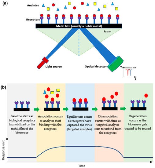
Plasmonic optical biosensors are classified into two types of platforms; one which uses a thin metal-based film and another which uses nanostructure-based inorganic plasmon resonance [21]. The most common plasmonic biosensor used is the surface plasmon resonance (SPR) which is a metal-based film sensor, mostly made of gold, used to characterize biomolecular interactions [22][23][24][22,23,24]. The angle at which the SPR is formed as light is focused on the metal film and reflected onto a detector. The output collected is due to energized plasmonic electrons formed from the collective refractive index (RI) of the oscillations of the electrons in the conduction band and the oscillations of the electric field formed by the light. The angle at which the SPR is measured relies on the RI of the material attached to the metal film. Hence, any alteration in the angle of the incidence or the RI of the material affects the resonance measured while detecting the analytes [25][26][25,26]. Decreasing the size results in a blueshift which holds a high frequency, while increasing the surrounding dielectric constant results in a red shift which holds a low frequency [27][28][29][27,28,29]. As shown in Figure 2a, the targeted analytes in a sample bind to the biological receptors restrained on the film to form a different RI which is usually known and indicates the presence of the targeted analyte [30]. This SPR biosensor can detect viruses and then be reused after applying proper chemical treatment practices as shown in Figure 2b [31][32][31,32].
Biology 11 00621 g002 550
Figure 2.
(
a
) A schematic diagram of the mechanism of plasmonic optical biosensors, and (
b
) Stages of SPR sensor from detecting analytes to detaching to be reused.
The efficiency of the plasmonic optical biosensor could be further enhanced when combined with surface-enhanced Raman scattering (SERS). SERS is used as a non-invasive, label-free diagnostic sensor that can attain abundant information from one measurement. Yet, due to the weak signal formed when detecting analytes at low concentrations, SERS is considered inappropriate [33]. Hence, by using plasmonic nanostructures for making the substrates for SERS, the signal becomes amplified by numerous orders of magnitude even though fabricating substrates for SERS is difficult [34]. This was proven in 2017 when Elsayed et al. fabricated a low-cost silicon substrate using silicon nanowires covered with silicon nanoparticles as substrates for SERS. Results showed detection levels reaching 10–11 M, which is a significant increase in the order of magnitude. The enhancement factor of silver NPs reaches 6–8 × 10⁵ but when deposited on the silicon nanowires, it reaches 1011 [35].

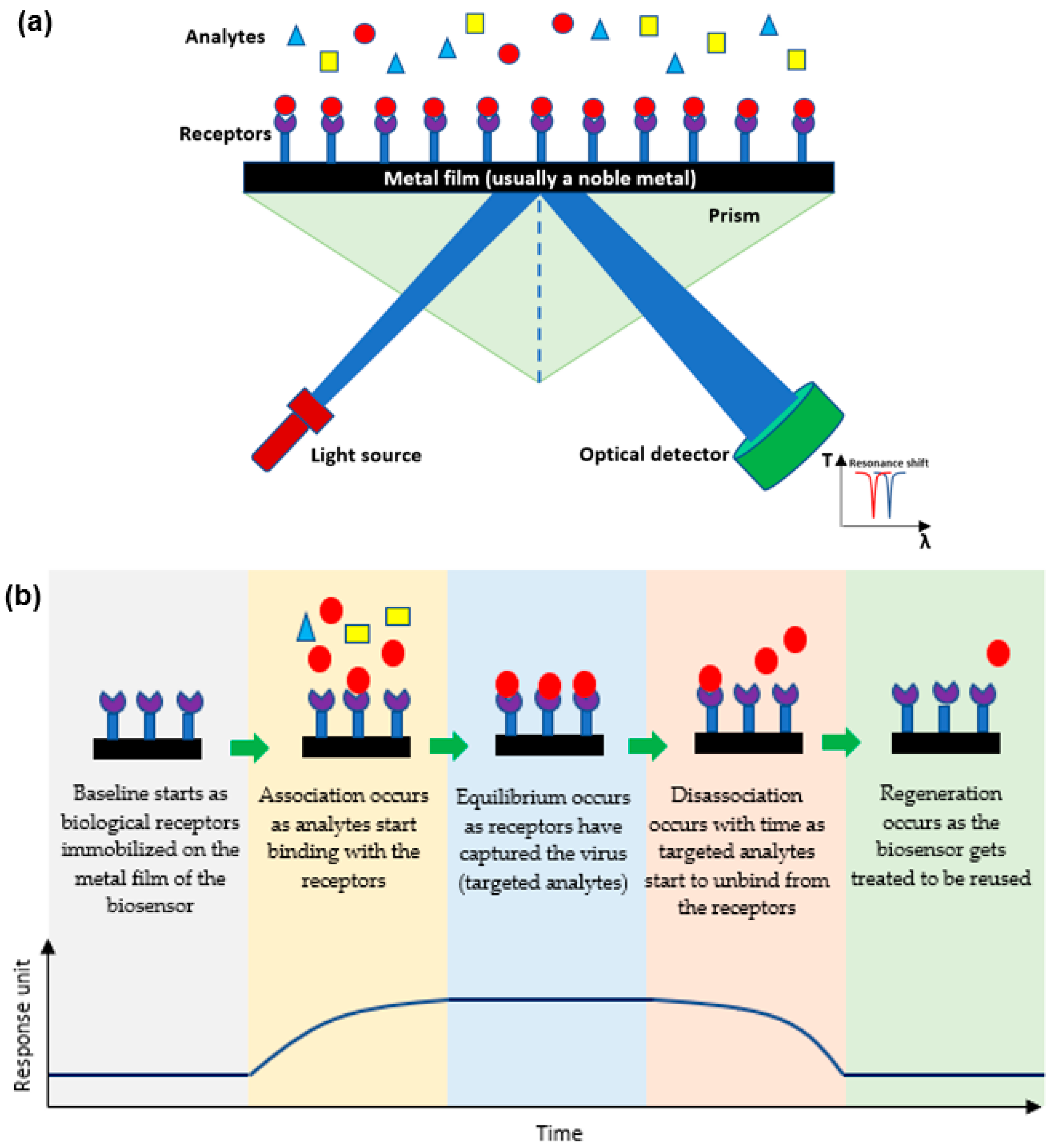
1.2. Determining the Efficiency of a Plasmonic Biosensor

To determine the efficiency of a biosensor, its limit of detection (LOD) and specificity(s) should be measured and attuned for its required application. The Clinical and Laboratory Standards Institute (CLSI) provides a guideline known as EP17 along with the protocols needed to determine the LOD [36] (p. 2).

2. Applications of Plasmonic Optical Biosensors

2.1. The Use of Plasmonic Biosensors for Viral Detection

In 2019, an unanticipated disease known as the coronavirus (SARS-CoV-2) was discovered in Wuhan, China, causing a global pandemic. In less than two years, this novel virus affected millions and resulted in the death of more than four million people worldwide as per the WHO [37][40]. Furthermore, the coronavirus has significantly affected the world’s economy and resulted in a noticeable change in the social lifestyle of people to avoid getting infected with the fast-spreading, contagious virus. Hence, immediate detection methods of the virus with accurate, reliable, and instant results were needed to limit the virus from outspreading any further. Infectious viruses have been detected over the years directly by targeting the virus itself or indirectly by targeting the secondary responses of the virus [38][41]. Targeting a virus directly involves targeting the entire virion, the antigens of the virus, or the single or double-stranded RNA or DNA of the virus. Indirect detection methods include serological testing for precise antibodies released due to a primary response to an antigen (IgM) or a secondary response due to previous exposure to the same type of antigen (IgG) [39][42]. Some of the most common methods in detecting infectious viruses include immunofluorescence assays, hemagglutination assays, viral plaque assay, viral flow cytometry, enzyme linked immunosorbent assay (ELISA) [40][43], chest computed tomography (CT) [41][44], and nucleic acid amplification test (NAAT) including polymerase chain reaction (PCR) and real-time quantitative PCR (RT-qPCR) [42][43][44][45][45,46,47,48]. Although those methods have shown successful outcomes, they have also displayed substantial limitations that deferred their usage in future viral detection as shown in Table 1.
Table 1.
Advantages and limitations of common viral detecting methods.

Current Methods for Virus Detection

Advantages

Limitations

Refs.

Immunofluorescence

Assays

Numerous, simultaneous samples can be analyzed and stored for some time.

Fluorescent molecules bound to primary antibody is limited.

Low sensitivity may result in false negatives.

[46][49]

. When detecting environmental analytes, biosensors usually include whole microorganisms, DNA, enzymes, and antibodies as recognition receptors [55][100]. Environmental monitoring using biosensors rather than conventional analyzing tools is of great benefit since they are portable, miniatured, compact, and has high selectivity to different matrices using low input of sample preparation which can be used on an on-going basis for regular environmental analysis [56][101]. Hence, biosensors can be used as monitoring tools in the environment to assess the biological quality of ecological molecules including organic and inorganic pollutants [57][102]. In 2017, a MIR sensor was fabricated by using doped silicon structured as nanowires with 10 nm radius using a numerical analysis technique known as Finite-difference time-domain method (FDTD). The simulation done by the 2D FDTD showed a total-field scattered-field source with a 3 μm wavelength around the plasmonic resonance used for exciting the nanowire.

2.3. The Use of Plasmonic Biosensors for Food Analysis

Diseases and malnutrition occurring due to the quality of products made food safety of great priority to diminish the health risks [58][118]. Conventional methods used to ensure food safety like PCR, ELISA, high-performance liquid chromatography (HPLC), and liquid chromatography-mass spectrometry (LCMS) are accurate but costly, time-consuming, and laborious [59][124]. Hence, using automated optical biosensors is an optimum solution for analyzing food by using a highly sensitive and selective low-cost analytical tool [60][125]. SPR among different types of optical biosensors has undergone great development to enable the detection of various pathogens found in food. Plasmonic biosensors were further upgraded for better analysis via coupling of various methods (Raman, fluorescence, and luminescence) to have less LOD and higher sensitivity. Hence, as shown in Table 2, SPR biosensor is considered much more efficient than traditional methods in monitoring and analyzing food.
Table 2.
SPR in comparison to other techniques for monitoring food.

Component

Other Methods

SPR

Refs.

Heavy metals

Atomic Absorption Spectroscopy

  • Destructive technique and single sample analyzed

Inductively Coupled Plasma Mass Spectrometry

  • Costly and destructive technique

X-ray Fluorescence Spectrometry

  • Radioactive technique

  • Low-cost

  • Quick measurement

  • Highly sensitive

  • Non-destructive

[61][126[62],127]

Hemagglutination Assays

Food Allergens

Low-cost instruments.

Results within hours.

ELISA

Has standardization as it is recognized in labs worldwide.

  • Varying as per the type of kit with LOD is 2.5 mg/L

Little specificity.

Requires trained personnel. Analysis needed by qualified individuals.

Difficult inter-laboratory comparison of results due to the several controlled variables.

  • Highly sensitive

  • LOD of 57.8 ng mL−1

[47][50]

[

61

][62][126,127]

Viral Plaque

Assay

Citrinin

(Mycotoxin)

Available in most labs.

Rapid results.

HPLC and LC-MS

  • Time-consuming and costly

Absence of standardization.

Involves costly repeat runs for accurate results.

  • Highly effective and selective

  • Simple, quick, and highly sensitive

[48][51]

[63][64][128,129]

Viral Flow

Cytometry

Pesticides

Rapid results.

Numerous cells

analyzed instantly.

LC-MS/MS

  • Complicated

  • Requires sample pre-treatment prior to analysis

Requires highly trained personnel.

  • Highly precise

Requires ongoing maintenance by service engineers.

  • Less response-time

  • Low-cost and low LOD

[49][52]

[65][66][130,131]

ELISA

Accurate/fast results.

β-Lactoglobulin

Very sensitive process.

Easily automated.

ELISA and LC-MS

  • Inconsistency and costly

Expensive preparation method. Requires trained personnel.

  • Speedy and detects in real time

  • Resistance to environmental factors

[40][43]

CT

Combined assessment.

Short acquisition time.

Expensive preparation method.

Requires trained personnel.

Exposure to gamma rays.

[41][44]

NAAT

Very sensitive process.

Accurate and reliable

Requires trained personnel.

Expensive detection kits.

Time-consuming (2–3 days). False-positive cases.

[43][44][45][46,47,48]

Hence, to overcome those drawbacks, biosensors were pursued as viral diagnostic tools since they are highly-sensitive, affordable, robust, automated, and have a low fluid consumption with faster reaction time [50][53]. Viruses are detected using plasmonic biosensors that have planar, optofluidic, nanoparticle-based, quantum dot enhanced fluorescent local SPR, or nanowire-based structures as shown in Figure 3 [31]. Further description of each method will be discussed later.
Biology 11 00621 g003 550
Figure 3. Common structures of plasmonic biosensors including (a) planar structure plasmonic biosensor where the base is a metal layer covered by a virus capturing layer to detect the viruses, (b) nanoparticle-based structure plasmonic biosensors bind to metal then a capturing molecule to capture viruses, (c) nanowire-based plasmonic biosensors with nanowires to entrap the targeted viruses, (d) optofluidic structure plasmonic biosensors with a virus capturing layer bind to a capturing molecule for higher detection of viruses, and (e) quantum dot fluorescence-based plasmonic biosensors with the capturing molecule bind to quantum dots which show visible changes among binding with viruses.

2.2. The Use of Plasmonic Biosensors for Environmental Evaluation

Plasmonic biosensors are great candidates when analyzing environmental contaminants. The amount of pollution increasing at a fast pace requires fast, highly specific, and cost-effective analytical tools that can be used for monitoring pollutants in our environment. Providentially, great initiatives have been made for controlling environmental pollution and several scientific researches were conducted and are still in progress to satisfy the concern of society regarding the environment and the pollution overtaking it [51][52][53][54][96,97,98,99]. Biosensors are great analytical techniques that can use a biological mechanism to detect analytes in the environment using chemical sensors for environmental evaluation [52][53][54][97,98,99]

[

67

]

[

68][132,133]

Tetrodotoxin

(Fish toxin)

LC-MS/MS, ELISA, and HPLC

  • Costly and time-consuming

  • Speedy

  • Low LOD

[69][70][134,135]

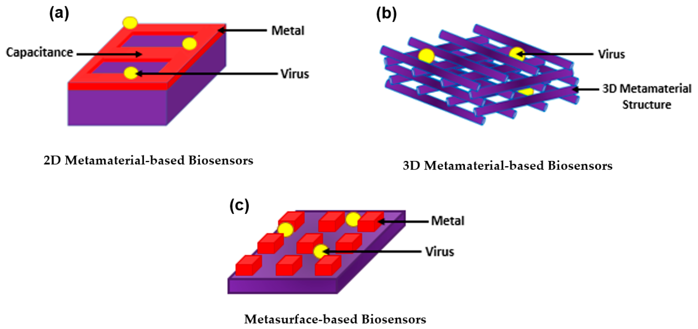
3. Introducing Metamaterials to Plasmonic Biosensors

With all the exquisite properties of plasmonic biosensors, the sensitivity has been further improved by introducing metamaterials. Metamaterial-based biosensors provide different geometric structures, each having its own sensing properties, which expands and improves the use of conventional plasmonic biosensors [68][133]. In the 1960s, a Russian physic named Victor Veselago initiated a theoretical concept for materials with simultaneous negative permittivity and permeability where light propagates in an opposite direction to that of the flow of energy, giving an uncommon refraction of light. Materials that follow this concept are known as left-handed materials [69][134]. In 1999, a theory was made by Pendry et al. declaring that microstructures having extremely small nonmagnetic conducting sheets in comparison to the wavelength of radiation reveal a magnetic permeability that is highly effective and can be further modified to display changing magnetic permeability together with the imaginary component [70][135]. This substance was named in the same year by Rodger Walser as “metamaterials” which he defined as “macroscopic composites having a synthetic, three-dimensional, periodic cellular architecture designed to produce an optimized combination, not available in nature, of two or more responses to specific excitation” [71][136]. The following year, Smith et al. confirmed the use of left-handed metamaterial using a microwave regime by experimenting with interspaced nonmagnetic conductive split-ring resonators with continuous wires [72][137]. Since then, metamaterials have been used, manipulated, and geometrically enhanced to have tuned properties that can be used in different applications including sensors, biological imaging, and spectroscopy [73][74][75][76][77][78][79][138,139,140,141,142,143,144]. The use of metamaterials was further explored as biosensors as they were categorized based on their structure into three main groups; 2D metamaterial-based biosensors, 3D metamaterial-based biosensors, and meta-surface-based biosensors as shown in Figure 4. The breakthrough of metamaterials with their improved sensitivity allowed the successful detection of several viruses including HIV, Zika virus, avian influenza virus, CPMV, and PRD1 [80][81][82][83][84][145,146,147,148,149]. Even further, metamaterials became a tool for novelty in label-free point-of-care viral detection.
Biology 11 00621 g014 550
Figure 4.
Common categories of metamaterials as (
a
) 2D metamaterial-based biosensors; (
b
) 3D metamaterial-based biosensors; and (
c
) metasurface-based biosensors.

4. Lab-on-a-Chip (LoC) for Plasmonic Biosensors

Molecular and serological testing is critical for diagnosing patients. Hence, the need for having faster and more reliable diagnostic tools for immediate disease analysis and prevention of the spreading of pathogens and diseases is always pursued and high in demand [85][152]. LoC is considered the best device used for Point-of-Care testing (POCT) as it is based on biosensors that are designed for their application and can be upgraded with the most recent advances using microfluidics [86][87][88][153,154,155]. In this section, plasmonic biosensors are focused on due to their various LoC applications. The main target of the plasmonic-based LoC devices is to use planar technology to make an integrated photonic circuit that has good sensing capabilities [89][90][91][92][93][156,157,158,159,160]. This technology has a few unique proficiencies which integrates many different sensors on the same chip to detect different pathogens. In addition, the utilization of planar technology supports the mass production need and provides cost-effective solutions. Hence, silicon-based photonics are known to be a major technology platform for such applications [94][95][96][161,162,163]. Hence, plasmonics have been recently introduced at a wide range for such applications due to their greater sensitivity and improved selectivity [97][98][113,164]. The integration of plasmonic materials such as gold, silver, and aluminum along with silicon photonics standard technology has been a challenge with two folds; the first one is technology-related where metals may cause contamination for the conventional standard silicon photonics technology, while the second is the ability to efficiently couple the light from dielectric waveguides with modal field ranges from few microns, in case of optical fiber, to few hundreds of nanometers, for silicon waveguide, to a plasmonic mode with tight surface confinement and modal field in the range of few nanometers only. The impact of the former challenge has been reduced over time by optimizing the fabrication technology and using materials that have both plasmonic effects and compatibility of the silicon technology such as TiN and ITO [99][100][101][165,166,167]. Good coupling can be achieved between a silicon waveguide and the plasmonic slot mode over a wide range of wavelengths using an orthogonal coupling scheme similar to Otto configuration [96][163]. In 2015, the ability of doped silicon to support plasmonic mode in the mid-infrared wavelength range was introduced for the first time [98][164]. The main mechanism was to control the doping level of the silicon to achieve a plasmonic wavelength within the mid-infrared range by doping for silicon. For instance, a doping ranging from 10−19 to 5 × 1020 cm−3 can achieve a plasmonic behavior starting from ~10 to ~3 microns, respectively. Other III-VI semiconductors have been recently utilized for such applications [101][167]. The III-V materials have superior advantages over the silicon such that the on-chip detector can be fabricated using a compatible material from the same material group. The MIR range also has the advantage of providing unique absorption peaks for sensing gases or liquids for biomedical applications. This added advantage can help increase the selectivity of the proposed sensing system. Hence, a plasmonic biosensor made only from a dielectric can be apprehended in the MIR using the planar fabrication technology and yet with high sensitivity and selectivity at the same time [102][103][104][105][103,115,168,169].