Your browser does not fully support modern features. Please upgrade for a smoother experience.
Please note this is a comparison between Version 2 by Dean Liu and Version 1 by Inga Zinicovscaia.
Among produced metal nanoparticles, silver nanoparticles are widely used in everyday life products, cosmetics, and medicine. It has already been established that, in nanoscale form, many even inert materials become toxic.
- nanoparticles
- silver
- toxicity
- occupational
- brain
1. Introduction
Nanoparticles are found in numerous spheres of life. Their application in medicine, food and cosmetic industries, and everyday life is expanding [1]. Due to their unique electrical, optical, chemical, and antimicrobial properties, silver nanoparticles (AgNPs) are the most common type of NPs used in consumer products [2]. At the same time, according to publications in toxicology, a wealth of information has already been accumulated confirming that contact with NPs entails serious negative consequences for cells, tissues, and organs, such as the development of oxidative stress, the accumulation of DNA disorders, the induction of apoptosis and inflammation, and the disturbance of the structure and functions of tissues and organs [3,4,5][3][4][5].
Almost every person in the modern world is constantly in contact with NPs, through products or objects containing nanoparticles, or through air, water, and soil. The environment begins to suffer greatly from pollution by various nano-sized substances as a result of emissions from enterprises and vehicle exhaust, sewage discharges, and waste. Due to the widespread use of NPs, including AgNPs, in various industries, employees in the workplace face an increased risk of health problems owing to prolonged (chronic) contact with small doses of NPs that enter the body from the air in contaminated work areas, as well as with water and/or food. Occupational pathology has already accumulated bodies of evidence on the development of diseases caused by NPs in industrial workers, which cannot be explained by other reasons [6,7][6][7]. In women of reproductive age employed in industrial fields, contact with NPs is associated with risks for their unborn children due to the ability of NPs to penetrate the placental barrier, which leads to the transfer of NPs from mother to child [8,9][8][9].
. General Toxicity of AgNPs for Cells, Tissues, and Organs
2. General Toxicity of AgNPs for Cells, Tissues, and Organs
According to numerous publications, the main mechanism of the cellular and molecular toxicity of AgNPs is oxidative stress [10,11][10][11]. Thus, 20 nm spherical AgNPs reduced mRNA levels of sodium dismutase 1 and glutathione reductase in male Wistar rats [9]. In the presence of AgNPs, the formation of reactive oxygen species [10], the level of gene expression and the content of antioxidant proteins decrease, and DNA damage [11] increases. The prolonged exposure of adult rats to citrate-stabilized silver NPs at a low dose of 0.2 mg/kg resulted in a decrease in the level of expression of structural proteins, in particular, myelin in myelin sheath cells. AgNPs were also found to increase the body weight and body temperature of animals [12]. The level of developmental regulators (for example, neuronal) also decreases; however, the levels of expression of apoptosis regulators increase; therefore, ultimately, the cell often undergoes apoptosis. At the tissue level, inflammation, swelling, and, as an extreme outcome, necrosis, can develop [13]. In zebrafish, AgNPs caused a decrease in brain and muscle acetylcholinesterase activity, as well as in liver and gill catalase activity. Contact with AgNPs also led to morphological changes such as the fusion of secondary lamellae, curvature, dilated marginal channel, and epithelial lifting [14]. The administration of male CD-1 mice with AgNPs of 10 nm size, compared with NPs with a size of 40 and 100 nm, resulted in overt hepatobiliary toxicity [15]. Some reports indicate that AgNPs are genotoxic and mutagenic, and the degree of their impact directly depends on the dose of NPs and inversely depends on their size [16,17,18][16][17][18]. After exposure of BEAS-2B cells to AgNPs of different primary particle sizes (10, 40, and 75 nm), a cytotoxic effect was observed only at a particle size of 10 nm [19]. The same finding was reported in [20]. Coating agents used to stabilize NPs in a solution can also affect general toxicity, including genotoxicity. For example, AgNPs coated with citrate had more noticeable toxic effects on L5718Y cells than those coated with polyvinylpyrrolidone [21]. The study by Recordati et al. [15] showed that the coating of NPs had no relevant impact on animals. Citrate-coated silver nanopowder was toxic to human skin HaCaT keratinocyte cells, whereas polyvinylpyrrolidone-coated silver nanoprism or nanoparticle powder did not exhibit any toxic effect [22]. In a study by Gliga et al. [19], the coating-dependent difference in the cytotoxicity of AgNPs was not detected. El Badawy and al. [23] demonstrated a direct correlation between the toxicity of AgNPs and their surface charge. Thus, negatively charged citrate-coated AgNPs were less toxic to microorganisms than positively charged branched polyethyleneimine-coated AgNPs. The low toxicity of citrate-coated AgNPs was observed in comparison with polyvinylpyrrolidone- and gum arabic-coated AgNPs [24]. It should be noted that the results of evaluating the mutagenicity of AgNPs may also depend on the chosen object of study; for example, Prokhorova et al. [25] did not find chromosomal abnormalities in plant cells after exposure to AgNPs. Studying the effect of AgNPs on freshwater invertebrates with different life strategies—namely, Hydra vulgaris, Daphnia carinata, and Paratya australiensis—the authors described Daphnia carinata as the most sensitive species, followed by Paratya australiensis and Hydra vulgaris [26]. Toxic effects of AgNPs on different organisms in the study by Ivask et al. [20] varied by about two orders of magnitude, with the lowest observed for crustaceans and algae and the highest for mammalian cells. The results of studies of the effects of NPs on the reproductive function and development of animal organisms are also ambiguous and often contradictory. The bulk of studies demonstrated that AgNPs could cause defects in the neurological, cardiovascular, reproductive, and immune systems of the embryos and fetus; however, in some studies, no adverse effect on the development of AgNPs was found, even at high doses [27]. The development of oxidative stress, the occurrence of morphological defects, slowing heart rate, the appearance of pericardial edema, delayed hatching from eggs, and increased mortality were observed in studies on zebrafish embryos exposed to AgNPs [28,29,30][28][29][30]. Morphological changes in the ovary and testis were observed in rat offspring exposed to AgNPs [31]. Developmental exposure to AgNPs results in long-term gut dysbiosis, body fat increase, and neurobehavioral alterations in mouse offspring [32]. At the same time, it should be mentioned that silver ions (Ag+) provoke similar toxic effects at concentrations hundreds of times lower than those applied for AgNPs [29,30][29][30]. It was suggested by some authors that the compounds used for NP coating significantly affected the level of AgNP toxicity. Thus, after contact with polyvinylpyrrolidone-coated AgNPs, zebrafish larva did not differ in morphology and behavior from control ones; however, when citrate was used as a coating agent, some disturbances were detected [33]. In a study by González et al. [34], in which the level of AgNPs in solution was close to their environmental concentrations in water (0.03–3 ppm), no negative changes in the survival, hatching, or morphology of Danio rerio were found. According to a number of studies, after a single oral administration, reproductive dysfunctions in males and females (inhibition of spermatogenesis, histopathological disorders in ovaries, etc.) were observed in rodents exposed to AgNPs. It is noteworthy that the smaller the dose and size of AgNPs were, the less pronounced were the effects [35]. Undoubtedly, the dose–response principle also works in nanotoxicology.2. Effect of AgNPs on the Brain and Behavior
3. Effect of AgNPs on the Brain and Behavior
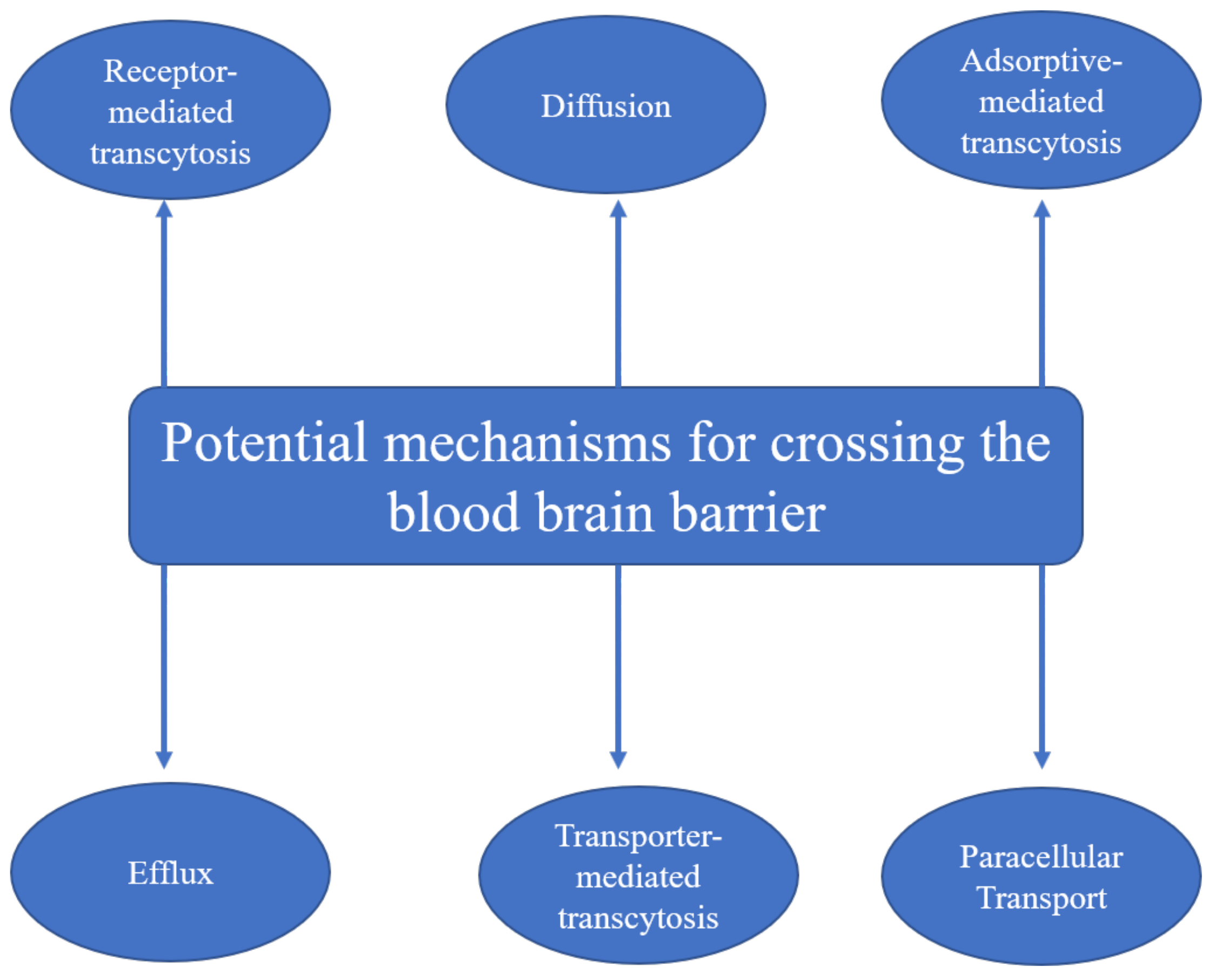
AgNPs along with other metal NPs are able to cross not just the placental barrier but the blood–brain barrier as well [8,9][8][9]. Tang et al. [36] proposed two mechanisms of AgNP penetration across the blood–brain barrier, i.e., the transcytosis of endothelial cells of the brain–blood capillary and the reduction in the close connection between endothelial cells, or dissolution of endothelial cell membranes. The main mechanisms of nanoparticles for crossing the blood–brain barrier according to the literature are summarized in Figure 1.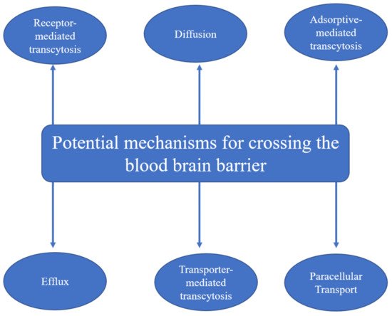
 Therefore, upon contact with NPs, there is a risk of biochemical, histological, and functional disorders in the brain, including cognitive dysfunctions. Studies of the effect of NPs on brain functions have received much attention in recent years. After a single dose injection of AgNPs, the parameters of oxidative stress and the antioxidant potential of the brain on gene expression and the level of protein (superoxide dismutase and glutathione reductase) activity were altered [37][40]. The shape of astrocytes was disturbed, cerebral capillaries were deformed, and edema was developed in adjacent areas [10,13,38][10][13][41]. In addition, the permeability of the blood–brain barrier increased in direct proportion to the dose of AgNPs received by animals [10].
After seven daily injections, impairments of working memory were noted in rats: The animals meaningfully more often made mistakes by repeatedly looking into the arm of the maze they had just examined; however, referential memory and the long-term understanding of the structure of space were not disturbed [38][41]. After three weeks of injections, the social behavior of the mice differed significantly from the normal behavior in the study by Greish et al. [42] The mice administrated with AgNPs preferred to stay in an empty chamber rather than to familiarize themselves with new animals. In experimental animals, motor coordination and balance were impaired, but the swimming speed in the Morris test was preserved, which revealed the absence of spatial memory formation in individuals treated with AgNPs. The emotional state, the level of anxiety, and the ability to conditioned-reflex learning, in which fear is the motivation, were considered in the study by Antsiferova et al. [43]. After long-term (1–6 months) oral contact with small doses of AgNPs (50 µg) through drinking water, the authors recorded and interpreted the test results as two attempts of the brain to adapt to the effects of AgNPs. Thus, after 2 months of contact, anxiety and fear increased, and after 4 months, they decreased with a simultaneous increase in exploratory behavior. After continued contact with AgNPs (up to 6 months), the impairment of long-term memory and conditioned-reflex learning was observed. The fading effect was noted in the research by González et al. [34]. Although several days after exposure to AgNPs, zebrafish larvae were hyperactive at changes in illumination, no fluctuations in their activity were detected after 5 days.
The influence of prenatal contact with AgNPs on the brain and behavior is described in a limited number of studies. After regular injections of AgNPs to future mothers during pregnancy, the spatial memory of their offspring was impaired, while conditioned-reflex learning and the emotional state of young rats did not differ from the offspring of the control group [44]. The experimental offspring demonstrated signs of depressive-like behavior, i.e., passivity and lack of interest in food [45]. After zebrafish embryos were exposed to AgNP solution, their avoidance motor response to the touch was disrupted; the membrane potentials of their motoneurons were decreased, and the expression of many genes connected with neurogenesis was low [46].
In studies of the effect of AgNP coating material (BSA, polyethylene glycol, and citrate) on cognition, spatial memory, and neurotransmitter levels in the rat hippocampus, only rats administrated with citrate-coated NPs maintained long-term spatial memory. For other NPs and Ag+, the induction of peripheral inflammation, which was reflected by alterations in the level of serum inflammatory mediators, was observed [47]. Wu et al. [48] showed a significant reduction in GAP-43 mRNA and protein expression in the hippocampus of offspring exposed to uncoated AgNPs, suggesting cognitive impairments in rats.
However, in some studies on adult animals, after subchronic contact with AgNPs, no behavioral disturbances were noticed. In the study by Liu et al. [49], no significant differences between experimental and control mice in the formation of spatial and working memory were found, and secondary neurogenesis in the hippocampus was not impaired. In the research by Dabrowska-Bouta et al. [12], pathological changes in the structure of myelin sheaths and a decrease in the level of three myelin-specific proteins were detected in the brain of experimental rats treated with AgNPs or Ag+. However, neither motion nor exploratory behavior or memory was impaired in animals treated with silver nanoparticles present in the two forms. After an intranasal introduction of AgNPs, experimental mice coped with the recognition of a new object in the same way as the control ones; however, their spatial memory was presumably impaired, while the ability for spatial learning itself did not decrease [50]. After AgNPs were injected into lactating mice, their grown offspring, which contacted with AgNPs through milk, did not differ from control animals in terms of their emotional state, social interactions, and locomotion [51].
Therefore, upon contact with NPs, there is a risk of biochemical, histological, and functional disorders in the brain, including cognitive dysfunctions. Studies of the effect of NPs on brain functions have received much attention in recent years. After a single dose injection of AgNPs, the parameters of oxidative stress and the antioxidant potential of the brain on gene expression and the level of protein (superoxide dismutase and glutathione reductase) activity were altered [37][40]. The shape of astrocytes was disturbed, cerebral capillaries were deformed, and edema was developed in adjacent areas [10,13,38][10][13][41]. In addition, the permeability of the blood–brain barrier increased in direct proportion to the dose of AgNPs received by animals [10].
After seven daily injections, impairments of working memory were noted in rats: The animals meaningfully more often made mistakes by repeatedly looking into the arm of the maze they had just examined; however, referential memory and the long-term understanding of the structure of space were not disturbed [38][41]. After three weeks of injections, the social behavior of the mice differed significantly from the normal behavior in the study by Greish et al. [42] The mice administrated with AgNPs preferred to stay in an empty chamber rather than to familiarize themselves with new animals. In experimental animals, motor coordination and balance were impaired, but the swimming speed in the Morris test was preserved, which revealed the absence of spatial memory formation in individuals treated with AgNPs. The emotional state, the level of anxiety, and the ability to conditioned-reflex learning, in which fear is the motivation, were considered in the study by Antsiferova et al. [43]. After long-term (1–6 months) oral contact with small doses of AgNPs (50 µg) through drinking water, the authors recorded and interpreted the test results as two attempts of the brain to adapt to the effects of AgNPs. Thus, after 2 months of contact, anxiety and fear increased, and after 4 months, they decreased with a simultaneous increase in exploratory behavior. After continued contact with AgNPs (up to 6 months), the impairment of long-term memory and conditioned-reflex learning was observed. The fading effect was noted in the research by González et al. [34]. Although several days after exposure to AgNPs, zebrafish larvae were hyperactive at changes in illumination, no fluctuations in their activity were detected after 5 days.
The influence of prenatal contact with AgNPs on the brain and behavior is described in a limited number of studies. After regular injections of AgNPs to future mothers during pregnancy, the spatial memory of their offspring was impaired, while conditioned-reflex learning and the emotional state of young rats did not differ from the offspring of the control group [44]. The experimental offspring demonstrated signs of depressive-like behavior, i.e., passivity and lack of interest in food [45]. After zebrafish embryos were exposed to AgNP solution, their avoidance motor response to the touch was disrupted; the membrane potentials of their motoneurons were decreased, and the expression of many genes connected with neurogenesis was low [46].
In studies of the effect of AgNP coating material (BSA, polyethylene glycol, and citrate) on cognition, spatial memory, and neurotransmitter levels in the rat hippocampus, only rats administrated with citrate-coated NPs maintained long-term spatial memory. For other NPs and Ag+, the induction of peripheral inflammation, which was reflected by alterations in the level of serum inflammatory mediators, was observed [47]. Wu et al. [48] showed a significant reduction in GAP-43 mRNA and protein expression in the hippocampus of offspring exposed to uncoated AgNPs, suggesting cognitive impairments in rats.
However, in some studies on adult animals, after subchronic contact with AgNPs, no behavioral disturbances were noticed. In the study by Liu et al. [49], no significant differences between experimental and control mice in the formation of spatial and working memory were found, and secondary neurogenesis in the hippocampus was not impaired. In the research by Dabrowska-Bouta et al. [12], pathological changes in the structure of myelin sheaths and a decrease in the level of three myelin-specific proteins were detected in the brain of experimental rats treated with AgNPs or Ag+. However, neither motion nor exploratory behavior or memory was impaired in animals treated with silver nanoparticles present in the two forms. After an intranasal introduction of AgNPs, experimental mice coped with the recognition of a new object in the same way as the control ones; however, their spatial memory was presumably impaired, while the ability for spatial learning itself did not decrease [50]. After AgNPs were injected into lactating mice, their grown offspring, which contacted with AgNPs through milk, did not differ from control animals in terms of their emotional state, social interactions, and locomotion [51].