Cancer is a major cause of death worldwide. With the advantages of simplicity, rapid response, reusability, and a low cost, aptamer-based electrochemical biosensors have received considerable attention as a promising approach for the clinical diagnosis of early-stage cancer.
- aptamer
- aptasensor
- electrochemical
- cancer diagnostic
1. Introduction
2. Redox-Active Molecules
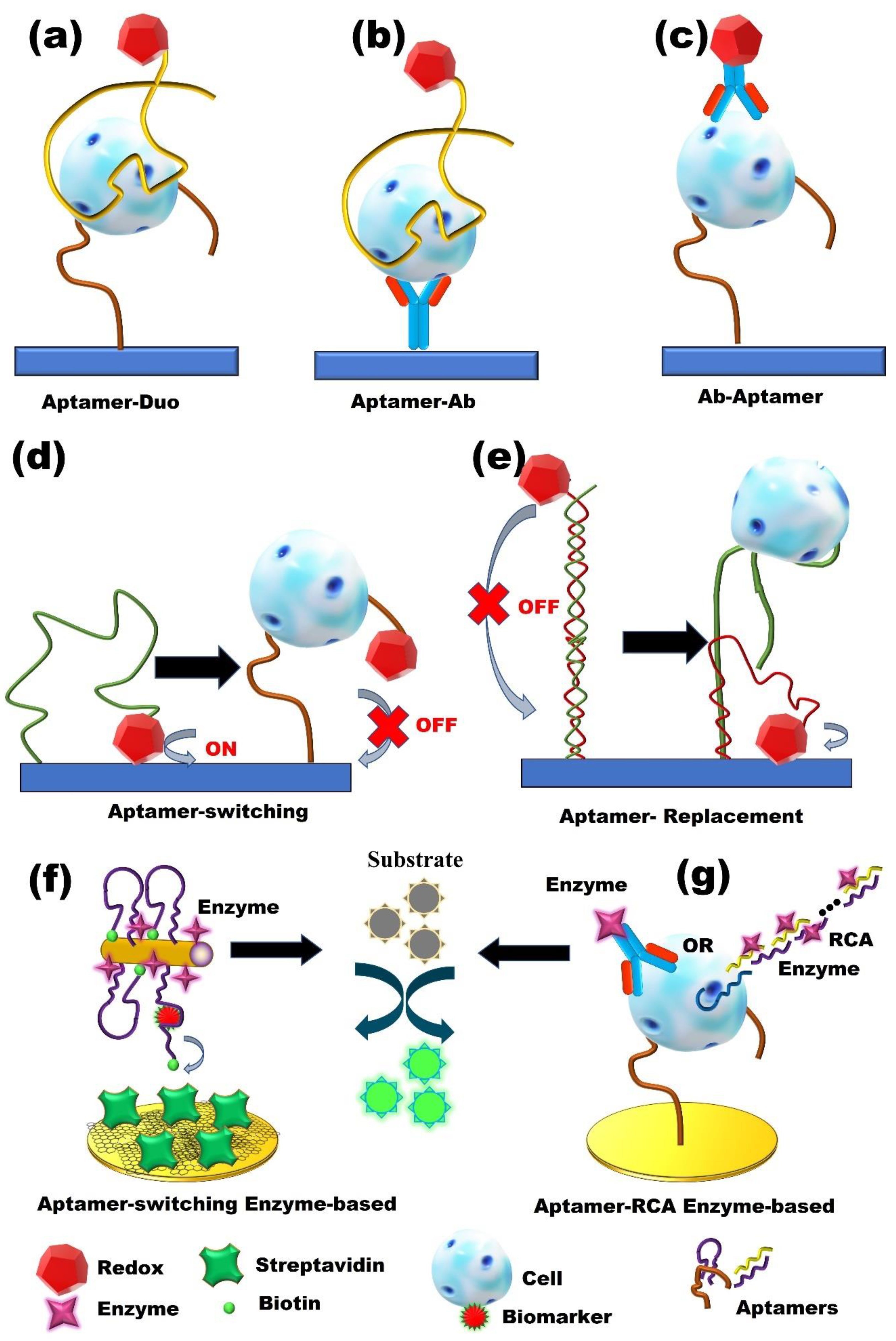
A simple way to generate an electrochemical signal is through the use of redox-active labels [27][28]. Using this strategy, aptamers can be incorporated to develop enhanced aptasensors. Aptamers can fold their flexible single-stranded chains into three-dimensional (3D) structures upon binding to a target molecule and can easily be immobilized on a conductive surface. These features enable redox-active molecules to be anchored to aptamers, allowing for the identification of the formation of aptamer–target complexes by probing the electron transfer features of the redox probes of rigidified complexes [29]. Generally, redox-active molecule-based electrochemical aptasensors include two subclasses: “signal-on” or “signal-off”. Due to the conformational change in aptamers in the signal-on mechanism, redox-active molecules are brought close to the electrode surface, and removed from the electrode surface (2. Redox-Active Molecules
3. Enzyme-Based Aptasensors
Although the application of redox-active molecules is a simple method to generate an electrochemical signal, electrochemical aptasensors suffer from low sensitivity [28]. Therefore, the development of signal-amplification strategies to enhance sensitivity is critical. To date, a wide variety of amplification strategies have been designed. Among them, enzymes (biocatalysts) show the advantage of enhancing through enzymatic electrochemical processes (3. Enzyme-Based Aptasensors
4. Nanomaterials-Based Aptasensors
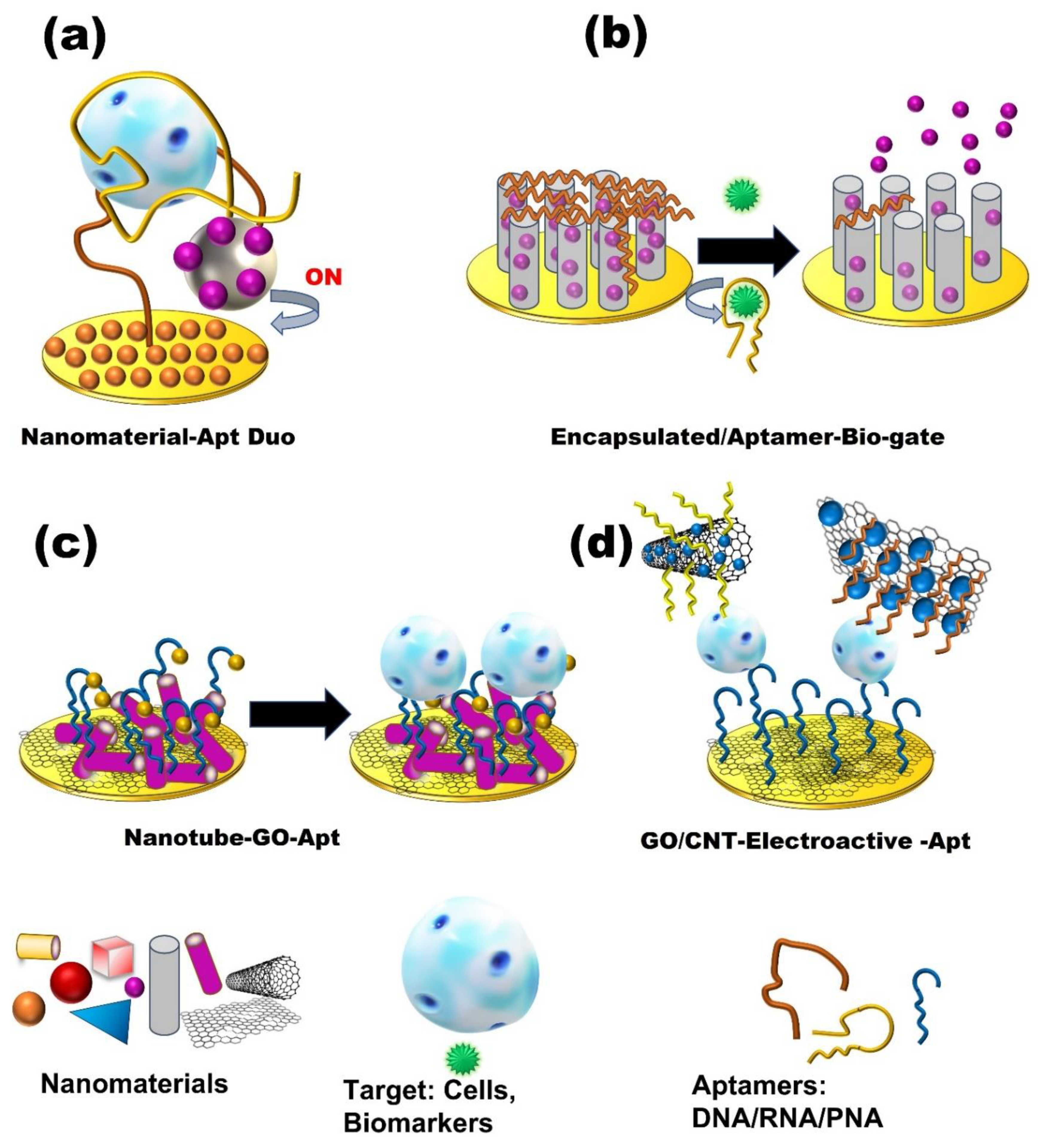
Owing to the unique characteristics of nanomaterials, such as their small size, increased surface-to-volume ratio, biocompatibility, and chemical stability, along with the excellent selectivity of aptamers as recognition elements, the combination of nanomaterials and aptamers can promote new innovations for the detection of cancer cells [38]. Different strategies have been described to conjugate aptamers with nanomaterials [39] (4. Nanomaterials-Based Aptasensors


Cancer Type | Target | Technique | Sample | Assay Time | LOD | Linear Range | Reference | |||||||||||||||||||
|---|---|---|---|---|---|---|---|---|---|---|---|---|---|---|---|---|---|---|---|---|---|---|---|---|---|---|
Breast Cancer | EGFR | DPV | Serum | 30 min | 50 pg/mL | 1–40 ng/mL |
[41] |
[46] |
||||||||||||||||||
ER | DPV | Buffer | 10 min | 0.001 ng/mL | 0.001–1000 pg/µL |
[42] |
[47] |
|||||||||||||||||||
Exosomes | CV | buffer | 1 h | 96 particles/μL. | 1.12 × 10²–1.12 × 10⁸ particles/μL |
[43] |
[48] |
|||||||||||||||||||
Exosomes (MCF-7 cells) | ECL | Blood serum sample | 120 min | 7.41 × 10⁴ particle/mL | 3.4 × 10⁵ –1.7 × 10⁸ particle/mL |
[44] |
[49] |
|||||||||||||||||||
HER2 | stripping voltammetry | Human serum | 20 min | 26 cells/mL | 50 to 20,000 cells/mL |
[32] |
[30] |
|||||||||||||||||||
HER2 | EIS | Buffer | - | 0.047 pg/mL | 0.01 to 5 ng/mL |
[45] |
[50] |
|||||||||||||||||||
HER2 | CV, EIS | Serum | 2 h | 1 pM | 1 pM–100 nM |
[46] |
[51] |
|||||||||||||||||||
HER2 | EIS | Serum sample | 40 min | 50 fg/mL | 0.1 pg/mL–1 ng/mL |
[43] |
[48] |
|||||||||||||||||||
HER2 | CV, DPV, EIS | PBS buffer | 5–10 min | 0.001 ng/mL | 0.001–100 ng/mL |
[47] |
[52] |
|||||||||||||||||||
MCF-7 | CC, CV, EIS | Serum | 25 min | 47 cells/mL | 0–500 cells/mL |
[48] |
[53] |
|||||||||||||||||||
MCF-7 | SWV, CV | Human plasma | 2 h | 328 cells/mL | 328–593 cells/mL |
[49] |
[54] |
|||||||||||||||||||
MCF-7 | CV, DPV | Human serum | 60 min | 20 cells/mL | 50–106 cells/mL |
[50] |
[55] |
|||||||||||||||||||
MCF-7 Exosomes | PEC | Buffer | 110 min (total) | 1.38 × 10³ particles/μL | 5.00 × 10³ to 1.00 × 10⁶ particles/mL |
[51] |
[56] |
|||||||||||||||||||
MDA-MB-231 | DPV | Blood Serum | 30 min | 5 cell/ mL | 10–1 × 10³ cell/mL |
[52] |
[57] |
|||||||||||||||||||
MUC1 | DPV | Serum sample | 25 min | 0.79 fM | 1 fM–100 nM |
[53] |
[58] |
|||||||||||||||||||
MUC1 | SWV, CV | Buffer | 1 h | 0.33 pM | 1.0 pM–10 µM |
[54] |
[59] |
|||||||||||||||||||
MUC-1 | EIS | PBS buffer | 2 h | 38 cells/mL | 100 to 5.0 × 10⁷ cells/mL |
[27] |
[33] |
|||||||||||||||||||
Nucleolin | DPV | Buffer | 1 h | 8 ± 2 cells ml/mL | 10–106 cells/mL |
[55] |
[60] |
|||||||||||||||||||
Nucleolin | ECL | Buffer | 10 min | 10 cells | 10–100 cells |
[56] |
[61] |
|||||||||||||||||||
Nucleolin | EIS | Buffer | - | 40 cells/mL | 103–107 cells/mL |
[57] |
[62] |
|||||||||||||||||||
Nucleolin | CV, EIS | Phosphate buffer | 30 min | 4 cells/mL | 1 × 101–1 × 10⁶ cells/mL |
[58] |
[63] |
|||||||||||||||||||
OPN | CV, SWV | Synthetic human plasma | 60 min | 1.3 ± 0.1 nM | CV: 25 to 100 nM | SWV: 12 to 100 nM |
[59] |
[64] |
||||||||||||||||||
OPN | CV | PBS buffer | 60 min | 3.7 ± 0.6 nM | 25–200 nM |
[60] |
[65] |
|||||||||||||||||||
PDGF-BB, | MCF-7 cells | CV, SWV | PBS buffer | - | PDGF-BB: 0.52 nM | MCF-7: 328 cells/mL | PDGF: 0.52–1.52 nM | MCF-7: 328 to 593 cells/mL |
[49] |
[54] |
||||||||||||||||
Lung Cancer | CEA, NSE | CV, DPV | Serum | 1 h | CEA: 2 pg/mL | NSE: 10 pg/mL | CEA: 0.01–500 ng/mL | NSE: 0.05–500 ng/mL |
[61] |
[66] |
||||||||||||||||
CEA | DPV, EIS | Human serum | 85 min (total) | 1.5 pg/mL | 5 pg/mL to 50 ng/mL |
[62] |
[67] |
|||||||||||||||||||
CEA | EIS | Buffer, serum | - | Buffer: 0.45 ng/mL | Serum: 1.06 ng/mL | 0.77–14 ng/mL |
[63] |
[68] |
||||||||||||||||||
Lung tumor | EIS | Blood plasma | ~25 min | - | - |
[64] |
[69] |
|||||||||||||||||||
Lung cancer tissues (proteins) | SWV | Blood plasma | 1 h | 0.023 ng/mL | 230 ng/mL to 0.023 ng/mL |
[65] |
[70] |
|||||||||||||||||||
VEGF165 | CV, EIS | Lung cancer Serum samples | 40 min | 1.0 pg/mL | 10.0–300.0 pg/mL |
[66] |
[71] |
|||||||||||||||||||
Lung cancer tumor | CV, DPV, SWV, EIS | Human blood | - | - | - |
[14] |
||||||||||||||||||||
Lung/Breast/ others cancer | VEGF | DPV | Buffer | 45 min | 30 nmol/L | 0–250 nmol/L |
[35] |
[40] |
||||||||||||||||||
CEA | DPV | Spiked Serum | 50 min | 0.9 pg/mL | 3 pg/mL to 40 ng/mL |
[29] |
[35] |
|||||||||||||||||||
CEA | DPV, EIS, CV | Human serum | 1 h | 0.34 fg/mL | 0.5 fg/mL to 0.5 ng/mL |
[38] |
[43] |
|||||||||||||||||||
CEA | DPV, CV, EIS | Serum | 1 h | 0.31 pg/mL | 1 pg/mL–80 ng/mL |
[67] |
[72] |
|||||||||||||||||||
CEA | EIS | Buffer/Blood sample | 1 h 30 min | 0.5 pg/mL | 1 pg/mL–10 ng/mL |
[68] |
[73] |
|||||||||||||||||||
CEA | DPV | Buffer | 1 h | 40 fg/mL | 0.0001–10 ng/mL |
[69] |
[74] |
|||||||||||||||||||
CEA | PES | Serum | 60 min | 0.39 pg/mL | 0.001–2.5 ng/mL |
[70] |
[75] |
|||||||||||||||||||
VEGF165 | CV | Buffer | 1 h | 30 fM | 100 fM to 10 nM |
[71] |
[76] |
|||||||||||||||||||
MUC 1 | CV, SWV, EIS | Buffer | 120 min | 4 pM | 10 pM to 1 μM |
[72] |
[77] |
|||||||||||||||||||
CEA | CV, EIS | Buffer | 1 h | 3.4 ng/mL | 5 ng/mL–40 ng/mL |
[73] |
[78] |
|||||||||||||||||||
CEA | CV | PBS/spiked human serum | 40 min | 6.3 pg/mL | 50 pg/mL to 1.0 μg/mL |
[11] |
||||||||||||||||||||
CEA | DPV | Buffer/spiked human serum | 45 min | 0.84 pg/mL | 10 pg/mLto 100 ng/mL |
[74] |
[79] |
|||||||||||||||||||
CEA and CA153 | PEC | Serum samples | 20 min | CEA: 2.85 pg/mL | CA153: 0.0275 U/mL | CEA: 0.005–10 ng mL, CA153: 0.05–100 U/mL |
[75] |
[80] |
||||||||||||||||||
Prostate Cancer | PSA | EIS | Buffer | 2 h | 0.5 pg/mL | 0.05 ng/mL to 50 ng/mL |
[5] |
|||||||||||||||||||
PSA | EIS | Buffer | 2 h (total) | 1 pg/mL | 1 × 10² pg/mL–1 × 102 ng/mL |
[76] |
[32] |
|||||||||||||||||||
PSA | DPV | Serum samples | 40 min | 0.25 ng/ mL | 0.25 to 200 ng/mL |
[77] |
[81] |
|||||||||||||||||||
PSA | SWV, EIS | Spiked human serum | - | EIS: 10 pg/mL | EIS: 10 pg/mL to 10 ng/mL |
[78] |
[82] |
|||||||||||||||||||
PSA | DPV | Blood serum | 30 min | 50 pg/mL | 0.125 to 128 ng/mL |
[79] |
[83] |
|||||||||||||||||||
PSA | PEC | Human serum | - | 0.34 pg/mL | 0.001 to 80 ng/mL |
[80] |
[84] |
|||||||||||||||||||
PSA | DPV | Human serum | 30 min | 0.064 pg/mL | 1 pg/mL to 100 ng/mL |
[81] |
[85] |
|||||||||||||||||||
PSA | DPV, EIS | Serum | sample | 40 min | 1.0 pg/ mL | DPV: 0.005–20 ng/mL | EIS: 0.005–100 ng/mL |
[82] |
[86] |
|||||||||||||||||
PSA | EIS | Human serum | 2 h 30 min | 0.33 pg/mL | 5 to 2 × 10⁴ pg/mL |
[83] |
[87] |
|||||||||||||||||||
PSA | CV, SWV, EIS | Buffer | 30 min | 0.028 * and 0.007 ** ng/mL | 0.5–7 ng/mL |
[84] |
[88] |
|||||||||||||||||||
PSA | PEC | PBS buffer/ spiked Serum | 40 min | 4.300 fg/mL | 1.000 × 10⁻⁵ to 500.0 ng/mL, |
[85] |
[89] |
|||||||||||||||||||
PSA | SWV, EIS | Serum sample | 4 h (total) | 2.3 fg/mL | 10 fg/mL–100 ng/mL |
[86] |
[90] |
|||||||||||||||||||
PSA | PEC | Human serum | 90 min | 0.52 pg/mL | 1.0 | pg/mL to 8.0 ng/mL |
[87] |
[91] |
||||||||||||||||||
PSA | ECL | Human serum | 60 min | 0.17 pg/mL | 0.5 pg/mL to 5.0 ng/mL |
[88] |
[92] |
|||||||||||||||||||
PSA | DPV | Spiked Urine Blood serum | 60 min | 280 pg/mL | 1 to 300 ng/mL |
[89] |
[93] |
|||||||||||||||||||
PSA | DPV | Human serum | 30 min | 6.2 pg/mL | 0.01–100 ng/mL |
[90] |
[94] |
|||||||||||||||||||
PSA, SAC | SWV | 50% | Human serum | PSA: 2 h | SAC: 1 h | PSA: 2.5 fg/mL, SAC: 14.4 fg/mL | PSA: 1 fg/mL to 500 ng/mL | SAC: 1 fg/mL to 1 μg/mL |
[91] |
[95] |
||||||||||||||||
Blood cell cancer | Ramos cell | LSV | Human serum | 3 h | 10 cells/mL | 1 × 101–1 × 10⁶ cell/mL |
[37] |
[42] |
||||||||||||||||||
Breast/ Liver cancer | HeLa, MCF-7, HepG2. | PEC | Buffer | 4 h 20 min (total) | 19 cell/mL (HeLa) | 50–5 × 10⁵ cell/mL (HeLa) |
[92] |
[96] |
||||||||||||||||||
Breast/ Prostate cancer | CTC | HER2, PSMA, and MUC1 | LSW | Spiked in Blood | 1 h | 2 cells/sensor | 2–200 | cells/sensor |
[93] |
[97] |
||||||||||||||||
PDGF-BB | DPV | PBS buffer | 40 min | 0.65 pM | 0.0007–20 nM |
[94] |
[98] |
|||||||||||||||||||
PDGF-BB | CV, EIS | ID water, 5% trehalose | 40 min | CV: 7 pM | EIS: 1.9 pM | CV: 0.01–50 nM | EIS: 0.005–50 nM |
[95] |
[99] |
|||||||||||||||||
PDGF-BB | DPV | PBS buffer | 2 h | 0.034 pg/ mL | 0.0001 to 60 ng/mL |
[96] |
[100] |
|||||||||||||||||||
PDGF-BB | EIS | PBS buffer | 2 h | 0.82 pg/ mL | 1 pg/mL to 0.05 ng/mL |
[97] |
[101] |
|||||||||||||||||||
CAT | HER2 | EIS, CV | Diluted human serum | 2 h 20 min | (total) | 15 fM | 0.1 pM to 20 nM |
[98] |
[102] |
|||||||||||||||||
Cervical cancer | HeLa | EIS | Buffer | 2 h | 90 cells/mL | 2.4 × 10²–2.4 × 10⁵ cells/mL |
[99] |
[103] |
||||||||||||||||||
Colon cancer | MUC-1 | EIS, CV | Buffer | 120 min | 40 cells/mL | 1.25 × 10²–1.25 × 10⁶ cells/mL |
[100] |
[104] |
||||||||||||||||||
CEA | PES | Human serum | 1 h | 1.9 pg/mL | 0.01 ng/mL to 2.5 ng |
[101] |
[105] |
|||||||||||||||||||
CEA | PEC | Serum | 90 min | 4.8 pg/ mL | 10.0 pg/mL–5.0 ng/mL |
[102] |
[106] |
|||||||||||||||||||
inflammation-associated carcinogenesis | TNF-α | SWV | Human blood | 4 h | 10 ng/mL | 10–100 ng/mL |
[34] |
[39] |
||||||||||||||||||
Leukemia, blood cancer | CCRF-CEM | SWV | Buffer | 40 min | 10 cells/mL | 1.0 × 10²–1.0 × 10⁶ Cells/mL |
[103] |
[107] |
||||||||||||||||||
K562 cells | EIS | Buffer | 40 min | 30 cells/mL | 1 × 10²–1 × 10⁷ cells/mL |
[104] |
[108] |
|||||||||||||||||||
Liver cancer | HepG2 | EIS | Buffer | 2 h | 2 cells/mL | 1 × 10²–1 × 10⁶ cells/mL |
[22] |
|||||||||||||||||||
HepG2 | DPV, CV, EIS | PBS buffer | 60 min | 15 cells/mL | 1 × 10²–1 × 10⁷ cell/mL |
[36] |
[41] |
|||||||||||||||||||
MEAR | DPV, CV, EIS | Diluted human blood | 60 min (Total) | 1 cell/mL | 1−14 Cells/mL |
[33] |
[38] |
|||||||||||||||||||
HepG2 | CV | buffer | 2 h | 2 cells/mL | 1 × 10²–1 × 10⁶ cells/mL |
[22] |
||||||||||||||||||||
AFP | EIS | PBS/ diluted human serum | 30 min | 0.3 fg/mL | 1 fg/mL to 100 ng/mL |
[105] |
[109] |


