Iron Deficiency in Heart Failure: Comparison
Please note this is a comparison between Version 1 by Niels Grote Beverborg and Version 3 by Vicky Zhou.

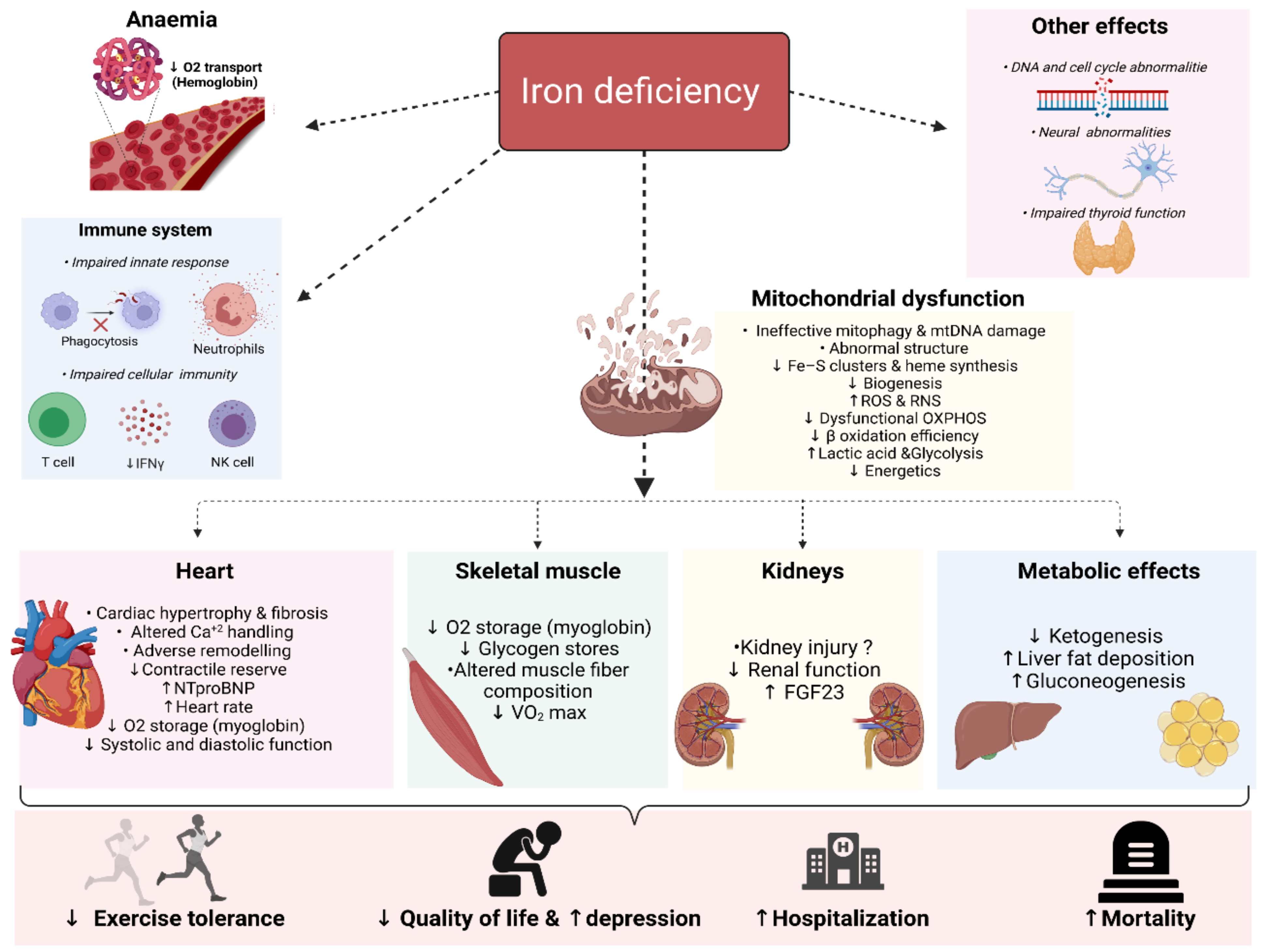
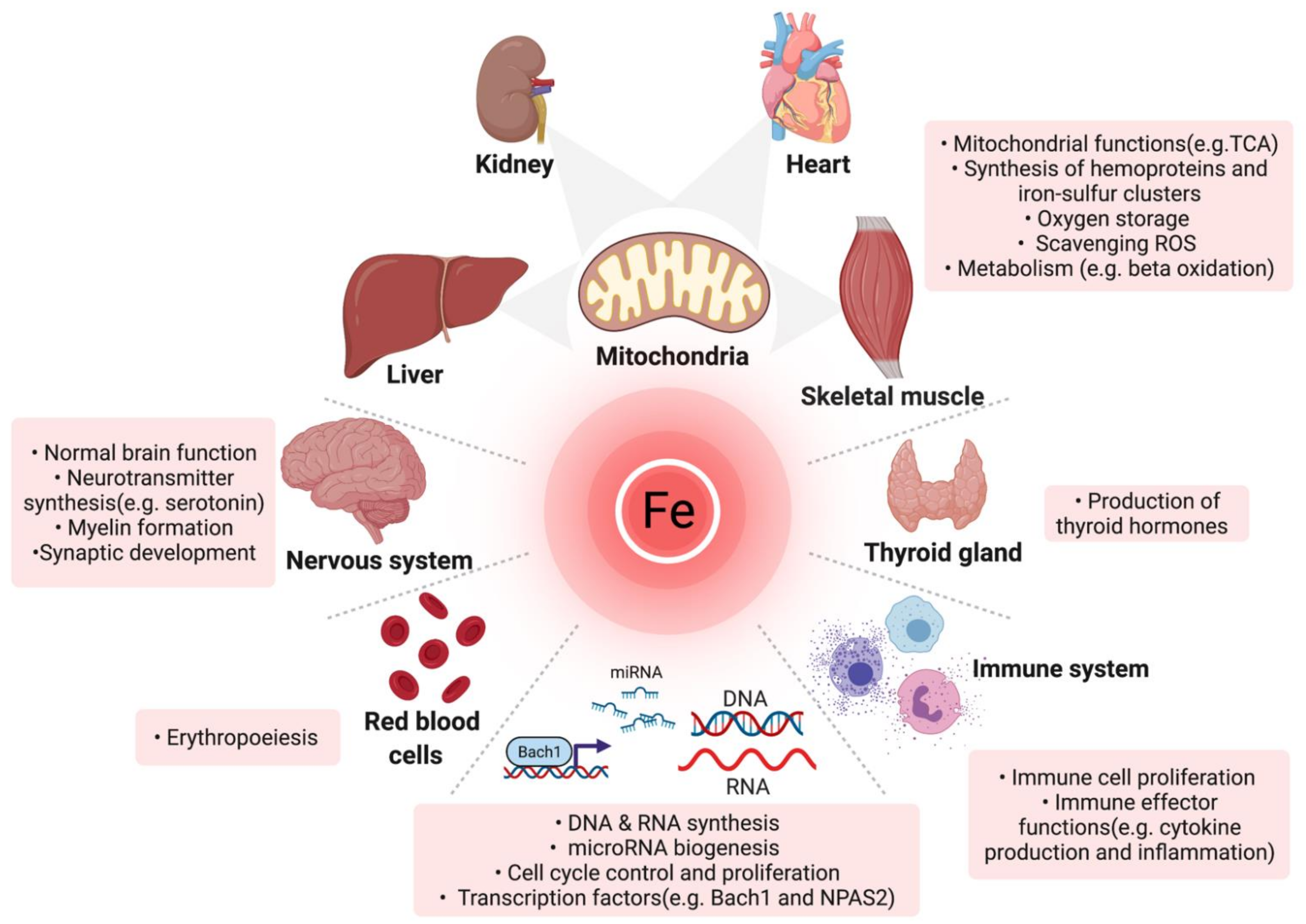
Iron is an essential micronutrient for a myriad of physiological processes in the body beyond erythropoiesis. Iron deficiency (ID) is a common comorbidity in patients with heart failure (HF), with a prevalence reaching up to 59% even in non-anaemic patients. ID impairs exercise capacity, reduces the quality of life, increases hospitalisation rate and mortality risk regardless of anaemia. Intravenously correcting ID has emerged as a promising treatment in HF as it has been shown to alleviate symptoms, improve quality of life and exercise capacity and reduce hospitalisations. However, the pathophysiology of ID in HF remains poorly characterised. Recognition of ID in HF triggered more research with the aim to explain how correcting ID improves HF status as well as the underlying causes of ID in the first place. In the past few years, significant progress has been made in understanding iron homeostasis by characterising the role of the iron-regulating hormone hepcidin, the effects of ID on skeletal and cardiac myocytes, kidneys and the immune system. In this review, we summarise the current knowledge and recent advances in the deleterious systemic and cellular consequences of ID in HF. 

  • iron deficiency
  • iron metabolism
  • heart failure
  • pathophysiology

1. Introduction

1.1. Physiologic Roles and Regulation of Iron

Iron is an essential cofactor for the normal functioning of many enzymes participating in vital cellular and organismal functions, making it indispensable for every living cell [1][2][3][35,36,37]. In addition to its important role in oxygen transport and storage as a constituent of hemoglobin and myoglobin respectively, iron is crucial for many enzymes and proteins involved in oxidative metabolic processes (e.g., mitochondrial respiratory chain, oxidative enzymes and protection against oxidative stress), microRNA biogenesis, the function of the thyroid gland, central nervous system and immune system [1][2][4][35,36,38]. Furthermore, iron is crucial for the synthesis and degradation of proteins, lipids (e.g., β-oxidation of fatty acids), carbohydrates, DNA and RNA [4][5][6][7][8][38,39,40,41,42]. Of note, iron is particularly important for cells either with high energy demand (cardiomyocytes, hepatocytes, neurons, renal and skeletal cells) or high mitogenic activity (e.g., haematopoietic and immune cells) [9][10][26,43]. Accordingly, these cells are more sensitive to Iron deficiency (ID) [9][26]. An overview of the functions and proteins that require iron are outlined in Figure 1 and Table 1 [10][11][12][43,44,45], respectively.
Jcm 11 00125 g001
Figure 1. Overview of the multifaceted roles of iron in diverse organs and molecular processes. TCA: tricarboxylic acid cycle; miRNA: microRNA, ROS: reactive oxygen species (Created with BioRender.com, accessed on 24 November 2021).
Table 1. Overview of proteins that require iron to function properly.
Function Protein
Oxygen transport Hemoglobin
Oxygen storage Myoglobin
Lipid and cholesterol biosynthesis NADPH-cytochrome P450 reductase, fatty acid desaturases, cytochrome P-450 subfamily 51 and Cytochrome P450 Family 7 Subfamily A Member 1
Oxygen sensing and regulation of hypoxia Hypoxia-inducible factor prolyl hydroxylases
Synthesis catecholamines and neurotransmitters Tryptophan hydroxylase, tyrosine hydroxylase, monoamine oxidase and aldehyde oxidase
Host defence, inflammation and production of nitric oxide Myeloperoxidase, NADPH oxidase, indoleamine 2,3- dioxygenase, nitric oxide synthase and lipoxygenases
DNA synthesis, replication and repair Ribonucleotide reductases, DNA polymerases, DNA glycolsylases, DNA primases, DNA helicasess and DNA endonucleases. Dihydropyrimidine dehydrogenas
Collagen synthesis Proline hydroxylase
Electron transport and respiratory chain Cytochrome C oxidase, Cytochrome b, cytochrome c1, Cytochrome oxidase P540, NADH dehydrogenase, aconitase, citrate synthase, Succinyl dehydogease, cytochrome reductase, Complex I-III, rieske protein, NADH ferrocyanide oxidoreductase
Adrenoxin Steroid hydoxylation
Antioxidant defence Catalase
Response to oxidative stress Glutathione peroxidase 2, lactoperoxidase
Amino acid metabolism Tryptophan pyrrolase, Phenaylalanine hydroxylase, deoxyhypusine hydroxylase
Carnitine biosynthesis α-ketoglutarate (αKG)-dependent oxygenases
Synthesis of thyroid hormone Thyroid peroxidase
Drug detoxification Cytochrome P450 , NADPH cytochrome P450 reductase
Prostaglandin thromboxane synthesis, inflammation and response to oxidative stress Cyclooxyenase
microRNA biogenesis DiGeorge Syndrome Critical Region Gene 8
Ribosome function and tRNA modification ABCE1, CDKRAP1, TYW1 and CDKAL1, Methylthiotransferase
Haeme biosynthesis Ferrochelatase
Apoptosis and oxygen transport in the brain Neuroglobin
Purine metabolism and synthesis Xanthine oxidase, amidophosphoribosyltransferase
Besides its crucial roles in the body, iron is a potentially harmful element to cells given its chemical reactivity and its propensity to generate reactive oxygen species through participating in Fenton’s reaction [1][10][35,43]. Iron is almost always linked to either ferritin intracellularly or extracellularly to transferrin as free iron ions are toxic [1][35]. In addition, iron status is tightly regulated both systematically by hepcidin and cellularly through iron-regulatory proteins. A detailed compredescription of iron metabolism is beyond the scope of this review. A comprehensive overview of cellular and systemic iron metabolism can be found elsewhere [12][13][14][15][45,46,47,48].

1.2. Definition of Iron Deficiency

ID ensues when iron supply is insufficient to meet the body’s needs or to cover the iron lost physiologically or pathologically [5][39]. ID may manifest itself in two distinct forms with intertwined pathophysiology, namely functional and absolute ID. Absolute ID (AID) reflects depleted iron stores, while functional ID (FID) is characterised by reduced availability of iron despite sufficient or overly abundant iron stores due to suboptimal iron trafficking induced by hepcidin. It is crucial to note that studies in HF differ in defining ID [16][17][18][19][10,49,50,51]. The most widely used definition of ID, which is also adopted by the European Society of Cardiology (ESC), is a ferritin level <100 μg/L (reflecting AID) or ferritin (100 to 300 μg/L) with a transferrin saturation (TSAT) <20% (reflecting FID) [20][8]. This definition of ID has, however, been criticised as it has never been validated against a gold standard and remains a subject of considerable debate, especially in patients with acute HF [17][18][21][22][23][24][49,50,52,53,54,55].
The ESC definition of ID (also called the FAIR-HF definition [25][22]) is limited by relying heavily on ferritin levels, thereby labelling patients with TSAT ≤20%, but a ferritin >300 μg/mL as iron sufficient, while labelling those with isolated hypoferritinaemia (ferritin <100 μg/mL with a TSAT >20%) as iron deficient. This latter category was found to be iron sufficient when compared to bone marrow staining, the golden standard for diagnosing ID [23][54]. Although ferritin is one of the most widely used biomarkers to detect iron deficiency [26][56], serum ferritin levels can be profoundly influenced by several factors such as inflammation, infection and malignancy, making it falsely elevated in an inflammatory state such as HF and thus does not correlate with iron availability [27][57]. HWerein showed that, compared to bone marrow iron staining, the ESC definition of ID has a sensitivity of 82.4% and a specificity of 72% for detecting ID in patients with HF [23][54]. Serum iron (≤13 μmol/L) and TSAT (≤19.8%) were significantly better cutoffs than the FAIR-HF definition, with areas under the curves (AUC) of 0.922 and 0.932, respectively. Adding ferritin to either definition did not result in a significant increase in the AUC, suggesting that ferritin does not contribute to more accurate identification of truly iron deficient HF patients, and as such, both serum iron ≤13 μmol/L and TSAT ≤19.8% are good indicators of ID as standalone. Prognostically, these two definitions are independently associated with a higher incidence of all-cause mortality, while isolated hypoferritinemia did not [23][54]. Several studies corroborated these findings [16][17][19][23][28][29][30][10,49,51,54,58,59,60]. More recently, it was found that persistent ID (defined as a serum iron ≤13 µmol/L) was associated with poor prognosis, while its resolution was associated with improved outcomes. Similar trends were found when defining ID as TSAT <20%, but not when defined as per the FAIR-HF criteria [17][49]. Remarkably, Cleland et al. found that higher ferritin levels (and not lower levels) were significantly associated with a higher risk of all-cause or cardiovascular mortality, further questioning the correlation between ferritin and iron availability in patients with HF. Additionally, subgroup analysis of individual patient data meta-analysis (n = 839) pooled from four double-blind, randomised controlled trials (RCTs) showed that although intravenous ferric carboxymaltose (FCM) generally reduces recurrent cardiovascular hospitalisations and cardiovascular mortality, patients with TSAT <20.1% benefit more from FCM iron than those with TSAT >20.1% even if ferritin levels were low [31][61]. Similarly, in the IRON-CRT trial, it was found that HF patients with TSAT <20% benefit more from FCM iron than if TSAT was >20% in terms of cardiac contractility and left ventricular ejection fraction (LVEF) [32][62]. However, similar interaction was not found in the AFFIRM-AHF trial [33][27]. The aforementioned findings confirm the accuracy of TSAT <20% and serum iron ≤13 µmol/L in identifying truly iron deficient HF patients while questioning the diagnostic and prognostic usage of ferritin in detecting ID in HF.
On the other hand, these two definitions of ID might have their own limitations, as serum iron is subjected to circadian variations [34][63], and TSAT might be falsely elevated in malnutrition and advanced stages of renal insufficiency [18][35][36][37][50,64,65,66]. Recent studies proposed serum soluble transferrin receptor (sTfR) as the most auspicious novel ID-related biomarker since circulating sTfR levels reflect the iron demand of the body in addition to the erythroid proliferation rate quantitatively [38][67]. In a similar approach to theour bone marrow study, Sierpinski et al. found that ID defined as serum sTfR of ≥1.25 mg/L is more accurate in identifying ID when compared to bone marrow staining in clinically stable patients with HF [24][55]. Of note, adding sTfR to multivariable models for predicting 3-year all-cause mortality in patients with HF abolishes the prognostic value of serum ferritin and TSAT after adjusting for all other prognosticators. These findings suggest that elevated serum sTfR is a better surrogate for depleted intracellular iron. In line with these findings, Leszek et al. found that only serum sTfR significantly correlated to myocardial and mitochondrial iron status, but not ferritin, serum iron or TSAT [39][68], indicating that sTfR reflects tissue iron demands more accurately. Nevertheless, the lack of assay standardisation restricts its implementation in clinical routines [37][66].
To summarise, defining ID in HF using classical biochemical iron parameters appears to be not straightforward. Mounting evidence suggests that ferritin should not be taken into consideration when diagnosing ID in patients with HF but may be used as a safety parameter to avoid the administration of iron to patients with potential iron overload; TSAT or serum iron alone are better indicators of systemic ID, while sTfR might outperform them all. Although the current ESC definition of ID performed thus far reasonably good in general, it is broad and unspecific in identifying those who are truly iron deficient; the high prevalence of ID in HF might have precluded the importance of choosing a more accurate definition to identify those who are truly iron deficient and need IV iron. Identifying truly iron-deficient patients is crucial as inaccurate diagnoses of ID [17][49] and unnecessary treatment with FCM might dilute the benefits of IV iron and lead to increased risks such as hypophosphatemia [40][69]. Furthermore, in light of existing evidence indicating different mechanisms leading to myocardial and systemic ID [41][70] (as discussed below) as well as poor accuracy of systemic biomarkers in detecting myocardial ID, which might be a major driver behind clinical improvements upon iron supplementation, future studies should evaluate other ID-related surrogates in order to identify HF patients that might benefit from iron supplementation on a systemic and cellular level.

2. Deleterious Biological Consequences of Iron Deficiency

The functional and clinical impairments as well as the benefits of correcting ID observed in non-anaemic iron-deficient HF patients point towards the important role of iron in nonhematopoietic tissues [42][43][44][45][46][47][16,20,21,23,185,186]. The underlying mechanisms between the worst symptomatic status and prognosis remain poorly characterised. Recent years sparked an avalanche of research seeking to explain the mechanisms by which ID contributes to these adverse effects. In order to understand the consequences of ID in HF patients beyond anaemia, consideration should be directed towards the roles of iron beyond mere erythropoiesis. In the following sections, the cellular and subcellular effects of ID are discussed. A summary of the deleterious effects of ID is shown in Figure 2.
Jcm 11 00125 g003
Figure 2. Overview of the biological consequences of iron deficiency. NTproBNP: N-terminal pro-b-type natriuretic peptide; OXPHOS: Oxidative phosphorylation; RNS: reactive nitrogen species; ROS: reactive oxygen species and FGF23: Fibroblast growth factor-23 (Created with BioRender.com, accessed on 24 November 2021).

2.1. Mitochondrial Function and Metabolic Effects

Cells with high energy demand such as cardiomyocytes, hepatocytes, nephrons and skeletal myocytes are abundant with mitochondria. Mitochondria are the major intracellular sites of iron utilisation and accumulation as they are the sites where synthesis of haeme and iron–sulphur clusters takes place [48][49][187,188]. Beyond its biosynthetic role in haeme and iron–sulphur clusters, iron was also shown to be crucial for mitochondrial biogenesis as ID affects both iron-containing and non-iron-containing mitochondrial proteins, indicating a reciprocal relationship between adequate iron content and mitochondrial function [50][189].
Mitochondria are the primary combustion machinery in cells for burning fuel such as glucose, fatty acids and ketone bodies. The final step to producing adenosine triphosphate (ATP) from these nutrients is oxidative phosphorylation (OXPHOS), for which sufficient iron (in addition to other pathways) is vital. This fact highlights how crucial iron is for proper energetics in all cells.
Besides producing ATP, mitochondria are also involved in controlling cellular Ca2+ [51][190], generating reactive oxygen species, cellular death, synthesis of pyrimidine, amino acids and lipids [48][187]. Accordingly, deficiency in iron impairs mitochondrial function at many levels. ID has been linked to morphological changes in the mitochondria, such as an increase in size and a decrease in cristae [52][191], as well as functional changes such as reduced production of ATP [53][192], mitochondrial DNA damage [52][191], increased gluconeogenesis [48][54][187,193], increased lactic acid production [46][47][55][185,186,194], reduced mitochondrial biogenesis and impaired mitophagy [56][50][179,189], increased mitochondrial cytochrome c release (and hence apoptosis) and reactive nitrogen species expression [57][58][59][60][61][62][195,196,197,198,199,200]. All of this culminates in mitochondrial damage. As such, ID may worsen HF by causing mitochondrial damage [63][64][201,202], and its correction augments mitochondrial function.
Oxidative Stress
Mitochondria are the primary source of production and scavenging of both reactive oxygen species (ROS) and reactive nitrogen species (RNS) [65][66][203,204]. Not only can iron excess lead to oxidative stress via Fenton-type reactions but ID was also shown to promote oxidative and nitrosative stress [66][204]. This is thought to be related to the reduced antioxidant activity (e.g., catalase enzyme) [67][205] and increased superoxide production as a result of mitochondrial dysfunction. In the heart samples of HF patients undergoing transplantation, myocardial ID is associated with reduced expression of key protective enzymes that scavenge ROS, such as catalase, glutathione peroxidase and superoxide dismutase 2 [68][183]. This may point towards ROS and/or RNS induction as an adverse consequence of ID, which is seen as one of the underlying mechanisms leading to myocardial remodeling and HF progression [69][206]. Toblli et al. showed that even without completely correcting anaemia, IV iron sucrose reversed the anaemia-induced cardiac remodeling, prevented cardiac fibrosis and improved cardiac function by mitigating oxidative/nitrosative stress and inflammation in the heart [70][207]. Furthermore, in another rat model of HF, intravenously administering iron resulted in higher tissue activity of the antioxidant superoxide dismutase [71][208]. A recent study found similar results in a mice model of myocardial infarction [72][209]. Several studies showed that ID participates in the induction of oxidative stress in many organs, including the liver and the kidneys [73][74][210,211]. All in all, ID makes cells more prone to oxidative/nitrosative damage, and its correction may ameliorate these adverse effects of ID.

2.2. Heart

Since the heart has the highest energy expenditure of all organs [75][212], intact performance of cardiomyocytes is inextricably linked to mitochondrial function, for which sufficient iron is vital. This makes cardiomyocytes especially susceptible to the adverse effects of ID. Depriving human cardiomyocytes of iron leads to impaired contractile function with reduced activity of respiratory complexes I, II and III [53][192]. Remarkably, these adverse effects of ID can be reversed by iron supplementation. Similarly, several animal studies showed that systemic ID, even without anaemia [76][213], is associated with structural changes in the heart, including cardiac hypertrophy, irregular sarcomere organisation, mitochondrial swelling, left ventricular(LV) dilation , LV hypertrophy, lung congestion and cardiac fibrosis [57][77][78][79][195,214,215,216]. In addition to structural remodelling, the hearts of mice with IDA exhibit a hypoxic phenotype and altered Ca2+ handling, with a metabolic shift towards lactic acid-producing glycolytic metabolism [46][80][185,217]. Furthermore, hearts of mice models with isolated myocardial ID without anaemia develop cardiomegaly, impaired contractile function, shifts towards anaerobic respiration, dysfunctional oxidative phosphorylation and impaired mitophagy despite normal systemic iron levels [81][56][178,179].
Failing human hearts are characterised by reduced total iron content [40][54][82][69,193,218]. Systemic ID and/or myocardial ID is associated with worse cardiac function, diminished contractile reserve [83][219], decreased mitochondrial enzymatic activities of both oxidative phosphorylation and anti-oxidative enzymes [40][84][53][54][69,71,192,193]. When taken together, these studies highlight the importance of normal iron content to the heart and that its deficiency could play a causal role in the pathogenesis of systolic and diastolic myocardial dysfunction as well as HF progression independently of systemic iron status.
Replenishing iron prevents abnormalities of Ca2+ handling, improves cardiac function and survival in rat models with HF [85][180]. In the Myocardial-IRON Trial, it was found that administering FCM intravenously in iron-deficient HF patients resulted in significant improvement in cardiac magnetic resonance sequences, indicating myocardial iron repletion [86][220]. This correction of ID was accompanied by improved right and left ventricular ejection fraction on the 7th day already [87][221]. Several other studies corroborate these positive echocardiographic effects of IV on the heart [79][88][89][90][91][216,222,223,224,225]. Remarkably, in HFrEF patients receiving cardiac resynchronisation therapy (CRT), the presence of ID is associated with diminished reverse remodelling and lesser likelihood of functional improvement after CRT implementation, suggesting that adequate myocardial iron content is a prerequisite to derive optimal benefits from CRT implantation with respect to reverse cardiac remodelling and improved cardiac function [92][93][226,227]. In line with these findings, the recent IRON-CRT trial showed that IV iron repletion reverses myocardial remodelling and boosts cardiac performance and contractility in patients receiving CRT, which were also accompanied by improvements in quality of life and exercise capacity [32][62]. These results demonstrate the incremental potential of IV iron in reverse cardiac remodelling in addition to guideline-directed therapies that induce reverse remodelling [94][228]. Whether IV iron can prevent myocardial remodelling inflicted by either ischaemic or non-ischaemic damage is not known. It is worth noting that although beneficial effects of IV iron on the failing heart are indubitable, some research indicates that mitochondrial iron chelation might also have beneficial effects for patients with HF [95][82][96][97][177,218,229,230]. Further work is needed to delineate the effects of IV iron both systemically and cellularly.

2.3. Skeletal Muscles

Exercise intolerance is a cardinal symptom of HF, with impaired oxidative metabolism, decreased blood perfusion to skeletal muscles and oxygen delivery implicated as potential causes [98][231]. Mitochondrial dysfunction inflicted by ID is not limited to the heart but extends to other organs, especially those with high energy demands such as skeletal muscles. Intuitively, decreased exercise capacity in ID is linked to defective O2 delivery due to anaemia. However, deficiency of iron impairs skeletal muscle function also by anaemia-independent pathways, which is oxidative metabolism and oxygen storage in myoglobin [10][99][43,232]. Several animal studies have shown that impaired exercise capacity is directly linked to ID due to diminished mitochondrial energy metabolism [10][100][43,233]. Even when hemoglobin levels were kept constant, iron-deficient animals showed a significantly lower exercise capacity accompanied by impaired oxidative metabolism, indicating a direct relationship between ID and impaired physical performance irrespective of anaemia [101][102][234,235]. A meta-analysis in athletes with isolated ID without anaemia showed that iron therapy improves systemic iron status and their aerobic capacity as evaluated using maximal oxygen consumption (VO2 max) [103][236]. Furthermore, in a knockout mice model of Tfr1 specifically to skeletal muscles, Barrientos et al. showed that isolated muscle ID had profound systemic metabolic effects besides impaired mitochondrial respiration in muscles, suggesting that muscle ID may have unrecognised effects on systemic energy homeostasis [104][237].
In patients with HF, results of the FAIR-HF, CONFIRM-HF and EFFECT-HF randomised controlled trials all indicate that iron repletion improved exercise capacity irrespective of attained hemoglobin levels [25][45][105][22,23,24]. Melenovsky et al. showed that iron-deficient HF patients exhibit more severe skeletal muscle myopathy than their iron-sufficient counterparts as assessed using Phosphorus-31 magnetic resonance spectroscopy (31P MRS). By using the same approach, Charles-Edwards et al. showed that in iron-deficient HFrEF, IV iron isomaltoside significantly shortens phosphocreatine regeneration after exercise despite no change in haemoglobin levels, indicating enhanced skeletal muscle energetics already by 2 weeks [106][238]. Boosting skeletal muscle energetics seems to be an important mechanism by which correcting ID leads to beneficial outcomes in patients with HF.

2.4. Kidneys

The kidneys are another high-energy-demanding organ system with high mitochondrial content [107][239]. From an energetics perspective, reabsorption and secretion of solutes in the nephrons occur either passively or actively (e.g., Na+/K+-ATPase), with the latter requiring energy for proper functioning. As such, iron, due to its crucial role in the mitochondria, is important for kidney function [108][240]. Although ID is mostly seen as a consequence of renal diseases, several studies suggest that iron itself influences kidney function [109][241]. A recent Mendelian randomisation study investigated the causal effect of serum iron levels on kidney function in the general population. They found a 1.3% increase in estimated glomerular filtration rate per standard deviation increase in serum iron (95% confidence interval 0.4–2.1%; p = 0.004), indicating a protective effect of higher iron levels on renal function [110][242]. In accordance with these findings, feeding rats an iron-deficient diet resulted in an increase in malondialdehyde (an indicator of increased lipid peroxidation) in the kidneys, suggesting that ID can also adversely affect the kidney through oxidative stress and mitochondrial dysfunction [73][210]. In children, ID was shown to be associated with tubular and glomerular damage accompanied by increased oxidative stress markers [111][243]. Whether ID has similar direct effects on the kidneys in patients with HF is unknown.
In patients with HF, Toblli et al. showed that IV iron in anaemic HF patients with CKD resulted in a significant improvement in renal function [112][244]. Similarly, in a sub-analysis of the FAIR-HF trial, it was found that patients in the FCM group had an improved kidney function as evaluated by estimated Glomerular Filtration Rate (eGFR) [113][245]. This improvement in renal function was observed in all pre-specified subgroups, including HF patients with preserved renal function and those without anaemia. Remarkably, correcting ID might be associated with a reduction in fibroblast growth factor 23 (FGF23) [114][115][246,247], which was linked with worse clinical outcomes in both HF and renal disease [116][248]. This suggests that the benefits of IV iron on the kidneys may extend beyond its effects on the energetics of renal mitochondria. Future studies should look into the interaction between FGF23, iron status, heart and kidneys since FGF23 has been shown to decrease hepcidin expression [117][249], in addition to acting as a mediator between ID and its association with mortality in patients with HF [118][250].
Altogether, treating ID in HF patients may pose renoprotection properties. Data on the effects of ID on the kidneys are scarce. Further studies are therefore warranted. Moreover, given the established role of the kidneys in controlling systemic iron levels [109][241], it is yet to be ascertained whether impaired kidney function could increase urinary iron excretion and thereby cause ID in HF.

2.5. The Immune System

Beyond the cardiocentric perspective, iron homeostasis is also important for the immune system [10][43]. The relationship between iron and immunity is complex and bidirectional. As alluded to earlier, inflammation as an immune effector mechanism can lead to iron dysmetabolism, but iron dysmetabolism itself also causes adverse changes in immune function [119][120][129,251].
ID can affect the immune system in multiple ways. ID negatively affects both the growth and effector mechanisms of the immune system. Deleterious effects of ID include reduced neutrophil activity (myeloperoxidase is iron-dependent), reducing functions of nuclear factor kappa and HIFs, nitric oxide (NO) formation, a defective proliferation of T cells (especially T helper 1 cells) [121][252] and impaired interleukin 2 production [11][122][123][44,253,254]. Howden et al. showed that activated CD4+ and CD8+ T-cells upregulate their transferrin receptor, suggesting that iron is important for T-cell activation [124][255]. In mice fed an iron-deficient diet, altered T lymphocyte and natural killer (NK) cell activity was demonstrated, indicating impaired cellular mediated immunity [125][126][256,257].
Contrary to the overly simplistic view where ID is merely seen as a consequence of inflammation, studies showed that ID could perpetuate and amplify inflammation [127]. ID in mice induces and enhances inflammation when compared to mice with normal iron status [128][258]. A recent study in a rat post-myocardial infarction (MI) HF model showed that iron supplementation (FCM) reduces inflammation [71][208]. This dampening effect of iron is thought to be due to the important role of iron for immune cells to mount an effective immune response. In fact, more than 10 years ago, Toblli et al. showed that administration of iron sucrose intravenously in anemic HF patients led to a significant reduction in C-reactive protein [112][244]. Other studies corroborated this evidence [70][207]. When taken together, these results support the idea that ID itself can also have a direct adverse effect on the immune system and that its correction has advantages beyond merely stimulating erythropoiesis.

2.6. The Brain

Mental functions have biochemical bases, and hence dysregulation herein can lead to mental effects. A growing body of evidence suggests that iron is important for neurological functions as the brain is also a metabolically active organ, making it particularly susceptible to ID [129][259]. In addition to energy deficits of neural cells, ID can also impair synaptic plasticity, myelination and reduce the activity of multiple iron-dependent enzymes involved in dopamine and serotonin synthesis (monoamine oxidase, tyrosine hydroxylase and tryptophan hydroxylase) [130][131][260,261]. Several studies showed that brain ID leads to deficits in memory, learning, behaviour and emotional problems [132][262]. Low levels of serotonin due to ID may lead to a relapse of depression [133][263]. Psychological disorders such as depression are very common in HF patients [134][264]. Several studies reported a higher prevalence of depression in iron-deficient HF [9][135][136][26,265,266]. Whether systemic ID is associated with brain ID in HF patients is unknown. Moreover, the effects of IV iron in patients with HF on mental functioning have not been assessed yet.

2.7. Thyroid Gland

Accumulated evidence shows that thyroid dysfunction is linked with an increased risk for and worsening of HF [20][137][138][8,267,268]. ID impairs thyroid hormone metabolism by different mechanisms, including ineffective erythropoiesis, reduced thyroid peroxidase activity (haeme-containing enzyme) and increased hepatic inactivation of thyroid hormones [139][140][269,270]. Iron deficient HFpEF patients have a significantly higher prevalence of thyroid disease [135][265]. The therapeutic consequence of replenishing iron on thyroid function in HF has not been studied yet.

3. Novel Therapeutic Options for Targeting Iron Metabolism

Although iron replacement therapy remains to be the cornerstone of treating ID in HF, several experimental iron metabolism-targeting agents have been developed to treat IDA, many of which were evaluated in patients with CKD and cancer-related anaemia [141][271]. These treatment options increase iron absorption as well as the mobilisation of sequestered iron by either downregulating the synthesis and/or function of hepcidin or by stabilising HIFs as a result of Prolyl-4-hydroxylases (PHDs) inhibition [117][249].
Manipulation of the hepcidin–ferroportin axis seems an attractive target as the interplay between hepcidin and ferroportin is crucial for regulating iron status and aberrations in this pathway are centrally involved in the pathophysiology of FID [142][9]. Targeting hepcidin directly (e.g., LY2787106, Lexaptepid pegol or Anticalins), or indirectly by targeting inflammatory markers (e.g., IL-6), bone morphogenic protein 6 (BMP6) [LY3113593], was proposed as a treatment option for anaemia of inflammation as these agents were shown to increase intestinal iron absorption and mobilisation in phase 1 and/or 2 studies [117][249]. Moreover, blocking hepcidin’s interaction with ferroportin using ferroportin antibodies (LY2928057) reduces ferroportin internalisation, thereby increasing mobilisation of sequestered iron from the reticuloendothelial system [143][272]. Manipulation of the hepcidin pathway in inflammatory conditions such as HF might be a compelling option to treat ID in HF besides targeting inflammation, especially in those with FID as they have clear inflammatory components accompanied by high hepcidin levels [84][71]. In a recent phase 1/2 clinical trial, Pergola et al. showed that administration of ziltivekimab, anti-IL-6 ligand antibody, led to a dose-dependent improvement in serum iron, TSAT, inflammatory markers and reduction in hepcidin in CKD patients on haemodialysis with hyporesponsiveness to erythropoiesis-stimulating agents [144][123]. Whether targeting inflammation by reducing IL-6 activity and thereby improving clinical outcomes and iron metabolism can also be a therapeutic option to patients with HF is unknown.
Another promising approach is the manipulation of the prolyl hydroxylase domain/hypoxia-inducible factor (PHD/HIF) pathway, which seems to be deranged in iron-deficient patients with HF [145][46][12,185]. Several HIFs stabilisers are being developed, some of which have entered/finished phase 3 of clinical trials, including Vadadustat, Daprodustat and Roxadustat [146][273]. Meta-analyses of several randomised controlled trials showed that PHD inhibitors increase the levels of hemoglobin, serum transferrin and increase intestinal iron absorption while reducing levels of hepcidin in anaemic CKD patients [146][147][148][273,274,275]. These effects seem to be independent of inflammation [146][273]. Such activation of HIF signalling can provide a physiologic approach towards improved iron metabolism and may complement and/or reduce the need for IV iron in iron-deficient patients with HF. Besides being orally administered, targeting this pathway may have other non-erythopoetic benefits, such as lowering cholesterol levels as well as blood pressure [149][150][276,277]. However, there are many concerns regarding its safety as stabilising HIFs might lead to unwanted effects such as promoting cancer development since HIFs modulate the expression of various proteins that are involved in energy metabolism, angiogenesis, cellular growth and differentiation [117][249]. Large, carefully designed, long-term clinical trials are required to clearly understand the effects of systemic activation of the HIF pathway on iron-deficient HF patients.
Lastly, it should be noted that these agents can mainly increase absorption or mobilisation of sequestered iron, making them potentially ineffective for patients who have AID. Whether targeting the PHD/HIF or hepcidin–ferroportin axes have additive or possibly even synergistic effects with IV iron to correct ID, improve iron mobilisation in a more physiological manner and prevent relapses of ID remains to be investigated.

4. Conclusions

The consequences of ID per se reach far beyond those of anaemia. Energy deficit is one of the hallmarks of HF. Mounting evidence indicates that mitochondrial dysfunction is a prominent repercussion of ID, which further aggravates the existing deficit in energy in HF. Current standard-of-care pharmacological approaches to HF provide symptomatic and clinical benefits by reducing the workload on the heart instead of increasing its reserve. Targeting ID in HF can improve the care of HF patients as it addresses this shortcoming by augmenting mitochondrial function. This makes ID stand out as a therapeutic target in HF since it is relatively easy to diagnose and treat with potential anti-remodelling effects.
ScholarVision Creations