Somatic polyploidy was found in the tissues of all multicellular organisms (including algae, mosses, lichens, vascular plants, invertebrates, and vertebrates), which points to its adaptive value. In human and warm-blooded animals, polyploidy can be a part of normal postnatal morphogenetic programs and can be a manifestation of response to pathological stimuli and diseases. Extensive comparative studies of mRNA seq data suggest that polyploidy is associated with common, evolutionary conserved manifestations in transcriptome.
- polyploidy
- evolutionary conserved features
- developmental programming
- cardio-vascular diseases
- transcriptome
1. Ploidy-Associated Transcriptome Features Are Related to Stress Response, Metabolism, Morphogenesis, and Longevity
The association between polyploidy, adaptation to stress, carcinogenesis, and decrease in organ functional potential suggests that genome accumulation is involved in the regulation of gene expression. The analysis of gene expression in the human and mouse hepatocytes, cardiomyocytes, trophoblast cells, neurons, adipose mesenchymal stem cells, interstitial cardiac stem cells, drosophila epithelial cells, and various types of cancer cells indicate that polyploidy can exert both common and specific effects [1][2][3][4][5][6][7][8][9][7,69,73,78,83,84,110,111,112]. The common effects are the induction of biological pathways related to stress response (i.e., abiotic, biotic, hypoxic, oxidative, genotoxic, and inflammatory), response to DNA instability, and drug resistance [1][2][5][10][11][12][13][14][15][7,57,69,72,83,87,113,114,115]. Polyploidy also activates the signaling cascades involved in embryogenesis (including Notch, TGFb, Hippo, Myc, EGFR, and WNT) and the growth-related gene modules implicated in stemness, DNA synthesis, glycolysis, and ribosome biogenesis [1][4][7][8][9][11][16][17][18][19][20][1,7,11,72,74,77,78,110,111,112,116]. The ploidy-inhibited modules are mostly involved in apoptosis, immunity, and aerobic metabolism [17][18][21][11,74,117]. Importantly, the main ploidy-associated features are similar to those observed in the long-living animal species and mutants. For example, an enhanced response to hypoxia, the induction of DNA repair pathways, proliferation, morphogenesis, glycolysis, adaptation to stress, ribosome biogenesis, as well as the suppression of apoptosis and aerobic metabolism, were found in the mole rats and in the long-living mutants of the mouse, nematodes, drosophila, and yeast [22][23][118,119]. Somatic polyploidy increases the lifespan of cells in the tissues and cultures, mainly due to the increased resistance to apoptosis, DNA damage, and genetic instability [13][18][74,113].1.1. Ploidy-Associated Transcriptomic Features Are Evolutionary Conserved
The fundamental nature of the above-described properties of somatic polyploidy is supported by the fact that similar gene modules are enriched in the ohnologs of polyploid organisms, such as the yeast, Arabidopsis, amphibians, and bony fishes [24][25][26][5,120,121]. These modules include ribosome biogenesis, transcription, proliferation, glycolysis, adaptation to hypoxic and oxidative stress, and negative regulation of apoptosis [25][27][120,122]. Similarly, certain modules suppressed in the case of somatic polyploidy (aerobic respiration, signal transduction, transport, apoptosis, and immune response) in the same polyploid organisms contain the genes that have lost their duplicates (i.e., they are non-ohnologs). This fact emphasizes the common features between organismal and somatic polyploidy. Probably, the adaptation to stress, which is important in the case of somatic polyploidy, plays a role in the evolutionary fixation of organismal polyploidy and retention of ohnologs. Thus, the properties of polyploid genomic machinery are conservative in phylogenesis and ontogenesis and are aimed at improving survival under new conditions, plasticity, adaptability to stress, and increasing longevity. The involvement of genomic duplication in the regulation of developmental programs, life expectancy, and adaptation to stress indicates the importance of polyploidy in the physiological and pathological processes, which affect postnatal morphogenesis and adaptation (including developmental programming of widespread diseases, tissue regeneration, and carcinogenesis). Figure 1 illustrates the most important common features of polyploidy existing at various physiological conditions and in different biological contexts.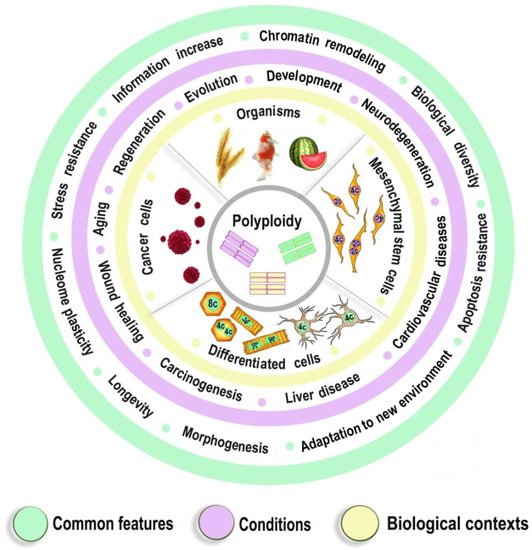
1.2. The Epigenetics of Ploidy-Associated Transcriptomic Features
The data from various fields of research indicate that polyploidy is associated with epigenetic changes at different levels of genome organization, which leads to chromatin remodeling and genome instability. The most obvious effect of polyploidy is the reduction in the nuclear surface-to-volume ratio resulting in a partial loss of nuclear lamina (NL) interactions with the lamina-associated domains (LADs), which increases the competition for NL contacts [28][29][123,124]. The LADs reside mostly in inactive chromatin (heterochromatin) and comprise a substantial part of the genome (about 35%) [30][125]. Detaching from the NL and moving inside the nucleus, LADs contribute to the opening of chromatin [28][123]. This is in agreement with the fact that the upregulation is stronger than the downregulation in the ploidy-associated changes in gene expression [31][126]. Polyploidy is also accompanied by a decrease in lamin B expression, regulating chromosome centromere attachment to mitotic spindle filaments and metaphase progression [32][127]. Inactivating of Lamin B decreases metaphase progression and increases genetic instability due to the impaired attachment of chromosomes to the mitotic spindle fibers [32][33][95,127]. Accordingly, the association between polyploidy and chromatin decompactization under stress was well documented for cardiomyocytes and hepatocytes [34][35][76,128]. At the lower levels of genome organization, polyploidy can alter global patterns of DNA methylation, microRNA expression, and histone modification in mammal, insect, and plant cells [16][36][37][31][38][39][1,2,98,126,129,130]. Polyploid cells show higher expression of bivalent genes, which harbor both activating (H3K4me3) and repressive (H3K27me3) chromatin domains [4][9][78,112]. This effect strikingly resembles strong enrichment of bivalent genes in the ohnologs in the case of organismal polyploidy (Figure 1A). The bivalent genes allow the cell to quickly change gene expression patterns, facilitating the formation of adaptive self-organizing regulatory networks [4][78]. Possibly, bivalent genes are the key regulators, which determine the common features of organismal and somatic polyploidy.2. Polyploidy Meets the Hallmarks of Developmental Programming of Adult Diseases in Slowly Renewing or Terminally Differentiated Organs
Growth retardation, inflammation, malnutrition in pregnancy, infancy, and childhood are associated with the increased risk of cardiovascular diseases, hypertension, stroke, type 2 diabetes, and neurodegenerative disorders [40][41][42][43][131,132,133,134]. This phenomenon was termed as Developmental Origin of Health and Disease (DOHAD) hypothesis [44][135]. The link between conditions of early postnatal development and human health decades later was suggested to be the consequences of developmental plasticity, the phenomenon when one genotype can give rise to a range of different morphological or physiological states in response to different environmental conditions during development [45][136]. This topic belongs to the preventive medicine considered as the medicine of the future [46][137]. It is well established that developmental programming operates via epigenetic changes in chromatin architecture, DNA methylation, microRNA expression, histone modifications, and others [47][48][49][50][51][52][138,139,140,141,142,143]. We Hhypotheticalsized that somatic polyploidy can be one of the epigenetic mechanisms of developmental programming in slowly renewing and terminally differentiated organs. This suggestion is based on the similarity between the properties of polyploidy and the mechanisms of developmental programming described in the literature [40][41][49][50][53][131,132,140,141,144]. Thus, polyploidy is similar to the mechanisms of developmental programming by the following hallmarks: (1) Promotes adaptive developmental plasticity; (2) Appears during critical developmental window; (3) Irreversible, therefore changes the organ structure and function permanently; (4) Decreases organ function and is involved in the trade-off between proliferation and function; (5) Responds to the main programming stimuli (including adverse growth conditions, growth retardation, pathologic functional load, inflammation, malnutrition, and others); (6) Regulates gene expression via the same epigenetic mechanisms as developmental programming; (7) Associated with the diseases that may arise as a result of ontogenetic programming (cardiovascular disease, hypertension, neurodegenerative disease, type 2 diabetes). Below we consider these points in more detail. Figure 2 demonstrates similar properties of polyploidy and developmental programming of adult diseases phenomenon.
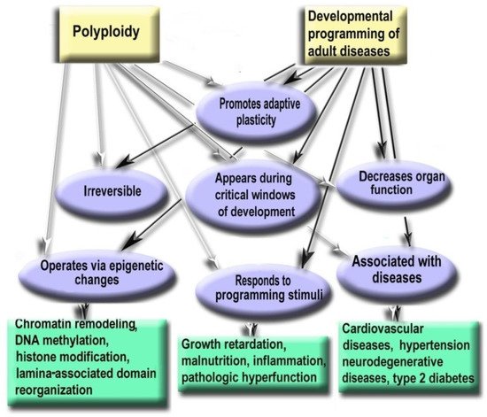
Figure 26. Polyploidy and developmental programming of adult diseases show similar properties.
- Polyploidy helps to cope with the adverse environments via the augmentation of stress resistance and adaptation through epigenetic mechanisms [4][9][54][3,78,112]. Furthermore, it is one of the most variable characteristics of somatic cells. The degree of polyploidization in homologous organs shows large across-species diversity. The percentage of cardiomyocytes with polyploid nuclei varies several folds in mammals of similar weight. For example, about 50% of human cardiomyocytes contain nuclei with 4, 8, 16, or even 32 genomes, whereas cardiomyocytes of the grey wolf or reindeer show only about 1% of cells with polyploid nuclei [55][56][71,82]. Accordingly, cardiomyocyte ploidy also varies between individuals of the same species. The mean ploidy in the normal human heart varies from about 4× to 10× [19][57][58][77,100,108]. Thus, polyploidy is characterized by the degree of biologic plasticity similar to the renowned factors of ontogenetic programming.
- Polyploid cells (e.g., cardiomyocytes, megakaryocytes, hepatocytes, pancreacytes, vascular epithelial cells, retina epithelium) appeared in the perinatal and early postnatal ontogenesis [59][9]. These periods are characterized by high biological plasticity and coincide in time with the critical periods of development [40][131[41],132].
- Cells of slowly renewing organs, including neurons of neocortex and cerebellum, cardiomyocytes, and hepatocytes, which accumulate additional genomes in infancy, childhood, and pre-pubertant period, retain the increased genome amount throughout their lives, regardless of environmental conditions [11][19][[60][957][59],72,77,100,145].
- Polyploidization is associated with a decrease in organ functional potential [55][56][61][62][71,82,104,109]. This decrease probably originates from the involvement of polyploidy in the trade-off between proliferation and function that is also a sign of the developmental programming of adult diseases factor [40][55][63][71,107,131].
- The level of ploidy, particularly in cardiomyocytes, responds to the well-established stimuli of developmental programming (including adverse growth conditions, increased functional load, inflammation, and malnutrition) similarly in the various species and various cells [41][50][55][59][9,71,132,141]. For example, in mammal hepatocytes, cardiomyocytes, retinocytes, and drosophila somatic cells, polyploidy is associated with the increased response to stress, activated pathways of morphogenesis and glycolytic metabolism, and the weakened aerobic metabolism and apoptosis [9][17][54][64][3,11,112,146].
- Polyploidy is associated with epigenetic changes at various levels of genome organization leading to chromatin remodeling and genome instability [28][29][33][95,123,124]. The association between polyploidy and chromatin decompactization under stress was well documented for cardiomyocytes and hepatocytes [34][35][76,128]. Polyploidy can alter global patterns of DNA methylation, microRNA expression, and histone modification in mammalian, insect, and plant cells [4][9][16][32][33][36][38][1,2,78,95,112,127,129]. Polyploid cells show higher expression of bivalent genes, which harbor both activating (H3K4me3) and repressive (H3K27me3) chromatin domains, allowing rapid switching between cellular programs [9][112]. Overall, ploidy-associated transcriptomic changes occur through the same epigenetic mechanisms as in the developmental programming of health and disease, including chromatin remodeling, DNA methylation, histone modification, and others.
- Excessive polyploidization can be associated with the diseases that usually originated from the developmental programming, including cardiovascular disease, hypertension, neurodegenerative disease, type 2 diabetes, metabolic syndrome, and others [18][19][40][41][42][43][9[,7459,77,131],132,133,134].
Experimental Studies Confirm the Role of Polyploidy in the Developmental Programming of Health and Disease
Recent experimental and clinical studies confirm that polyploidy can be involved in the developmental programming of adult diseases. The most convincing evidence was obtained for cardiovascular diseases that are the most susceptible to developmental programming. Thus, studies in sheep indicated that pre-term birth irreversibly increases the percentage of polyploid mononuclear cardiomyocyte and induces DNA damage, fibrosis, and lymphocytic infiltration [60][145]. In humans, pathologic hemodynamic load during postnatal growth permanently increases cardiomyocyte ploidy and decreases cardiac performance [33][57][65][66][67][68][69][79,92,95,100,101,147,148]. The inflammatory stress caused by gastroenteritis in the rat resulted in cardiomyocyte hyperpolyploidization, long-term atrophy, and cell remodeling [59][70][9,75]. The experimental model of gastroenteritis was used as gastroenteritis triggers developmental programming factors, including inflammation, growth retardation, and malabsorption, and as gastroenteritis is a major cause of diseases in toddlers, infants, and children [71][72][73][149,150,151]. Both types of neonatal gastroenteritis cause irreversible excessive polyploidization, long-term atrophy, and remodeling of cardiomyocytes [59][74][70][9,10,75]. Altogether, these data indicate that polyploidy can be involved in developmental programming as it is irreversible, responds to programming stimuli during the critical period of development, changes cell phenotype, and weakens cell function, thus meeting all basic criteria of developmental programming.