Extracellular Matrix and Its Artificial Substitutes: Comparison
Please note this is a comparison between Version 1 by Aleksandra Bandzerewicz and Version 2 by Amina Yu.

The existence of orderly structures such as tissues and organs is made possible by cell adhesion, i.e. the process by which cells attach to neighbouring cells and a supporting substance in the form of the extracellular matrix. The extracellular matrix is a three-dimensional structure composed of collagens, elastin and various proteoglycans and glycoproteins. It is a storehouse for multiple signalling factors. Tissue disruption often prevents the natural reconstitution of the matrix. The use of appropriate implants is then required. The possibilities of regenerating damaged tissues using an artificial matrix substitute are described, detailing the host response to the implant. An important issue is the surface properties of such an implant and the possibilities of their modification.

  • extracellular matrix
  • cellular receptors
  • cell adhesion
  • cell signalling
  • biomaterials
  • scaffolds

1. The Extracellular Matrix—Composition, Structure, Functions

1.1. Two Types of the Extracellular Matrix

Although the basic organisation of the  extracellular matrix (ECM) structure is the same throughout, two basic types of the matrix are distinguished by their location and composition: the interstitial matrix, which forms a three-dimensional porous network surrounding the cells (especially connective tissues), and the pericellular matrix, which is more compact and forms a layer adjacent to the cells [1][2][15,16] (see Figure 1).
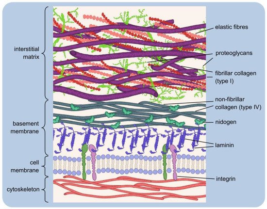
Figure 1. Simplified extracellular matrix structure: three-dimensional macromolecular network composed of various proteins and polysaccharides. The pericellular matrix forms a layer adjacent to the cells: integrins bind to polymerised laminin, which, in turn, is connected via nidogen to the type IV collagen. Interstitial matrix forms porous network of fibrillar collagens, elastic fibres, and proteoglycans.
The interstitial matrix can be equated with the “proper” matrix, as it forms the structural scaffolding for the cells. Its basic components are heterotypic fibrils, composed mainly of type I collagen with small amounts of type III and V collagens in variable proportions, both playing an important role in fibrillogenesis [2][16]. The collagens of the interstitial matrix are mostly secreted by fibroblasts [3][17]. Important components of this “amorphous three-dimensional gel” also include fibronectin and elastin, involved in the organisation of the structure [4][5][18,19].
A typical example of the pericellular matrix is the basement membrane, a delicate and flexible nanostructure that separates the epithelium from the deeper layers of connective tissues. It ensheathes smooth, skeletal, and cardiac muscle fibres, Schwann cells, and adipocytes. The basement membrane forms a specific boundary of many organs in mature tissues, often surrounding their functional units [2][6][7][8][16,20,21,22]. It is mainly composed of type IV collagen, laminins, nidogens and heparan sulfate proteoglycans (HSPGs): perlecan and agrin [7][21]. The basement membrane contains so-called matricellular proteins that do not contribute to its physical stability or structural integrity, although they may be connected to building components. Instead, they have regulatory functions and interact with surface receptors, proteases, hormones or other biologically active molecules. They may be tissue-specific in terms of function and structure [9][10][11][12][23,24,25,26]. Matricellular proteins include SPARC (secreted protein acidic and rich in cysteine, or osteonectin; characteristic of mineralising tissues, mainly bone), thrombospondin-1 (which is rich in platelet α-granules; when secreted, it causes, among other things, activation of TGF-β1, i.e., transforming growth factor-beta 1), and tenascin-C (the gene of this protein is expressed during embryonic life, while in adult tissues, tenascin-C is very poorly detectable, being present rather in the course of pathological processes [13][14][15][27,28,29]. The tasks of the basement membrane include regulation of tissue development, function, and regeneration by controlling the cellular response. It is a storehouse of growth factors and modulates their activity and concentration. It serves to maintain the phenotype of the cells it surrounds [16][30]. The interstitial matrix and the basement membrane are closely interconnected, ensuring the integrity of the tissue [2][16].
The functional equivalent of the basement membrane described above is a type of pericellular matrix that surrounds chondrocytes in articular cartilage [17][31]. It acts as a physical barrier that filters molecules entering and leaving the cells. Together with an adjacent thin layer of matrix, each chondrocyte forms a structural unit called a chondron [18][32]. The morphology of chondrons varies. They can take a discoid/ellipsoid/rounded shape and a variable orientation, which depends on the position, i.e., the depth of location in the cartilage. In some cases, a chondron comprises more than one cell (up to four) [19][33]. AIn this case, an essential component of the pericellular matrix is type VI collagen, although it generally constitutes a negligible percentage of the collagens of cartilage tissue [20][34]. However, because of its specific presence in the chondrocyte environment of articular cartilage, it often serves as a marker of chondrons [20][21][34,35]. A characteristic feature of articular cartilage is the small number of chondrocytes compared to the extensive extracellular (interstitial) matrix for which synthesis, organisation, and maintenance they are responsible [20][34].

1.2. Major Components of the Extracellular Matrix and Their Functions

1.2.1. Collagens

Collagen proteins account for up to 30% of all proteins in vertebrates and are major extracellular matrix components. The basic collagen macromolecules are composed of three same (homotrimers) or different (heterotrimers) polypeptide chains. They are characterised by the repetitive Gly-X-Y sequences, where X usually stands for proline and Y for 4-hydroxyproline. The intertwined chains form a specific triple helix structure [3][22][23][17,36,37].
Due to their supramolecular organisation, fibrillar (types I, II, III, V, XI, XXIV, and XXVII) and non-fibrillar collagens are distinguished. Characteristic for the non-fibrillar collagens is a disrupted continuity of the typical structure. Compared to fibrillar collagens, they contain shorter (although more numerous) helical (collagenous) domains interspersed with so-called telopeptides, for i.example., non-helical domains. As a result, they may occur in various forms, forming, e.g., network systems (types IV, VIII, X), anchor fibres (type VII), beaded filaments (type VI), or belong to the FACIT group (for examplei.e., fibril-associated collagen with interrupted triple helix, types XI, XII, XIV, XVI, XIX-XXII). The terminology and affiliation are not fully systematised. Collectively, collagens form a family of 28 proteins [24][25][26][27][28][29][30][38,39,40,41,42,43,44].
Historically, collagens were thought to have only a supportive function. Although their main function is indeed to form the structural scaffolding of cells (especially for types I, II, and III), it is known that their role is much broader [1][4][23][31][15,18,37,45]. Collagens are involved in regulating the course of cell adhesion (as ligands of cell receptors) [32][33][34][35][46,47,48,49], cell migration (contact guidance) [36][37][38][50,51,52] and tissue reconstruction and remodelling [23][26][31][39][40][37,40,45,53,54]. Not only is the physical deposition or movement of cells itself important, but also the processes conditioned by this,for example,.g., wound healing, immune response and so on, etc. Although collagens are present in most body tissues and affect their mechanical properties, their distribution varies, for examplee.g., type I collagen is characteristic of bone, skin and tendon, and type II collagen of cartilage tissue [41][42][55,56].

1.2.2. Elastin

Elastin is a hydrophobic fibrillar protein, which owes its characteristic elastic properties to extensive covalent cross-linking of the structure [43][57]. The monomer from which the mature insoluble protein is formed is tropoelastin, secreted by fibroblasts, smooth muscle cells, endothelial cells, respiratory epithelial cells, chondrocytes, and keratinocytes [44][45][46][47][48][58,59,60,61,62]. After secretion into the intercellular space, tropoelastin spontaneously associates into larger particles through interactions between hydrophobic domains in a process called coacervation [49][63]. Such precursors undergo oxidative deamination of lysine residues in tropoelastin. The process is catalysed by LOX family enzymes (lysyl oxidases). The result is the formation of allysine from lysine. Cross-linking occurs via the reaction between lysine and allysine residues (Schiff base reaction) or by aldol condensation of two allysine residues [50][51][52][53][64,65,66,67]. The final fibres are not composed of elastin alone. Elastin forms a core (about 90% of the whole structure), covered by an envelope of microfibrils composed mainly of glycoproteins from the fibrillin group (fibrillin-1 and -2) [44][54][55][58,68,69]. In this way, elastic fibres are formed, giving tissues susceptibility to stretching. They are a particularly important component of blood vessel walls, skin, lungs, heart, tendons, ligaments, bladder, elastic cartilage tissue (for example.g., auricle, larynx, epiglottis) and so on, etc. [1][54][56][15,68,70].
The gene expression and formation of elastic fibres occur at early development stages —prenatal and early childhood. De novo production of elastin in adult organisms is unlikely to occur, which is quite uncommon among the ECM components [44][57][58][59][60][58,71,72,73,74]. However, elastin has high metabolic stability and a half-life of approximately 70 years, making the limited synthesis time sufficient (by comparison, the half-life of type VII collagen is estimated to be approximately one month [61][62][75,76]. The adult organism cannot reconstitute elastic fibres that become damaged or degrade progressively with age. They are then repaired incorrectly and consequently do not perform their normal functions. The tissues become too stiff, leading to cardiovascular disease, lung disease or typical signs of ageing, such as loss of skin elasticity [1][53][63][64][15,67,77,78].

1.2.3. Proteoglycans

Proteoglycans are macromolecules of a complex three-dimensional structure. They are composed of a protein core covalently linked to one or more chains of glycosaminoglycans (GAGs), a type of linear, unbranched heteropolysaccharides. The glycosaminoglycan chains may belong to one or different types. Based on localisation, four basic groups of proteoglycans can be distinguished: intracellular and those occurring on the cell surface, in the pericellular space (basement membrane) or intercellular space [1][65][66][67][15,79,80,81].
GAG chains are built by repeating disaccharide units, where one residue is an amino sugar (N-acetylated hexosamine), and the other is uronic acid (D-glucuronic or L-iduronic acid). GAGs differ in the type of monosaccharide residues and the geometry of the linkages between the constituent units (α- and β-glycosidic linkages) and the degree of sulfation of the polysaccharide backbone and the position of this substitution. Based on the chemical structure of the chain, four basic groups of glycosaminoglycans are distinguished: heparan/heparan sulfate, keratan sulfate, chondroitin sulfate/dermatan sulfate, and hyaluronic acid [1][68][69][70][71][72][15,82,83,84,85,86]. Hyaluronic acid represents the simplest type of structure. It is the only one that does not contain sulfate groups (hydroxyl groups are not esterified with sulfate groups) and does not undergo complex modifications in the Golgi apparatus [70][73][74][84,87,88]. Unlike other GAGs, it does not form covalent bonds with proteins and, therefore, is not part of typical proteoglycans. Instead, it can exist in the form of non-covalent complexes with other protein components of the ECM [71][72][75][76][77][85,86,89,90,91].
Hyaluronan has excellent water retention ability. It is abundant in the skin, cartilage, brain, vitreous body, umbilical cord, and synovial fluid. Its physical and physiological properties depend on molecular weight and concentration in the tissue. When highly concentrated, hyaluronan molecules form a three-dimensional meshwork structure exhibiting remarkable viscoelasticity. The organised structure acts as a molecular sieve of proteins and other macromolecules. Hyaluronan is reported to modulate cellular behaviours via the reprogramming of cellular metabolism coupled to its production [78][92]. Hyaluronan activates signalling cascade by interacting with CD44 receptor. CD44 was originally identified as a hyaluronan and hyaluronic acid receptor but can bind to various other ligands. It also serves as a marker for stem cells of several types [79][93].
Glycosaminoglycan chains (and, therefore, the proteoglycans) are negatively charged. It is the result of carboxyl and sulfate residues in their structure [74][80][88,94]. Due to the strong negative charge, these molecules tend to elongate in solution under physiological conditions. This allows them to bind large amounts of water and form a gel. Such properties provide tissues with resistance to deformation by high physical forces, as exemplified by aggrecan, the most important cartilage proteoglycan [65][72][81][82][79,86,95,96]. The proteoglycan family also includes compounds, such as syndecans (trans-membrane receptors; they bind numerous ligands present in the ECM, mediate signal transduction, cell adhesion, migration et al.) [83][84][97,98], serglycin (the only known intracellular proteoglycan; found in leukocyte granules, regulates granulopoiesis) [85][86][87][99,100,101], perlecan and agrin (characteristic of the basement membrane, regulators of many cellular processes; agrin is involved in the formation of neuromuscular synapses) [88][89][90][91][92][93][102,103,104,105,106,107] and fibromodulin (involved in the collagen fibrillogenesis) [94][95][108,109].

1.2.4. Glycoproteins

Like proteoglycans, glycoproteins are composed of covalently linked protein and carbohydrate parts. However, the saccharide chains are much shorter, contain no (or few) repeating units, and are usually branched [96][97][98][2,110,111]. Glycoproteins often act as connectors in the ECM, as they have functional groups capable of binding other proteins, growth factors, or receptors [96][99][100][2,112,113]. Their participation is essential for many biological processes: fertilisation, immune and inflammatory response, blood coagulation, wound healing, and so onetc. [99][101][102][103][104][105][106][107][112,114,115,116,117,118,119,120]. The two most important glycoproteins are fibronectin and laminin. The glycoprotein family also includes fibulins [108][121], tenascin [109][122], fibrinogen [110][123], vitronectin [111][124], osteonectin [13][27], bone sialoprotein [112][125], and reelin [113][126].
The basic structural unit of fibronectin is a dimer composed of two nearly identical polypeptide chains linked by a pair of disulfide bonds. Each such chain is built by irregularly repeating amino acid units (types I, II, and III), forming a mosaic structure of the protein. The molecules consist of domains, for i.example., differently structured sections with different functions [114][115][116][127,128,129]. Fibronectin contains domains capable of interacting with the ECM proteins (for example.g., collagen), glycosaminoglycans, surface receptors and other fibronectin molecules. Due to these properties, fibronectin can simultaneously bind to cells and components of the surrounding matrix [115][117][118][119][120][121][128,130,131,132,133,134]. In the body, fibronectin exists in two forms: soluble plasma fibronectin (synthesised by hepatocytes and secreted into the blood) and insoluble cellular fibronectin (produced by fibroblasts, endothelial cells, chondrocytes, myocytes, and others). The insoluble form is a fibrillar cross-linked structure on the cell surface and in the ECM. It is responsible for cell adhesion, proliferation, migration, and the ECM protein deposition [115][122][123][124][125][126][128,135,136,137,138,139]. Both forms of fibronectin are encoded by one gene, while structural differences result from alternative mRNA splicing [127][128][140,141].
Laminins are a group of large, multi-domain glycoproteins of a heterotrimeric structure. The three subunits (α, β, and γ chains) connected by a pair of disulfide bonds form a characteristic Latin cross-shaped structure (a Y-shape/rod shape form is also possible [129][130][131][142,143,144]). The three shorter arms (their globular N-terminal domains) are mainly involved in laminin polymerisation and network self-assembly. At the same time, the longer one mediates cell–cell interactions by binding to receptors [132][133][134][135][136][145,146,147,148,149]. Proteins of the laminin family are an integral part of the basement membrane and play an essential role in forming and maintaining its structure. A critical step in developing the basement membrane is the polymerisation of laminin [136][137][138][149,150,151]. This process is initiated by binding laminin molecules to the cell surface. A connection is formed between the long arm of the protein and the receptors—cognate integrin and dystroglycan. As a result, there is a local increase in the concentration of laminin, and after exceeding a critical value, polymerisation occurs. The structure, thus, formed binds to nidogens and HSPGs (perlecan). The entire network is further stabilised by polymerising type IV collagen [138][139][140][141][142][143][144][145][151,152,153,154,155,156,157,158]. The basement membrane layer built up by the complex network of the described components is called lamina densa (the middle layer between the lamina lucida and the lamina fibroreticularis [146][159].

1.3. The Dynamic Structure of the Extracellular Matrix

The structure of the extracellular matrix undergoes continuous remodelling, during which changes in its composition and overall architecture occur. Cells embedded in the ECM are actively involved in its reorganisation. In addition to synthesising and secreting building components, they are also the source of enzymes that degrade these components. Remodelling processes are complex and must be tightly regulated to maintain environmental homeostasis [5][147][148][149][19,160,161,162].
Protein-degrading enzymes belong to the class of hydrolases and are called proteases (proteinases). Depending on the mechanism of catalysis, they can be divided into several families, including serine proteases (serine residue in the enzyme active site), cysteine proteases (cysteine residue) or metalloproteases (they require the presence of a metal cation in the active centre). These enzymes can be secreted by the cell into its external environment or remain anchored in the cell membrane [150][151][163,164].
The main group of enzymes involved in ECM degradation are the zinc-dependent matrix metalloproteinases (MMPs). More than 20 representatives of this group are known, capable of degrading different types of collagen, gelatin, elastin, laminin, fibronectin and many others [152][153][154][165,166,167]. The sources of MMPs are mainly connective tissue cells (fibroblasts, osteoblasts), inflammatory cells (macrophages, neutrophils, mast cells), and endothelial cells [152][155][165,168]. MMPs are secreted in the form of zymogens, inactive precursors that must undergo biochemical modifications to be activated [5][152][155][19,165,168]. Through controlled degradation of ECM proteins, metalloproteinases facilitate cell migration and trigger the release of growth factors [156][157][158][169,170,171]. They participate in tissue remodelling, an interesting example of which is postpartum uterine involution. In addition, they regulate angiogenesis (blood vessel formation), wound healing, embryonic development, and so onetc. [152][159][160][165,172,173]. In pathological states, their abnormal and/or increased activity contributes to the course of cardiovascular, cancer, autoimmune diseases, and so onetc. [152][161][162][163][165,174,175,176].
The proteolysis occurring in tissues relates not only to the extracellular matrix per se but also concerns the so-called ectodomain shedding, i.e., proteolytic cleavage of cell surface proteins. Modification, degradation, and changes in the activity of these proteins are one of the mechanisms of the cell’s response to changes in microenvironment conditions [164][165][177,178]. Enzymes of the ADAM (a disintegrin and metalloproteases) family, also known as adamalysins, are mainly involved in this process. They have various functions, primarily engaged in intercellular interactions and signal transduction [5][166][167][19,179,180]. The release of biologically active extracellular domains of multiple proteins (cytokines, adhesion molecules, growth factors) from the cell membrane can contribute, e.g., to inflammation (physiological and pathological), as occurs as a result of ADAM17 enzyme activity. The pro-inflammatory action of this sheddase consists of a modification of the cell surface and enrichment of its environment with active soluble molecules [168][169][170][171][181,182,183,184]. The structure and function of ADAM group proteins are similar to the metalloproteinases found in snake venom, responsible for the typical effects of snakebites (haemorrhage, tissue necrosis) [172][185].

1.4. The Extracellular Matrix as a Storehouse of Growth Factors

The ECM significantly influences the cell’s most important natural biological processes: growth, proliferation, and programmed death [173][186]. In addition to mediating interactions and activating relevant mechanisms by contact with its building proteins, the ECM serves as a storehouse of growth factors (and proteases and protease inhibitors). These molecules can be released by proteolytic degradation of the matrix, and the degradation itself regulates the rate, site and intensity of such activation. The fact that growth factors are stored in the vicinity of cells favours increased specificity of their action [5][174][175][176][19,187,188,189].
Growth factors are generally not freely dispersed in the extracellular space but bind, for example, to heparan sulphate proteoglycans. HSPGs then participate in the matrix storage function by preventing the movement and proteolysis of growth factors. They allow their controlled release when necessary. However, another role of HSPGs is also to bind to such molecules to activate them. Then, they act as a coreceptor in ligand–receptor interactions [174][177][178][179][187,190,191,192]. The type of interaction of HSPGs with growth factors depends on the localisation of these proteoglycans. They may remain anchored to the cell membrane or form a structural component of the ECM [180][193].
A well-studied group is the fibroblast growth factors (FGF), which include 22 proteins with key functions in cell development, morphogenesis, tissue repair processes, and angiogenesis. They are among the neurotrophic factors, for i.example., those that stimulate and regulate neurogenesis. Some are being investigated for involvement in the development of depression [179][181][192,194]. FGF molecules are mainly bound by heparan sulfate and heparin chains [182][183][195,196]. Proteolytic release of FGF allows subsequent binding of FGF ligands to receptors on the cell surface. This stimulates cell signalling [96][2].

2. Artificial Substitutes of the Extracellular Matrix

2.1. Host Response to Implantation

The body’s first biological reaction to an implant is forming a layer of water on its surface. This happens in just a few nanoseconds. Water molecules form a mono- or bilayer, and the way they are ordered is strongly dependent on surface properties at the atomic level. Water molecules can dissociate on a highly reactive substrate, resulting in the hydroxylation of the implant surface, for i.example., it becomes covered with -OH groups. Water molecules can also be strongly bound but not dissociate. Both of these cases occur as a result of contact with a hydrophilic surface. If the surface is hydrophobic, its interactions with water are much weaker. Therefore, the strength of water-binding determines hydrophobicity or hydrophilicity to the surface. It influences the value of the wetting angle formed between the solid and the plane tangent to the droplet deposited on it. Hydrated ions, such as Cl, Na+, Ca2+, enter the formed water layer [184][185][403,404].
Once the aqueous layer covers the material’s surface, proteins from body fluids (extravasated blood/tissue fluid) reach it. In the first stage, mainly smaller proteins with the highest mobility are adsorbed, resulting from faster diffusion of small than large molecules. It is a transient state. A dynamic adsorption–desorption equilibrium is established at the contact surface, as proteins with larger size and a stronger affinity for the implanted material, arriving late, can force the desorption of smaller, weak-bound molecules. This phenomenon is called the Vroman effect. It should be kept in mind that fluids in contact with the implant, such as plasma, contain hundreds of different proteins competing for access to the surface. Therefore, the adsorption–desorption process is much more complex and depends on factors, such as the protein concentration in the fluid. The higher the concentration, the greater the primary surface dominance [185][186][404,405].
Proteins usually have an asymmetric structure in which domains of different chemical nature can be distinguished. They have a more or less ellipsoidal shape (globular proteins) [187][406]. As a result of adsorption, conformational changes of the molecule can occur if it is sufficiently susceptible. It is the effect of binding to the substrate with a privileged side in a given case. As a result, the molecule adopts a certain orientation where part of it invariably contacts the body fluid [188][189][190][407,408,409]. Structurally stable proteins do not readily undergo conformational changes. Their adsorption may occur along the longest axis (“side-on”). Otherwise, this axis is perpendicular to the implant surface (“end-on”) [188][407]. The issue is not insignificant in the context of establishing a dynamic adsorption–desorption equilibrium, as the ability to structurally reorient increases the possibility of contact with the substrate [188][191][407,410].
A major problem with implantation is the foreign body response (FBR), a complex process involving different cell types. Neutrophils are the first to reach the implant site and adhere (via proteins) to the protein-coated surface of the material. Activated neutrophils attempt to degrade the implant by secreting factors, such as proteolytic enzymes or reactive oxygen species. They release chemokines that attract other immune cells, mainly monocytes [192][193][194][411,412,413]. These, in turn, reaching their target, differentiate into macrophages [195][414]. The number of macrophages at the implantation site increases due to their progressive proliferation. They replace the initial wave of nucleophiles and release further pro-inflammatory factors. It may lead to implant damage and/or the release of toxic substances into the surrounding tissue environment [196][197][415,416]. Macrophages may fuse into foreign body giant cells (FBGCs) due to chronic cytokine activity. FGBCs can adhere to the material’s surface for an extended time, leading to collagen deposition and fibrous encapsulation (approximately 3–4 weeks after implantation). As a result, the implant is isolated from the surrounding tissues. It prevents integration and vascularisation and ultimately leads to implant loss [193][198][412,417]. The fibrous layer is usually thinner on porous than on solid materials [199][200][418,419]. The presence of mast cells, degranulating upon activation, is also characteristic at the implant site. Among other things, histamine is released from the granules. Histamine dilates blood vessels, improves their permeability and facilitates the arrival of other immune cells. Pro- and anti-inflammatory cytokines and angiogenic or profibrotic factors are also secreted [201][202][420,421].
Immunosuppressive drugs are used to weaken the body’s immune response and prevent implant rejection. A more recent solution is to incorporate anti-inflammatory agents into the implanted material. They must be released in a controlled manner and at an appropriate rate. An additional requirement is to promote angiogenesis [203][204][422,423]. For years, biomaterials engineering has been focused on obtaining biologically inert materials, for i.example,,., minimising the interaction with the organism and reducing the immune response. The contemporary trend is the generation of biomimetic materials, for example,,i.e., mimicking the natural solutions of the organism and stimulating the desired responses. These include enhancing or inhibiting the normal functioning of immune cells [205][206][207][424,425,426].

2.2. Influence of Material Properties on Cell Adhesion

Cells do not experience direct contact with the implanted material but are only ‘informed’ of its physicochemical properties via proteins deposited on the surface. One of the more important characteristics of the material is the wettability of its surface, which, in the case of an aqueous environment, can be equated with hydrophilicity. It is assumed that the ability of cells to adhere increases on hydrophilic surfaces and decreases on hydrophobic surfaces, even though it is hydrophobic surfaces that are generally considered to be more protein-adsorbent [208][427].
The surface protein layer that forms shortly after implantation consists mainly of albumin, fibrinogen, immunoglobulin G, fibronectin, vitronectin et al. The first interactions are usually dominated by albumin due to its relatively small size (66 kDa) and high-concentration in plasma [192][209][210][411,428,429]. It binds much more readily to hydrophobic than hydrophilic surfaces but does not promote cell adhesion. The strong adsorption of albumin reduces the likelihood of being replaced by larger adhesion-promoting proteins, such as fibronectin and vitronectin [211][212][213][430,431,432]. The ability of fibronectin to displace surface-bound albumin is limited on hydrophobic surfaces. As a result of the strong binding of albumin molecules, changes in their secondary structure occur and the degree of denaturation increases [214][433]. Proteins tend to denature as the contact time with the material increases, which occurs when albumin adsorbs onto a hydrophobic material. The binding energy of the adsorbed phase then increases, and, as a result, the probability of desorption decreases [187][406].
Adsorption occurs more readily if there is a charge difference between the protein molecules and the material surface [215][434]. Furthermore, the affinity of the protein for the material may show greater specificity than the distinction between hydrophobicity/hydrophilicity and be based on the recognition of specific functional groups [210][214][429,433]. Additionally, the cells themselves, depending on the type, show a different preference for the functionality of the surface groups [216][217][218][219][435,436,437,438].
Adhesion of cells to the implant surface is made possible by integrins recognising and binding to specific amino acid sequences in the polypeptide chain of the adsorbed protein. It mimics the formation of integrin connections with the ECM proteins under natural conditions. The best known among the pro-adhesive sequences is the tripeptide RGD (arginine-glycine-aspartic acid), present, e.g., in the structure of fibronectin [220][221][439,440]. One way to modify the material to increase biocompatibility is the coating of tripeptide RGD on its surface in the form of immobilised proteins or short synthetic polypeptide ligands. In addition to RGD, the collagen peptide GFOGER (glycine-phenylalanine-hydroxyproline-glycine-glutamate-arginine) and the laminin-specific sequences IKVAV (isoleucine-lysine-valine-alanine-valine) and YIGSR (tyrosine-isoleucine-glycine-serine-arginine), among others, have been identified [222][223][224][225][226][441,442,443,444,445].
Functionalisation of the implant surface with peptides containing the RGD sequence has drawbacks. Integrins that recognise RGD may require the presence of other peptides (synergistic effect) to form a bond. The biological activity of short synthetic peptides is less than that of a whole protein. In turn, modification of these peptides (e.g., by chain elongation) can also result in an undesirable change (increase/reduction) in their activity. Another problem is that cells adhere too strongly to the surface, reducing their movement ability [227][228][229][446,447,448].
An interesting conclusion is provided by the study of cell adhesion on materials exhibiting extreme wettability types. Superhydrophobic surfaces are characterised by a water contact angle value higher than 150°, while superhydrophilic surfaces are around 0°. Although the type of cell determines the contact behaviour, only a few show good adhesion to a surface if the material is superhydrophobic. If the surface has highly hydrophilic and hydrophobic regions, cells will usually selectively attach to the superhydrophilic areas [230][449].
A significant feature of an implant is the topography of its surface, which, like the chemical composition, influences the interactions with integrins and ultimately stimulates the cellular response [231][450]. The shape of the natural matrix at the micro- and nanoscale is understood to be the structure formed by the ECM proteins and the neighbouring cells. For synthetic materials, it is the degree of roughness, the type and size of patterns on the surface. Modifications of these features at the nanoscale affect the activity of the adsorbing proteins by forcing specific changes in their conformation. However, the detailed investigation of such relationships is complicated because the cellular response is always a resultant of the influence of different stimuli. In addition, modifications of topography may be accompanied by changes in surface chemistry [232][233][234][235][236][451,452,453,454,455].
Techniques to create micropatterns on substrates can be divided into two main types: (1) coating portions of the material with an agent that promotes selective adhesion or (2) applying a layer that blocks adhesion and subsequently removing it without harming the cells embedded around it [237][456]. The resulting pattern geometry influences the subsequent formation of cells, for example.g., it promotes cell elongation. Furthermore, it supports/inhibits the spreading of cells on the surface. It is related to facilitating/hindering their movement, respectively, depending on the continuity of the pattern [238][457]. The size of the contact area between cells can influence their differentiation, for i.example., result in different types of daughter cells [239][458]. Discontinuities in topography are the cause of local differences in surface free energy. If the cell can detect it, it will modify the contact orientation by reorganising its cytoskeleton. Mechanical signals transmitted to the cell nucleus affect changes at the level of gene transcription and consequently determine cell behaviour. However, the mechanisms underlying the cellular response are still poorly understood [240][241][459,460].
In addition to patterns characterised by uniformity of shape and size, cell adhesion is influenced by the surface roughness, understood as the overall three-dimensional topography of the substrate, regardless of its regularity. The surfaces of the used materials are rarely smooth at the molecular level, while roughness is not uniformly describable in this case. Cells must be able to recognise a rough surface to react in a certain way, which is dependent on the cell type, as the primary determining factor is the size of the cell. It means that a cell will recognise a surface as smooth if the peak-to-peak distance is greater than the size of the cell [242][243][244][461,462,463].
Video Production Service