Carbonic Anhydrase IX for Cancer Immunotherapy: Comparison
Please note this is a comparison between Version 1 by Eloah Rabello Suarez and Version 2 by Peter Tang.

Carbonic anhydrases are metalloenzymes that reversibly catalyze the hydration of carbon dioxide, generating bicarbonate ions and protons. Several tumors, such as clear cell renal cell carcinoma (ccRCC), glioblastoma, triple-negative breast cancer, ovarian cancer, colorectal, and others overexpress carbonic anhydrase isoform IX (CAIX). The CAIX enzyme is constitutively overexpressed in the vast majority of clear cell renal cell carcinoma (ccRCC) and can also be induced in hypoxic microenvironments, a major hallmark of most solid tumors. CAIX expression is restricted to a few sites in healthy tissues, positioning this molecule as a strategic target for cancer immunotherapy.

  • chimeric antigen receptor
  • antitumor monoclonal antibodies
  • clear cell renal cell cancer
  • hypoxic tumors
  • immunotherapies
  • immune checkpoint inhibitors
  • carbonic anhydrase

1. Introduction

Carbonic anhydrases are metalloenzymes that reversibly catalyze the hydration of carbon dioxide, generating bicarbonate ions and protons [1]. Several tumors, such as clear cell renal cell carcinoma (ccRCC), glioblastoma, triple-negative breast cancer, ovarian cancer, colorectal, and others [2] overexpress carbonic anhydrase isoform IX (CAIX). This transmembrane enzyme differs from most other CAs by having its catalytic site located in the extracellular domain, responsible for tumor microenvironment acidification [1]. In consequence of the low pH, cathepsin B and other proteolytic enzymes are activated, creating a favorable environment for cancer cell migration and metastasis. An acidic pH also impairs the tumoricidal function of cytotoxic T cells and natural killer cells (NK), favoring the occurrence of minimal residual disease and recurrence [3].
CAIX expression occurs when tumor growth exceeds vascularization due to hypoxia. In this condition, the inhibition of an enzyme called prolyl-hydroxylase occurs since this enzyme uses oxygen as a co-substrate, resulting in a dissociation between the hypoxia-inducible factor 1α (HIF-1α) and von Hippel Lindau (pVHL) protein. This process results in HIF-1α accumulation and subsequent dimerization with HIF-1β, activating the transcription of several hypoxia response genes, including CAIX [4]. A mutation in the pVHL-coding gene present in about 95% of clear cell renal carcinoma (ccRCC) cases can also be responsible for HIF-1α accumulation, leading to the CAIX constitutive expression found in this cancer type [5][6][7][5,6,7]. In addition to tumors, CAIX expression is restricted to a few healthy tissues, such as intrahepatic biliary ducts, gastric mucosa, and duodenum [8], highlighting its potential for developing cancer-targeted therapies.
Immunotherapy with monoclonal antibodies has emerged in the last decades as a modality of cancer treatment with less toxicity when compared to conventional chemotherapy and radiotherapy treatments, increasing the survival rate for several patients. More recently, adoptive cell therapies, especially those driving T cells or NK cells against the tumor using the expression of chimeric antigen receptors (CAR) against tumor-associated antigens (TAAs), are being positioned as powerful strategies against cancer. The CAR acts independently of the expression of antigens via MHC for T cell activation, and neither needs an external co-stimulatory signal, transposing several mechanisms of tumor immune evasion [9][10][9,10].

2. Anti-CAIX Monoclonal Antibodies: Preclinical and Clinical Efficacy

This section will present antitumor responses and adverse effects of different anti-CAIX mAbs available, used alone or in combination with either radioisotopes or cytokines. Table 1 and Table 2 summarize chronologically the primary data of preclinical and clinical studies based on anti-CAIX mAbs, respectively.

2.1. Murine G250 IgG1 mAb—Isolated and Associated with Radionuclides

Murine G250. IgG1 mAb (mG250) was one of the first anti-CAIX antibodies developed and tested for ccRCC detection and treatment. Preclinical studies performed in vivo and ex vivo in perfusion kidneys containing ccRCC and clinical trials have shown the potential of the molecule as a bioimaging agent, conjugated with 99mTc, 125I, or 131I-mG250 antibodies [11][12][13][11,12,13]. Phase I and II clinical trials with 131I-mG250 using different doses of 131I and 10 mg of G250 in a single dose injection at doses greater than 30 mCi/m2 induced important hematotoxicity and hepatotoxicity. The maximum tolerated dose (MTD) of 90 mCi/m2 was used in 45% of the patients (15/33). Two patients had a 30–35% reduction in the sum of the diameters in lung metastases without new injuries, and 51% presented stable disease. However, all patients developed human antimouse antibodies (HAMA) within four weeks, excluding the possibility of retreatment [14]. Radioimmunotherapy using two other radionuclides (111In e 177Lu) conjugated to mG250 was also tested against human ccRCC xenografts in mice. Treatment with 177Lu-benzyl-isothiocyanate-1,4,7,10-tetraazacyclododecane-tetraacetic acid (DOTA)-mG250 almost tripled the median survival when compared to 111In-DOTA-mG250 and 177Lu-DOTA conjugated with an unspecific antibody, demonstrating the superior performance of the radionuclide Lutetium 177 conjugated with mG250 for the treatment of human ccRCC xenografts [15]. The mG250 mAb without radioisotope conjugation had its efficacy tested for treating human colorectal carcinoma cells (HT-29) in a murine subcutaneous model. Here, one of the groups treated with mG250 injected ten days after tumor implantation responded with three-fourths tumor volume shrinkage compared to the control group [16].
Table 1.
Anti-CAIX monoclonal antibodies-based preclinical studies reporting antitumor responses.

Author

Antibody Type

Tumor Type

Dosage

Response

mIL2-XE114-mTNFmut: 50%

ADCC: antibody-dependent cell cytotoxicity, Bz: benzyl, DOTA: 1,4,7,10-tetraazacyclododecane-tetraacetic acid, DTPA: diethylenetriaminepentaacetic acid, I: iodine, IL2: interleukin-2, In: indium, IFN: interferon, Lu: lutetium, MAG3: mercaptoacetyltriglycine, MOPC21: unspecific control antibody, NSCLC-PDX: non-small cell lung cancer patient-derived xenograft, PBMCs: peripheral blood mononuclear cells, RCC: renal cell cancer, Re: rhenium, TNF: tumor necrosis factor, TNFmut: low potency mutated tumor necrosis factor, TPL: triptolide, Y: yttrium.
Table 2. Anti-CAIX monoclonal antibodies-based clinical trials reporting antitumor responses and adverse effects on renal cell cancer.

Author

Phase

Treatment

Clinical Response

Adverse Effects (≥3 Grade)

Surfus et al. (1996) [17]

cG250

Divgi et al. (1998) [14]

I/II

RCC and breast carcinoma cell lines

mG250 (10 mg single i.v. infusion) combined with 131I (30, 45, 60, 75, and 90 mCi/m2)

cG250: 0.5 µg/mL,

IL2: 100 U/mL

ADCC with PBMCs (effector to target rate 100:1) after 4 h

RCC—SK-RC-13: cG250 48%; cG250 + IL2 50%;

SK-RC-30: cG250 25%; cG250 + IL2 65%;

Breast cancer—BT-20: cG250 38%; cG250 + IL2 28%

1/33 CR; 17/33 SD—2 months after treatment

19/33 grade 3 (thrombocytopenia, hematotoxicity, hepatoxicity); 3/33 grade 4 (thrombocytopenia and hematotoxicity); 33/33 HAMA

Liu et al. (2002) [18]

cG250

Steffens et al. (1999) [29]

RCC and chronic myelogenous leukemia

I

cG250: 1 µg/mL, IL2 10 IU/mL;

cG250 (5 mg single i.v. infusion) combined with 131

IFNγ, IFN-2a, IFN-2b 1000 IU/mL

l (222–2775 MBq/m2)

6/12 PD; 1/12 SD—lasting 3–6 months; 1/12 PR—9 months or longer

ADCC with PBMC (effector to target rate 25:1) after 2 days

RCC—SK-RC-52: cG250 + IL2 42%; cG250 + IFN-? 33%; cG250 + IFN-?-2a or cG250 + IFN-α-2b 25%;

SK-RC-09: cG250 + IL2 28%; cG250 + IFN-?; cG250 + IFN-?-2a, and cG250 + IFN-α-2b < 10%;

Leukemia—K562: cG250 + IL2 60%; cG250 + IFN-? 30%; cG250 + IFN-?-2a or cG250 + INF-α-2b 43%

1/12 grade 3 (leukocytopenia); 2/12 grade 4 (thrombocytopenia and leukocytopenia); 1/12 HACA

Brouwers et al. (2004) [19]

Bleumer et al. (2004) [30]

131I-cG250, 90Y-SCN-Bz- DTPA-cG250, 177Lu-SCN-Bz-DTPA-cG250, or 186Re-MAG3 cG250

II

RCC

cG250 (25 mg/m2 weekly i.v. infusion for 12 weeks)

30 µg 131I-cG250,

30 µg

10/36 SD, 17/36 PD—week 16; 8/36 SD—week 24; 1/36 CR, 1/36 PR—week 38–44

90Y-SCN-Bz-DTPA-cG250,

60 µg

177

Lu-SCN-Bz-DTPA-cG250, or

35 µg

186

Re-MAG3-cG250;

Variable doses of radioisotopes

Best median survival (SK-RC-52 cells)

177

Lu-SCN-Bz-DTPA cG250: 294 days;

90

Y-SCN-Bz-DTPA cG250: 241 days;

186

Re-MAG3-cG250: 211 days;

131

I-cG250: 164 days;

Control groups < 150 days

* 33/36 grade 3 (pain, pulmonary, cardiovascular, constitutional symptoms, neurological, bone marrow, genitourinary, hemorrhage, hepatic, metabolic/laboratory); 5/36 grade 4 (pulmonary, hemorrhage)

Bauer et al. (2009) [20]

cG250-TNF and cG250

RCC

100 µg of cG250 or cG250-TNF

Bleumer et al. (2006) [31]

III

300 ng every 3 days

cG250 (20 mg by i.v. infusion for 11 weeks) combined with IL2 (1.8–5.4 MIU daily for 12 consecutive weeks)

In vivo tumor size after 78 days (SK-RC-52 cells)

1/35 PR, 11/35 SD, 23/35 PD—week 16; 1/35 PR, 7/35 SD, 4/35 PD—week 22

cG250-TNF + IFNγ: 60% decrease;

cG250-TNF: 50% decrease;

cG250 + IFNγ: no difference in tumor size

compared to negative control

17/35 grade 3 (constitutional symptoms, pain, pulmonary, blood/bone marrow, hepatic); 2/35 grade 4 (renal/genitourinary and metabolic/laboratory); 2/36 HACA

Zatovicova et al. (2010) [21]

VII/20

Davis et al. (2007) [32]

Pilot

Colorectal carcinoma

cG250 (10 mg/m2/week, first and fifth doses trace-labeled with 131I) and 1.25 × 10⁶ IU/m2/day IL2 for six weeks

100 μg twice a week

2/9 SD, 7/9 PD—after six-week cycle 1; 1/9 SD, 1/9 PD—after six-week cycle 2

In vivo tumor weight/volume reduction (HT-29 cells)

60%/73% treatment initiated

after 10 days of tumor implantation;

88%/93% treatment initiated

in the same day of tumor implantation

* 3/9 grade 3 or 4 (dyspnea and anemia)

Oosterwijk-Wakka et al. (2011) [22]

125

Davis et al. (2007) [33]I-cG250 + sorafenib, sunitinib, or vandetanib

RCC

I

cG250 (5, 10, 25, or 50 mg/m2 i.v. for 6 weeks) combined with 131l (200–350 MBq/m2) weeks 1 and 5

125I-cG250 185 kBq/5 μg

35 mg/kg of sunitinib,

50 mg/kg of sorafenib,

50 mg/kg of vandetanib

1/13 CR, 8/13 SD, 3/13 PD—first six-weeks cycle; 1/13 CR, 6/13 SD, 2/13 PD—second six-weeks cycle

In vivo tumor volume (NU-12 cells) decrease for continuous treatment (14 days)

Vandetanib: 57%, sunitinib: 49%, and

sorafenib: 37%,

all compared to

125

* 1/13 grade 3 (bone pain), 1/13 HACA

I-cG250 alone

Petrul et al. (2012) [23]

Siebels et al. (2010) [34]

BAY 79-4620

I/II

Colorectal cancer, gastric carcinoma, and NSCLC-PDX

cG250 (20 mg i.v. infusion; week 2–12) combined with LD-IFNα (3 MIU s.c. 3 times/week; weeks 1–12)

Variable

In vivo tumor regression (3 doses of every 4 days)

Colorectal cancer (dose 10 mg/kg): HT-29: 100%, Colo205: 85%;

Gastric carcinoma (dose 60 mg/kg): NCI-N87: 87%, MKN-45: 90%, SNU-16: 75%;

NSCLC-PDX: complete regression in 2/5, partial regression in 3/5

2/26 PR, 14/26 SD—week 16; 1/26 CR, 9/26 SD—24 weeks or longer

11/26 grade 3 (constitutional symptoms, pain, pulmonary, musculoskeletal, cardiovascular, secondary malignancy, lymphatics); 1/26 grade 4 (gastrointestinal)

Muselaers et al. (2014) [15]

Stillebroer et al. (2013) [35]

111In-DOTA-mG250 and 177Lu-DOTA-mG250

RCC

I

cG250 (10 mg i.v. infusion—three consecutive) combined with 131ln (1110–2405 MBq/m2)

13 MBq 177Lu-DOTA-mG250,

13 MBq nonspecific

177Lu-DOTA-MOPC21,

20 MBq

111

In-DOTA-mG250

17/23 SD—during the 3 months 1/23 PR—lasted 9 months

Median survival (SK-RC-52 cells)

177

Lu-DOTA-mG250: 139 days;

177

Lu-DOTA-MOPC21: 49 days;

111

In-DOTA-mG250: 53 days;

Control: 49–53 days

3/23 grade 4 (myelotoxicity); 4/23 HACA

Zatovicova et al. (2014) [16]

Muselaers et al. (2016) [36]

mG250

II

Colorectal carcinoma

cG250 (10 mg i.v. infusion) combined with 111In (185 MBq/m2); 177Lu (2405 MBq/m2) 9–10 days after infusion; 177

100 µg/dose

Lu (1805 MBq/m2) weeks 12–14

1/14 PR, 8/14 SD, 5/9 PD—after cycle 1; 1/14 PR, 4/14 SD, 1/14 PD—after cycle 2

In vivo tumor weight/volume reduction (HT-29 cells)

Treatment initiated after 10 days

of tumor implantation: 55%/73%;

Treatment initiated at the same day

of tumor implantation: 90%/93%

12/14 grade 3–4 (thrombocytopenia); 9/14 grade 3–4 (leukocytopenia); 2/14 grade 3 (fatigue and anorexia); 4/14 grade 4 (neutropenia)

Chang et al. (2015) [24]

In vitro: G10, G36, G37, G39, and G119;

In vivo: only G37 and G119 were tested

RCC

ADCC in vitro: 5 µg/mL,

In vivo: 10 mg/kg

CAIX-TPL-Lips: 90 days (statistically significant compared to saline control);

Chamie et al. (2017) [37]

ADCC in SK-RC-09 cells:

25:1 effector to target cells: 25% for G36 and G119; 15–20% for G10, G37, and G39;

50:1 effector to target cells: 45% for G10, G36, G37, and G119; 30% for G39

In vivo tumor weight (Day 29)/volume (Day 28)

III

cG250 (50 mg i.v.; week 1; 20 mg i.v. weeks 2–24)

reduction (SK-RC-59 CAIX+ cells):

85%/75% for G37, G119, mG37, and mG119

NR

72/864 grade 3 or 4—type not mentioned

Oosterwijk-Wakka et al. (2015) [25]

111In-cG250 and Sunitinib

RCC

0.4 MBq/5 µg 111In-cG250 three days after administration of 40–50 mg/kg of sunitinib

for 13 days

In vivo tumor growth reduction 20 days after the beginning of the treatment with sunitinib

NU-12: 60%;

SK-RC-52: not statistically significant compared to control

Yamaguchi et al. (2015) [26]

chKM4927 and chKM4927_N297D

RCC

10 mg/kg i.p. twice a week for three weeks

In vivo tumor volume (VMRC-RCW cells)

reduction after 32 days

chKM4927 and chKM4927_N297D:

60% compared to negative control

Lin et al. (2017) [27]

Anti-CAIX functionalized liposomes with TPL

Lung cancer cells

0.15 mg/kg once every 3–4 days for 8 times

via pulmonary

delivery

Median survival time (A549 cells)

Nontargeted TPL-lips: 71 days (not statistically significant compared to saline control);

Control group: 45 days

De Luca et al. (2019) [28]

IL2-Anti-CAIX(XE114)-TNFmut and

IL2-Anti-CAIX(F8)-TNFmut

Colon Carcinoma

30 µg i.v. four times every 24 h

Tumor volume reduction (CT26-CAIX cells) after 18 days

IL2-F8-TNFmut: 58%;

mIL2-F8-mTNFmut: 72%;

IL2-XE114-TNFmut: 63%;

PD: progressive disease, SD: stable disease, PR: partial response, CR: complete response, MTD: maximum tolerated dose, ND: not detected, NE: not evaluable, NR: no response, HAMA: human antimouse antibodies, HACA: human anti-chimeric antibodies. * All grade 3 and 4 toxicities were not related to the study medication. Doses highlighted in bold are related to clinical responses reported.

2.2. Humanized Chimeric Monoclonal Antibody IgG1 G250 (cG250)—Isolated or Associated with Cytokines

Due to mG250 toxicity, this antibody was adapted to an IgG1 chimeric humanized version using the variable region of the murine monoclonal antibody G250, being called cG250, WX-G250, or girentuximab (Rencarex®, Heidelberg Pharma AG, Ladenburg, Germany). Initial preclinical studies showed that the cG250 antibody could induce cytotoxicity in CAIX-positive cells [18]. In a clinical trial, 36 RCC patients received 50 mg of cG250 (12 infusions, equivalent to 25 mg/m2), without the development of human anti-chimeric antibodies (HACA) and with a poststudy median survival of 15 months, with two late clinical responses [30]. Most patients treated in this study developed other types of grade 3 adverse effects (AE), with a few grade 4 cases. A phase III clinical trial evaluating disease-free survival and overall survival in 433 patients treated with cG250 compared to 431 treated with placebo found no significant difference between the groups [37]. Davis et al. (2007) demonstrated a significant decrease in grade 3 or 4 AEs rate using 5 mg/m2 cG250 combined with 131l to treat patients with metastatic ccRCC or those presenting tumors not eligible for surgical resection [32].

2.3. Chimeric Monoclonal Antibody G250 (cG250) Conjugate with Radionuclides

The stability, biodistribution, and therapeutic effect of several radioisotopes conjugated to cG250 alone or with other drugs were tested in RCC, including 131I, 88/90Y, 177Lu, and 186Re. In vivo studies in mice with human RCC xenografts treated with 177Lu-SCN-Bz-DTPA cG250 yielded the most outstanding results, duplicating the median survival compared to control [19]. The safety of cG250 conjugated with 131I was evaluated in metastatic RCC patients, and the dose of 2220 MBq/m2 induced only grade I adverse effects without hepatic toxicity [29]. Posteriorly, 131I cG250 associated with IL2 was tested, with low grade 3 or 4 AE, but no complete or partial response was observed [32]. The cG250 antibody conjugated with 177Lu-SCN-Bz-DTPA and 177Lu-DOTA led to higher radiation doses into the tumor, 87 and 78%, respectively. These data associated with preclinical data using the same therapies suggested that these radionuclides were possibly better candidates for radioimmunotherapy than 131I-cG250 [15][19][15,19].

2.4. cG250 and Other Associations

The 125I-cG250 antibody was tested preclinically with three different types of tyrosine kinase inhibitors (TKI): sorafenib, sunitinib, and vandetanib in mice inoculated with NU-12 RCC cells. Best results were obtained when mice received a TKI daily for 14 days with 125I-cG250 infected intravenously in the middle point (seventh day). Vandetanib promoted the most effective association, followed by the groups treated with sunitinib and sorafenib, all compared to 125I-cG250 associated with vehicle only [22]. An antibody-uptake hindering occurs after the end of antiangiogenic therapy, limiting the association schemes [38][39][40,41]. Another study showed the combination of 111In-cG250 injected three days after administration of 40–50 mg/kg of sunitinib for 13 days to treat human RCC engrafted in mice, reducing in 60% the tumor growth compared to the group treated with 111In-cG250 alone [25].

2.5. Other Antibodies

Display libraries were further used to select new anti-CAIX antibodies with a therapeutic focus. Two selected anti-CAIX mAbs were reported by Ahlskog et al. (2009), named A3 and CC7, presenting high CAIX affinity [40][42].
Xu et al. (2010) questioned if antibodies selected against other CAIX epitopes could be more effective than G250 to recruit effector cells to the tumor site, antagonizing the proliferative effects and CAIX-mediated transformation. Researchers developed an anti-CAIX high affinity human monoclonal antibody panel and tested it against RCC to address this issue. Of all forty antibodies tested, only six exhibited different degrees of effectiveness by inducing surface-expressed CAIX internalization. The antibodies G119 and G36 allowed the internalization of CAIX in endosomes; G6, G39, G37, and G125 showed inhibition of CAIX activity of 40–50% [41][43]. Chang et al. (2015) tested the antitumor activity of some of these human anti-CAIX antibodies on ccRCC lines in vitro, including SK-RC-09 (high CAIX expression), SK-RC-52 (moderate CAIX expression), and SK-RC-59 (originally negative for CAIX). All monoclonal antibodies limited the migration of ccRCC cells, with G37 inducing the lowest percentage of migration, followed by G119 with almost the same rate of migration, classified as high and moderate, respectively, by the authors. In vivo tests in an orthotopic human ccRCC xenografts model indicated that G37 and G119 reduced tumor weight by 85% and tumor volume by 75%, the most outstanding results observed preclinically with an antibody used alone [24].

2.6. Fusion Proteins

De Luca et al. (2019) reported the characterization of fusion proteins targeting CAIX while simultaneously linked to IL2 and a low-potency TNF mutant (mut). Mice implanted with CAIX positive murine colon adenocarcinoma cells CT-26 treated with the fusion protein IL2-Anti-CAIX(XE114)-TNFmut and IL2-Anti-CAIX(F8)-TNFmut showed around 60% reduction in tumor volume compared to the control group injected with PBS after 18 days of treatment [28].

3. Current Insights

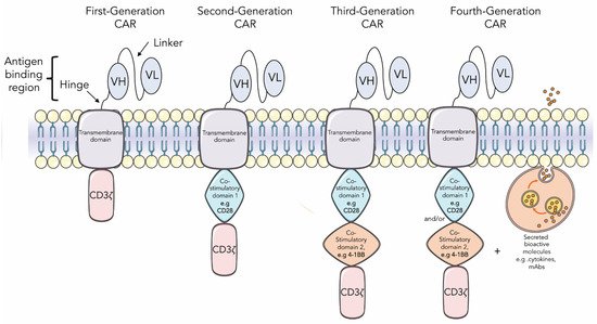
As shown in Figure 1, there are diverse generations of chimeric antigen receptors (CAR), which vary according to the extracellular, transmembrane, and intracellular co-stimulatory domains and the ability to secrete bioactive molecules such as cytokines or antibodies. The CAR is usually expressed in T cells or NK cells, directing the immune system to fight against the tumor [42][44].
Figure 1. Schematic representation of first, second, third, or fourth generations of chimeric antigen receptors (CAR). CARs are hybrid receptors that comprise an antibody-derived extracellular binding domain selected against a molecular target, usually in the form of a single-chain variable fragment (scFv), and a hinge/transmembrane domain fused to an intracellular signaling domain responsible for activating T cells. First-generation CARs have only one CD3ζ chain in the intracellular domain for activating T cells. Second- and third-generation CARs harbor one and two additional intracellular co-stimulatory domains, respectively. Fourth-generation CARs are CARs of second- or third-generation designed to induce expression of transgenic products constitutively or by induction, such as cytokines or monoclonal antibodies.