Hepatic Lipotoxicity in Acute Fatty Liver of Pregnancy: Comparison
Please note this is a comparison between Version 1 by Sathish Kumar Natarajan and Version 3 by Amina Yu.

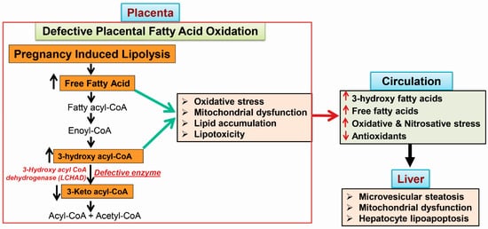
Acute fatty liver of pregnancy (AFLP), a catastrophic illness for both the mother and the unborn offspring, develops in the last trimester of pregnancy with significant maternal and perinatal mortality. AFLP is also recognized as an obstetric and medical emergency. Maternal AFLP is highly associated with a fetal homozygous mutation (1528G>C) in the gene that encodes for mitochondrial long-chain hydroxy acyl-CoA dehydrogenase (LCHAD). The mutation in LCHAD results in the accumulation of 3-hydroxy fatty acids, such as 3-hydroxy myristic acid, 3-hydroxy palmitic acid and 3-hydroxy dicarboxylic acid in the placenta, which are then shunted to the maternal circulation leading to the development of acute liver injury observed in patients with AFLP. 

  • acute fatty liver of pregnancy
  • 3-hydroxy fatty acids
  • lipoapoptosis
  • fatty acid oxidation

1. Maternal Liver Disease Associated with Fatty Acid Oxidation Defects

1.1. Acute Fatty Liver of Pregnancy

Acute fatty liver of pregnancy (AFLP) is an obstetric and medical emergency to the pregnant mother and unborn fetus that develops in the third trimester of pregnancy. Maternal AFLP is highly associated with carrying a fetus deficient in the long-chain hydroxy acyl-CoA dehydrogenase (LCHAD) enzyme [1][3]. The predominant mutation reported in the HADHA gene is at the exon 15, 1528G>C, resulting in a loss or dramatic decrease in LCHAD activity with normal enoyl-CoA hydratase activity [1][2][3,19]. Maternal and fetal demise in AFLP are predicted to be 10% and 45%, respectively. Women with AFLP initially show common symptoms and features of liver disease during their pregnancy that rapidly progress to renal failure, coagulopathy, ascites and hepatic encephalopathy. Accurate diagnosis of AFLP requires histological evidence of hepatic microvesicular steatosis [3][4][5][17,29,30]. Recently, the Swansea criteria have been commonly used following the publication of criteria observed in most women with AFLP [6][7][31,32]. However, questions were raised regarding the accuracy of the Swansea criteria in diagnosing AFLP without histological evidence of hepatic microvesicular steatosis [8][9][18,33]. Patients with AFLP show a dramatic increase in the circulating biomarkers for hepatocyte and biliary injury like alanine amino transferase (ALT), aspartate amino transferase (AST), alkaline phosphatase (ALP) and γ-glutamyl transpeptidase (GGT) with increased prothrombin time [10][8].
Ibdah et al. [11][25] reported a strong association between fetal LCHAD deficiency and development of maternal AFLP. In a series of studies, Ibdah et al. demonstrated that women who carry LCHAD deficient fetuses with documented mutations in the HADHA gene develop maternal AFLP [11][12][13][14][25,34,35,36]. Treatment of LCHAD deficient children includes consumption of a diet rich in carbohydrates and medium chain fatty acids to substitute for the fatty acid demand and energy requirements, especially during fasting periods [15][16][37,38]. Medium chain fatty acids are transported to the liver through an enterohepatic portal vein for the energy demand. However, long chain fatty acids are transported as triglycerides via chylomicrons through lymphatic circulation. Further, medium chain fatty acid oxidation enzymes are soluble enzymes in the mitochondrial matrix. The mutation in the LCHAD active site results in the accumulation of 3-hydroxy fatty acids in the placenta. Since the fetal part of the placenta is identical to the genetic makeup of the fetus. The accumulated 3-hydroxy fatty acids produced in the placenta are shunted to maternal circulation, leading to the acute liver injury observed in AFLP patients [17][18][10,39].

1.2. The Incidence of AFLP and LCHAD Mutations

The incidence of AFLP is estimated to be 1 in 10,000 pregnancies in the United States. However, the incident was reported to be more frequent in some unique populations. For example, a prospective cohort study from Southwest Wales, UK reported that AFLP occurs in five out of 4377 pregnancies [19][40], whereas a tertiary care center in India reported that AFLP occurs in one out of 3333 pregnancies [20][41], and in southern India the frequency was found to be one in 6691 pregnancies [8][6][9][18,31,33]. Similarly, an association with mitochondrial fatty acid oxidation has been reported in patients with preeclampsia, which is a common pregnancy disorder in the United States [11][21][22][23][25,26,27,28]. At least 40% of AFLP patients have a history of preeclampsia [10][24][8,42].
The incidence of LCHAD is highly prevalent in different ethnic groups. The prevalence of LCHAD deficiency is reported to be high in Baltic Sea countries compared to other populations in the world. Recently, the LCHAD variant 1528G>C was identified to be highly prevalent in the Kashubian population of northern Poland (one carrier in 57 individuals), southern Poland (one in 107), northern Pomerania (one in 207) and isolated regions of Poland (one in 187) [25][43]. The 1528G>C mutation in the HADHA gene corresponds to the amino acid change of Glu to Gln at position 474 of the mature LCHAD domain, which resides in the active site of the enzyme. This mutation affects and reduces LCHAD catalytic enzyme activity and decreases the protein stability [26][44]. The LCHAD mutation is also highly prevalent in the populations of Finland (one in 240), Netherlands (one in 680), Sweden (one in 540) and Estonia (one in 173) [27][11][25][28][29][30][31][32][33][34][35][9,25,43,45,46,47,48,49,50,51,52]. Due to the high incidence and prevalence of LCHAD mutation, the Swedish government mandated neonatal screening for LCHAD mutation in 2012 as a routine practice to minimize the incidence of AFLP [26][44].

1.3. 3-Hydroxy Fatty Acid Accumulation

Acute fatty liver of pregnancy is highly prevalent in mothers carrying LCHAD deficient fetuses. Biochemical hallmarks of LCHAD deficiency are the accumulation of long-chain 3-hydroxy fatty acids such as 3-hydroxy lauric acid, 3-hydroxy myristic acid, 3-hydroxy palmitic acid and 3-hydroxy dicarboxylic acid in the systemic circulation and increased excretion of 3-hydroxy dicarboxylic acids in the urine [36][37][38][53,54,55]. Several studies support the evidence for the accumulation of long-chain 3-hydroxy fatty acids (3-HFA) in patients with LCHAD deficiency and AFLP [36][38][39][40][41][42][43][44][45][46][53,55,56,57,58,59,60,61,62,63]. Children with the LCHAD deficiency are reported to develop sudden death with hypoglycemia, cardio-respiratory failure, acute cardiac failure and insufficiency, severe neonatal cardiomyopathy, hepatic dysfunction and acute liver failure, and skeletal myopathy with rhabdomyolysis [27][45][9,62]. Further, 34% of LCHAD deficient children die between four days and 10 years after birth [27][9]. This multi-organ damage is believed to be due to the lipotoxicity of toxic 3-hydroxy fatty acid intermediate accumulation.

2. Metabolic Phenotypes Associated with 3-Hydroxy Fatty Acid Accumulation

2.1. Hypoglycemia in AFLP and Hormonal Regulation

Fatty acid oxidation is an important source of energy especially for infants and children and has been reported to account for 80% of energy during initial hours of fasting [47][5]. Glucagon, epinephrine, norepinephrine, cortisol and growth hormones were secreted under the hypoglycemic condition to act on enhancing adipose tissue lipolysis [48][64]. This normal physiological function of peptide hormones enhances circulating free fatty acids levels, which then reach the liver for energy production via mitochondrial β-oxidation during hypoglycemic conditions. AFLP patients show severe hypoglycemic and hypoketotic states due to defective fatty acid β-oxidation. The levels of glucagon and stress hormones such as plasma cortisol were reported to be increased in children with LCHAD deficiency [49][65]. Further, itwe hasve shown increased circulating levels of long chain fatty acids like arachidonic acid, palmitic acid, myristic acid, oleic acid in patients with AFLP. AFLP patients also show increased maternal circulating long chain 3-hydroxy fatty acids such as 3-hydroxy myristic acid (3-HMA) and 3-hydroxy palmitic acid (3-HPA) due to the LCHAD defect and increased lipolysis [36][42][53,59]. Further, several case reports have shown an increased lipolysis in patients with LCHAD deficiency along with an increase in plasma dicarboxylic acid, long chain fatty acids and 3-hydroxy fatty acids after 4–6 h of fasting [27][49][50][9,65,66]. IRecent st wasudies have shown that lipid droplet accumulation is a protective event that packages non-esterified fatty acids as lipid droplets [51][52][53][67,68,69]. However, data on the hormonal regulation of hypoglycemia and peptide hormone-induced lipolysis during severe hypoglycemic conditions observed in AFLP patients are scarce and need further investigation.

2.2. Mitochondrial Trifunctional Protein (MTP)-Deficient Mice Develop Intra Uterine Growth Retardation (IUGR), Neonatal Hypoglycemia, and Sudden Death

Ibdah et al. generated mice that lack both mitochondrial trifunctional protein (MTP) α and β subunits [54][70]. Mitochondrial tri-functional protein (MTP) homozygous knockout mice developed hepatic lipotoxicity with diffuse hepatic enlargement and lipid accumulation associated with mitochondrial swelling and damage. These MTP null mice rapidly developed cardiac and diaphragmatic lesions leading to sudden death 96 h after birth. Further, MTP null mice also showed severe intrauterine growth retardation, neonatal hypoglycemia and reported to develop hepatic microvesicular steatosis, a pathophysiological hallmark of AFLP. These MTP homozygous defective fetuses accumulate long chain fatty acids and their metabolites due to the impairment in their mitochondrial β-oxidation [54][70]. The cause of sudden death in fetuses with MTP null mice is likely due to the cardiac and diaphragmatic damage and possibly cardiac arrhythmias. Further, cardiac arrhythmia and sudden death are also reported in children with other fatty acid oxidation defects such as defective carnitine palmitoyl transferase 2 (CPT2) and carnitine fatty acyl-CoA translocase and MTP deficiency [55][56][20,71]. MTP−/− mice were also reported to develop intrauterine growth retardation similar to the fetal phenotype observed in AFLP [54][70]. Thus, MTP null mice serve as a tool to study LCHAD deficiency and AFLP.

2.3. MTP Heterozygous Mice Develop Hepatic Insulin Resistance

Hepatic insulin resistance was reported in MTP heterozygous mice [57][72]. A fifty percent reduction in the mitochondrial fatty acid oxidation and glycogen levels were reported in the hepatocytes isolated from MTP heterozygotes compared to controls. MTP heterozygous mice also showed defective insulin-induced hepatic insulin signaling pathway activation. Insulin-induced activation of insulin receptor substrate phosphorylation and their downstream targets like protein kinase B, glycogen synthase kinase 3β, forkhead family of transcription factor class O1 (FoxO1) activation were shown to be blunted in the liver from MTP heterozygous mice compared to the liver from wild-type littermates, suggesting that defective fatty acid β-oxidation causes hepatic insulin resistance [57][72]. IThis report also suggests that mitochondrial fatty acid oxidation plays an important role in hepatic insulin signaling pathways for the maintenance of normal homeostasis of liver insulin sensitivity and glycogen production. The MTP heterozygous mouse was recently used as a model for non-alcoholic fatty liver disease [58][59][60][61][73,74,75,76].

3. Mechanisms of 3-Hydroxy Fatty Acid-Induced Lipotoxicity

3.1. Placental Damage in AFLP Patients

The placenta acts as a lung, liver, and kidney for the fetus and the fetal part of the placenta is identical to the genetic make-up of the fetus. High activity of fatty acid oxidation enzymes is shown in the placenta compared to the adult liver [62][63][64][65][77,78,79,80]. Placental injury was also observed in patients with AFLP. A case reprort of pregnant women who developed severe AFLP showed abruptio placentae (placental abruption), premature delivery and fetal demise [66][81]. Maternal floor infarction of the placenta was also reported in a mother who gave birth to an LCHAD deficient child [67][82]. Further, placenta from AFLP patients were reported to have abnormalities and increased oil red O staining, suggesting an increase in placental lipid droplet accumulation and lipotoxicity [68][83].
In AFLP, a fetus homozygous for an LCHAD mutation will have defective placental metabolism of long chain fatty acids resulting in the accumulation of 3-hydroxy fatty acids and long chain fatty acids. Accumulated 3-hydroxy fatty acids and other fatty acids enter the mother’s circulation and affect the maternal liver, resulting in AFLP. Further, LCHAD deficient children have been known to display mitochondrial damage as evidenced by increased mitochondrial swelling, and irregular mitochondrial cristae in the skeletal muscle [38][40][43][44][45][46][67][69][70][71][55,57,60,61,62,63,82,84,85,86]. The cytotoxic 3-hydroxy fatty acids are also known to inhibit mitochondrial processes like β-oxidation and oxidative phosphorylation enzymes resulting in decreased ATP production and increased reactive oxygen species; thereby resulting in mitochondrial damage [38][45][72][55,62,87]. Although the strong association between maternal AFLP and fetal LCHAD deficiency is well-documented, there are few case reports that suggest an association with other fetal fatty acid oxidation disorders. For instance, a published report linked maternal AFLP with pediatric carnitine palmitoyl transferase I deficiency [73][88]. A recent report also showed that AFLP developed in a mother carrying a fetus with HADHB homozygous mutation [74][89]. The possibility that AFLP can be associated with fatty acid oxidation defects other than LCHAD deficiency is intriguing and warrants further investigation to understand the underlying molecular mechanisms.

3.2. Subcellular Damage and Oxidative Injury

Subcellular damage and oxidative injury was evident in animal models of microvesicular steatosis. ItWe hasve shown that inhibition of mitochondrial β-oxidation using near-lethal doses of valproate developed hepatic microvesicular steatosis and oxidative stress in the liver. Hepatic mitochondrial membrane damage and dysfunction were also evident in this rat model of microvesicular steatosis [10][75][8,90]. Further, placental mitochondria isolated from patients with AFLP showed decreased respiration, altered mitochondrial calcium homeostasis, increased superoxide generation and increased mitochondrial swelling, suggesting a placental mitochondrial dysfunction compared to controls. ItWe hasve also shown a dramatic increase in the levels of oxidative injury biomarkers in placental mitochondria, peroxisomes, and microsomes in patients with AFLP compared to controls [76][77][1,2]. Further, circulating oxidative and nitrosative stress markers were increased in the maternal circulation of AFLP patients, suggesting that reactive nitrogen species act together with reactive oxygen species to damage cells. Concomitantly, circulating levels of antioxidants like tocopherols and retinol were dramatically decreased in patients with AFLP compared to controls suggesting an oxidative and nitrosative stress in the maternal systemic circulating in patients with AFLP [10][8]. The simultaneous presence of reactive oxygen species and nitric oxide could lead to the formation of peroxynitrite, a highly damaging oxidant.

3.3. 3-Hydroxy Fatty Acid-Induced Hepatocyte Lipoapoptosis

ItWe has beenve shown that long-chain fatty acids like palmitic acid and arachidonic acid levels were dramatically elevated in the systemic circulation of patients with AFLP. The lipotoxic role of saturated free fatty acid like palmitate was reported to induce caspase-dependent hepatocyte and cholangiocyte lipoapoptosis [52][78][79][80][81][68,95,96,97,98]. Recently, Itwe hasve also demonstrated that the signaling mechanism of palmitate-induced cholangiocyte lipoapoptosis is via the activation of mitogen-activated protein kinase (MAPK) and forkhead family of transcription factor class O3 (FoxO3) and its downstream targets like p53-upregulated modulator of apoptosis (PUMA) protein and pro-apoptotic microRNA 34a [52][82][83][68,99,100]. Further, it wase reported that the exposure of arachidonic acid to the hepatocyte similar to the concentration observed in AFLP patients showed an increased lipid droplet accumulation, mitochondrial reactive oxygen species and caspase 3 activation leading to hepatocyte lipoapoptosis. These results suggest that AFLP-related long-chain fatty acid accumulation in the maternal systemic circulation can induce hepatocyte lipoapoptosis in patients with AFLP [77][10][2,8]. Increased long chain 3-hydroxy fatty acid in patients with AFLP can also induce mitochondrial dysfunction and hepatocyte lipoapoptosis. TheOur unpublished preliminary data show that the treatment of long chain 3-hydroxy fatty acids (3-HFAs) such as 3-hydroxy myristic acid (3-HMA) and 3-hydroxy palmitic acid (3-HPA) to cultured hepatocytes results in caspase-dependent hepatocyte lipoapoptosis. However, interestingly, short chain 3-hydroxy octanoic acid (3-HOA) did not induce hepatocyte lipoapoptosis. Similar to mitochondrial fatty acid oxidation defects, mice lacking peroxisomal fatty acyl-CoA oxidase also show microvesicular steatosis, hepatocyte apoptosis and liver injury [84][101]. A schematic presentation of the sequence of events that occur in-patient with AFLP is shown in Figure 12. Experiments to elucidate the mechanism of 3-HFA-induced hepatocyte lipoapoptosis are currently underway in theour laboratory.
Figure 12. Schematic representation of the sequence of events that happen during acute fatty liver of pregnancy (AFLP). Fetal long chain 3-hydroxy acyl-CoA dehydrogenase (LCHAD) deficiency results in accumulation of 3-hydroxy fatty acids in the placenta, since the fetal part of placenta is identical to the genetic makeup of the fetus. Increased accumulation of placental free fatty acids and 3-hydroxy fatty acyl-CoA cause oxidative stress, mitochondrial dysfunction and placental lipotoxicity. Further, lipolysis induced in the third trimester of pregnancy would also trigger the accumulation of fatty acid intermediates, which are shunted from the placenta to the maternal circulation, where they can promote oxidative and nitrosative stress. These fatty acid intermediates reach the maternal liver resulting in microvesicular steatosis, hepatic mitochondrial dysfunction and hepatocyte lipoapoptosis.

Abbreviations

AFLPacute fatty liver of pregnancy
ALTalanine amino transferase
ASTaspartate amino transferase
ALPalkaline phosphatase
CPT2carnitine palmitoyl transferase 2
DHAdocosa hexaenoic acid
FoxOforkhead family of transcription factor class O
GGTγ-glutamyl transpeptidase
LCHADlong chain hydroxy acyl-CoA dehydrogenease
MAPKmitogen activated protein kinases
MTPmitochondiral trifunctional protien
PUMAp35-upregulated modulator of apoptosis
TCA cycletricarboxylic acid cycle
3-HFA3-hydroxy fatty acid
3-HMA3-hydroxy myristic acid
3-HOA3-hydroxy octanoic acid
3-HPA3-hydroxy palmitic acid
Video Production Service