A highly innovative approach to ultrasound imaging involves the targeting and real-time, in vivo visualization of physiologic processes with molecular-specific imaging. Molecular-targeted microbubbles have been developed as an extension of CEUS
[23][24][38,39]. This could be of particular interest in the metastatic setting, where the response to systemic therapy is currently determined by the change in tumor volume. However, changes in tumor physiology occur sooner than measurable tumor volume changes, which might allow for earlier assessment of tumor progression, response to systemic therapy and, ultimately, therapeutic decision making. Moreover, the use of molecular imaging techniques might better characterize features relating to intrinsic disease biology, such as angiogenesis, potentially leading to more individualized treatment decision making
[25][26][40,41]. In a recent report, Rojas et al. studied ccRCC in a xenograft model of immunodeficient mice treated with the anti-angiogenic vascular endothelial growth factor receptor (VEGFR) tyrosine kinase inhibitor sunitinib
[24][39]. They administered a microbubble contrast agent targeted to VEGFR-2 and subsequently imaged the tumors with CEUS after 1 week of treatment. They reported changes in VEGFR-2 expression at that time, as determined on ultrasound molecular imaging in the sunitinib-treated group, as opposed to changes in tumor volume, which only became apparent after 3 weeks. Moreover, after 1 week, response to therapy was detected in 92% of cases with ultrasound molecular imaging, whereas the detection rate was only 40% with volume measurements. Likewise, Ingels et al. studied the potential of ultrasound molecular imaging to track the response to sunitinib in a ccRCC mice xenograft model
[23][38]. These mice, harboring ccRCC, were randomized between treatment with sunitinib and control and were injected with both non-targeted microbubbles and microbubbles targeting VEGFR-1 and follicle-stimulating hormone receptor (FSHR). Both the VEGFR-1 and FSHR signal enhancement were significantly lower in the sunitinib group at all times of treatment, while there was no significant difference between the two groups for the non-targeted microbubble ultrasound signal. Thus, they confirmed the potential of ultrasound molecular imaging for the longitudinal assessment of treatment response to sunitinib. However, despite its potential for serial monitoring of disease, as well as longitudinal assessment of disease response to systemic therapy, ultrasound molecular imaging is still in the very early phases of development and further research endeavors will determine whether these techniques can provide additional value in clinical practice.
3. Elastography
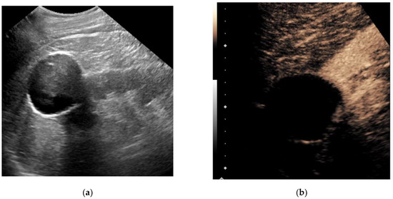
Equivalent to the use of palpation during physical examination, ultrasound elastography measures changes in tissue stiffness, which are often seen with diffuse parenchymal diseases and the associated changes in tissue architecture
[27][42]. Strain elastography provides a qualitative or semi-quantitative assessment of tissue elasticity using external compression–decompression cycles from the ultrasound transducer. Shear-wave elastography (SWE) involves a quantitative assessment of tissue stiffness by measuring the propagation speed of generated shear waves through tissues. This technique does not require external compression by a transducer, relying instead on a high-amplitude push pulse (also known as acoustic radiation force impulse or ARFI), thus making it less operator dependent
[28][43]. Strain elastography was shown to aid in the distinction of benign vs. malignant lesions, the distinction of RCC from AML and the distinction of RCC from transitional cell carcinoma
[27][29][30][42,44,45]. SWE had potential value in the differentiation of ccRCC versus oncocytoma, ccRCC versus chrRCC or papRCC and pseudotumor from ccRCC or AML, though failed to differentiate between ccRCC and AML
[28][31][43,46]. However, few studies have been conducted and these results lack validation, rendering clinical applications for renal mass characterization limited at this time.
4. Micro-Doppler Techniques
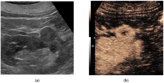
The presence of blood flow within a renal mass indicates solid tissue, as opposed to a renal cyst. The pattern of vascularity may help characterize indeterminate renal masses, such as differentiating malignancy from pseudomasses. Many companies are releasing novel micro-Doppler techniques with advanced clutter suppression. Some of these include Superb Micro-Vascular Imaging (Canon Medical Systems, Tochigi, Japan), Micro Vascular Imaging (GE Healthcare, Waukesha, WI, USA), Micro-Flow Imaging (Philips Healthcare, Bothell, WA, USA) and Micro Vascular Flow (Samsung Medison, Seoul, Korea). These techniques appear to improve the detection of slower flow within smaller vessels, increasing the ability to detect subtle vascularity within indeterminate renal masses that were previously below the detection threshold for traditional color and power Doppler techniques.
Leong et al. recently imaged 41 patients harboring 50 renal masses with Superb Micro-Vascular Imaging (SMI). They found that SMI had a higher diagnostic accuracy than standard color Doppler imaging and power Doppler imaging for the detection of vascularity within solid renal masses
[32][47]. They concluded that SMI might have potential in the detection of microvascularity within indeterminate solid renal masses. Subsequently, Mao et al. showed that SMI could distinguish significantly different patterns of vascularization between pathologically proven malignant and benign renal masses in a study on 53 patients
[33][48]. Conversely, conventional Doppler flow imaging could not discern these differences in vascularization.
Although the benefit of these micro-Doppler techniques includes intravenous contrast not being required, there has not been a direct comparison to CEUS for the detection of malignancy, which limits the current applications of these techniques. Future endeavors should specifically study whether the addition of these techniques to CEUS could improve the diagnostic accuracy and could potentially be a useful addition to current techniques in terms of characterizing indeterminate renal masses.