Antibody-Drug Conjugate Targeting c-Kit: Comparison
Please note this is a comparison between Version 1 by Kwang-Hyeok Kim and Version 3 by Rita Xu.

Lung cancer is the leading cause of cancer-related deaths. Small cell lung cancer (SCLC) accounts for 15–25% of all lung cancers. It exhibits a rapid doubling time and a high degree of invasiveness. Additionally, overexpression of c-Kit occurs in 70% of SCLC patients.

폐암은 암 관련 사망의 주요 원인입니다. 소세포폐암(SCLC)

은 전체 폐암의 15~25%를 차지합니다. 그것은 빠른 배가 시간과 높은 수준의

침습성을 나타냅니다. 또한, c-Kit의 과발현은 SCLC 환자의 70%에서 발생합니다. 본 연구에서는 SCLC

의 잠재적 치료제인 c-Kit을 표적으로 하는 항체-약물 접합체(ADC)를 평가하였다 .

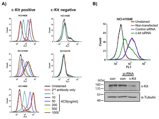
먼저, 우리는 c-Kit을 표적으로 하는 완전한 인간 항체인 4C9를 생성하고 특성화했으며,

결합 친화도는 KD = 5.5 × 9 M인 c-Kit을 발현하는 SCLC 세포에 특이적으로 결합합니다.

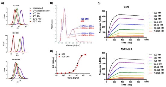
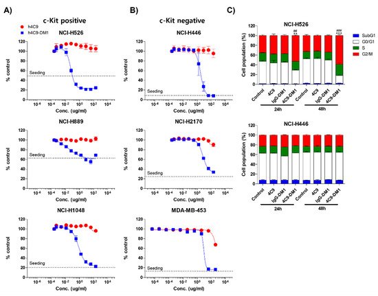
그런 다음, DM1을 사용하여 ADC를 개발했습니다. 페이로드로서의 미세소관 억제제. 4C9-DM1 효율적으로

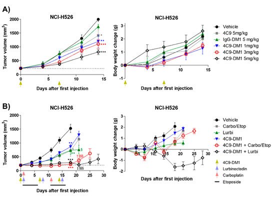
158 pM에서 4 nM 범위의 IC50으로 SCLC에서 세포자멸사를 유도했습니다. 이종이식 마우스 모델 을 사용한 생체내 분석 은 4C9-DM1 단독에 대해

45%(3mg/kg) 및 59%(5mg/kg)의 종양 성장 억제(TGI) 비율 을 나타냈습니다.

4C9-DM1 + ​​카르보플라틴/에토 포시드

또는 루르비넥티드의 조합 치료는 비히클 대조군과 비교하여 90% 초과의 TGI 비율을 초래했습니다. 종합

하면, 이러한 결과는 4C9-DM1이 SCLC 치료를 위한 잠재적 치료제임을 나타냅니다.

  • c-Kit
  • small cell lung cancer
  • monoclonal antibody
  • antibody-drug conjugate

1. Introduction1. 소개

Lung cancer is the leading cause of cancer-related deaths in the Western world and is classified into two groups: small cell lung cancer (SCLC) and non-SCLC (NSCLC) [1]. SCLC, a neuroendocrine tumor, is distinguished from NSCLC by its rapid tumor growth, high degree of invasiveness and early development of widespread metastases [2]. SCLC is distinctly different from extrapulmonary small cell carcinoma with respect to disease progression, prognosis, and etiology [3]. Without proper treatment, the life expectancy of SCLC patients is less than four months. Although the five-year relative survival rate has improved by 7% over the last few decades, it remains extremely poor [4].폐암은 서구 세계에서 암 관련 사망의 주요 원인이며 소세포폐암(SCLC)과 비소세포폐암(NSCLC)의 두 그룹으로 분류됩니다[ 1 ]. 신경 내분비 종양인 SCLC는 빠른 종양 성장, 높은 수준의 침습성 및 광범위한 전이의 조기 발달에 의해 NSCLC와 구별됩니다[ 2 ]. SCLC는 질병 진행, 예후 및 병인과 관련하여 폐외 소세포 암종과 분명히 다릅니다[ 3 ]. 적절한 치료가 없으면 SCLC 환자의 기대 수명은 4개월 미만입니다. 5년 상대생존율이 지난 수십 년 동안 7% 향상되었지만 여전히 매우 열악합니다[ 4 ].
A variety of molecular markers have been implicated in the pathogenesis and prognosis of SCLC [5][6]. Paracrine or autocrine signal transduction pathways are widely used to explain dysregulated SCLC growth [6]. In addition, tumor protein p53, retinoblastoma protein, NOTCH, MYC, and phosphatidylinositol 3-kinase (PI3K) are aberrantly mutated in SCLC; however, well-established etiological factors, such as EGFR mutations that occur in NSCLC, have not been identified [7][8][9][10]. SCLC has a very aggressive course and is characterized by genomic instability, increased vascularity, and a high metastatic potential [11]. Consequently, most SCLC patients already present with metastatic disease outside of the chest, at the time of diagnosis, which results in premature death [12]. In addition, most SCLC patients are current or former heavy smokers, which is associated with a high tumor mutational burden, with C:G > A:T transversions being the most common type of base substitutions [13][14]다양한 분자 마커가 SCLC 의 발병기전과 예후와 관련이 있습니다[ 5,6 ] . 측분비 또는 자가분비 신호 전달 경로는 조절되지 않은 SCLC 성장을 설명하는 데 널리 사용됩니다[ 6 ]. 또한, 종양 단백질 p53, 망막모세포종 단백질, NOTCH, MYC 및 포스파티딜이노시톨 3-키나제(PI3K)는 SCLC에서 비정상적으로 돌연변이됩니다. 그러나 NSCLC에서 발생하는 EGFR 돌연변이와 같은 잘 확립된 병인학적 요인은 확인되지 않았습니다[ 7 , 8 , 9 , 10 ]. SCLC는 매우 공격적인 경과를 보이며 게놈 불안정성, 증가된 혈관성 및 높은 전이 가능성을 특징으로 합니다[ 11]. 결과적으로, 대부분의 SCLC 환자는 진단 당시 이미 흉부 외부에 전이성 질환이 존재하여 조기 사망을 초래한다[ 12 ]. 또한 대부분의 SCLC 환자는 현재 또는 과거에 심한 흡연을 했으며 이는 높은 종양 돌연변이 부담과 관련이 있으며 C:G > A:T 변환이 가장 흔한 염기 치환 유형입니다[ 13 , 14 ]
The c-Kit proto-oncogene encodes a transmembrane tyrosine kinase growth factor receptor that belongs to the platelet-derived growth factor receptor (PDGFR) family [15][16]. Its ligand stem cell factor (SCF) is a hematopoietic growth factor that promotes the proliferation of multiple hematopoietic stem cells [17][18]. In addition, c-Kit activity is dysregulated in various cancers [19][20]. Previous studies reported that the expression of c-Kit containing oncogenic mutations is either dysregulated and/or up-regulated in various cancers, which results in SCF-independent c-Kit activation and an aggressive form of cancer [20]. Interestingly, a variety of evidence indicates that SCLC cell lines and tumors express both the c-Kit receptor and SCF mRNA, suggesting that these gene products constitute an autocrine loop that mediates tumor cell survival and growth [21][22]. Although SCLC is significantly correlated with smoking, it does not contain oncogenic c-Kit mutations. Immunohistochemical staining showed that overexpression of c-Kit occurs in 70% of SCLC patients [23][24]. Imatinib, which was developed to target BCR-ABL, platelet-derived growth factor receptor (PDGFR), and c-Kit, is currently used to treat chronic myeloid leukemia, acute lymphoid leukemia, gastrointestinal stromal tumor (GIST), and hypereosinophilic syndrome [25][26][27][28]. A variety of in vitro and in vivo studies demonstrated that imatinib exhibits therapeutic efficacy against SCLC [29][30]. However, in phase 2 clinical trials, imatinib failed to exhibit significant therapeutic efficacy as shown by a lack of objective responses [31][32][33]. Thus, an alternative approach to target c-Kit in SCLC is needed.c-Kit 원종양유전자는 혈소판 유래 성장 인자 수용체(PDGFR) 패밀리에 속하는 막횡단 티로신 키나제 성장 인자 수용체를 인코딩합니다[ 15 , 16 ]. 리간드 줄기 세포 인자(SCF)는 여러 조혈 줄기 세포의 증식을 촉진하는 조혈 성장 인자입니다[ 17 , 18 ]. 또한 c-Kit 활성은 다양한 암에서 조절되지 않습니다[ 19 , 20 ]. 이전 연구에서는 발암성 돌연변이를 포함하는 c-Kit의 발현이 다양한 암에서 조절 이상 및/또는 상향 조절되어 SCF 독립적인 c-Kit 활성화 및 공격적인 형태의 암을 유발한다고 보고했습니다. [ 20]. 흥미롭게도 다양한 증거는 SCLC 세포주와 종양이 c-Kit 수용체와 SCF mRNA를 모두 발현한다는 것을 나타내며, 이는 이러한 유전자 산물이 종양 세포 생존과 성장을 매개하는 자가분비 루프를 구성한다는 것을 시사합니다[ 21 , 22 ]. SCLC는 흡연과 유의한 상관관계가 있지만 발암성 c-Kit 돌연변이는 포함하지 않습니다. 면역조직화학염색에서 c-Kit의 과발현은 SCLC 환자의 70%에서 발생하는 것으로 나타났습니다[ 23 , 24 ]. BCR-ABL, 혈소판유래성장인자수용체(PDGFR), c-Kit 등을 표적으로 하기 위해 개발된 이마티닙은 현재 만성골수성백혈병, 급성림프성백혈병, 위장관기질종양(GIST), 과호산구성 증후군 치료제로 사용되고 있다. 25 ,26 , 27 , 28 ]. 다양한 시험관 내 및 생체 내 연구에서 이마티닙이 SCLC에 대한 치료 효능을 나타내는 것으로 나타났습니다[ 29 , 30 ]. 그러나 임상 2상에서 이마티닙은 객관적인 반응이 부족하여 유의한 치료 효능을 나타내지 하였다 [ 31,32,33 ] . 따라서 SCLC에서 표적 c-Kit에 대한 대안적 접근이 필요합니다.

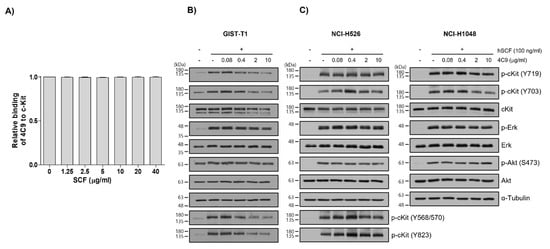
2. 4C9 Antibody Specifically Binds to c-Kit2. 4C9 항체는 c-Kit에 특이적으로 결합합니다.

First, researchers examined whether the 4C9 antibody specifically binds to c-Kit on cell surface. FACS analysis revealed that 4C9 binds to various SCLC cell lines, including NCI-H526, NCI-H1048, and NCI-H889, in a dose-dependent manner (Figure 1A). Interestingly, 4C9 antibody binding was saturated at 50 ng/mL in c-Kit-positive SCLC cell lines. In addition, the expression of c-Kit was higher in NCI-H889 cells compared with that in NCI-H526 and NCI-H1048 cells, which is consistent with a previous report [34]. However, 4C9 did not show cross-reactivity with NCI-H446 and NCI-H2170 cells, which are c-Kit-negative SCLC cell lines. Researchers further examined the specific binding of 4C9 to c-Kit using siRNA knockdown experiment. Western blot analysis showed that c-Kit siRNA efficiently decreased protein expression in NCI-H1048 cells (Figure 1B). FACS analysis demonstrated that c-Kit expression on the cell surface was also down-regulated by c-Kit siRNA. Taken together, 4C9 binds specifically to the extracellular domain of c-Kit on the cell surface.먼저 4C9 항체가 세포 표면의 c-Kit에 특이적으로 결합하는지 여부를 조사했습니다. FACS 분석은 4C9가 NCI-H526, NCI-H1048 및 NCI-H889를 비롯한 다양한 SCLC 세포주에 용량 의존적 방식으로 결합함을 보여주었다( 1A). 흥미롭게도, 4C9 항체 결합은 c-Kit-양성 SCLC 세포주에서 50ng/mL로 포화되었습니다. 또한, c-Kit의 발현은 NCI-H526 및 NCI-H1048 세포에 비해 NCI-H889 세포에서 더 높았는데, 이는 이전 보고서와 일치한다[ 34]. 그러나, 4C9는 c-Kit 음성 SCLC 세포주인 NCI-H446 및 NCI-H2170 세포와 교차 반응성을 나타내지 않았다. 우리는 siRNA 녹다운 실험을 사용하여 c-Kit에 대한 4C9의 특이적 결합을 추가로 조사했습니다. 웨스턴 블롯 분석은 c-Kit siRNA가 NCI-H1048 세포에서 단백질 발현을 효율적으로 감소시키는 것으로 나타났습니다( 그림 1B). FACS 분석은 세포 표면의 c-Kit 발현도 c-Kit siRNA에 의해 하향 조절된다는 것을 보여주었습니다. 종합하면, 4C9는 세포 표면에 있는 c-Kit의 세포외 도메인에 특이적으로 결합합니다.
Figure 1. Determination of specific binding of the 4C9 antibody to the surface of SCLC cells. (A) SCLC cell lines were incubated with 4C9 antibody in a dose-dependent manner and analyzed by FACS. (B) NCI-H1048 cells were transfected with control or c-Kit si-RNA and incubated for 72 h. Binding of 4C9 (1 μg/mL) to NCI-H1048 cells was determined by FACS. The si-RNA-mediated down-regulation of c-Kit protein expression was evaluated by Western blot analysis. Alpha-tubulin was used as a loading control.그림 1. SCLC 세포 표면에 대한 4C9 항체의 특이적 결합 측정. ( A ) SCLC 세포주를 용량 의존적 방식으로 4C9 항체와 함께 인큐베이션하고 FACS에 의해 분석하였다. ( B ) NCI-H1048 세포를 대조군 또는 c-Kit si-RNA로 형질감염시키고 72시간 동안 인큐베이션하였다. NCI-H1048 세포에 대한 4C9(1㎍/mL)의 결합을 FACS에 의해 결정하였다. c-Kit 단백질 발현의 si-RNA 매개 하향 조절은 웨스턴 블롯 분석에 의해 평가되었다. 알파 튜불린을 로딩 대조군으로 사용했습니다.
Next, researchers investigated whether the 4C9 antibody could inhibit the binding of SCF, a ligand for c-Kit. Competitive enzyme-linked immunosorbent assay (ELISA) results showed that the binding of 4C9 antibody to c-Kit was not affected by SCF, even at high concentrations (Figure 2A), suggesting that 4C9 antibody binds to c-Kit independent of SCF. In addition, researchers assessed whether 4C9 could inhibit SCF-mediated phosphorylation of c-Kit. Using GIST-T1 cells, pretreatment with 4C9 antibody resulted in decreased c-Kit phosphorylation in a dose-dependent manner; however, the phosphorylation levels of the ERK and Akt, downstream molecules of c-Kit, were not changed by treatment with 4C9 antibody (Figure 2B). Interestingly, total c-Kit levels decreased by 4C9 antibody treatment (Figure 2B). Hence, a decrease in phosphorylated c-Kit levels may result from decreased expression of c-Kit. A stability assessment of c-Kit indicated that the 4C9 antibody dramatically decreased total c-Kit levels in a time-dependent manner in both GIST cell lines and in some SCLC cell lines, which may be associated with ubiquitination-dependent degradation. Nevertheless, this needs further elucidation. In contrast to GIST-T1, 4C9 did not reduce SCF-mediated c-Kit phosphorylation or c-Kit stability in the NCI-H526 and NCI-H1048 cell lines (Figure 2C). Furthermore, the 4C9 antibody did not inhibit phosphorylation of ERK and Akt induced by SCF, which suggests that the 4C9 antibody does not function as an antagonist of SCF/c-Kit signaling.다음으로 우리는 4C9 항체가 c-Kit의 리간드인 SCF의 결합을 억제할 수 있는지 조사했습니다. Competitive enzyme-linked immunosorbent assay(ELISA) 결과는 c-Kit에 대한 4C9 항체의 결합이 고농도에서도 SCF의 영향을 받지 않는 것으로 나타났으며( 그림 2A), 이는 4C9 항체가 SCF와 무관하게 c-Kit에 결합함을 시사합니다. 또한 4C9가 c-Kit의 SCF 매개 인산화를 억제할 수 있는지 여부를 평가했습니다. GIST-T1 세포를 사용하여 4C9 항체로 전처리하면 용량 의존적으로 c-Kit 인산화가 감소되었습니다. 그러나 c-Kit의 하류 분자인 ERK 및 Akt의 인산화 수준은 4C9 항체 처리에 의해 변경되지 않았습니다( 그림 2B). 흥미롭게도, 총 c-Kit 수준은 4C9 항체 치료에 의해 감소했습니다(그림 2 B). 따라서 인산화된 c-Kit 수준의 감소는 c-Kit의 발현 감소로 인해 발생할 수 있습니다. c-Kit의 안정성 평가는 4C9 항체가 GIST 세포주와 일부 SCLC 세포주 모두에서 시간 의존적 방식으로 총 c-Kit 수준을 극적으로 감소시켰으며, 이는 유비퀴틴화 의존적 분해와 관련될 수 있음을 나타냅니다. 그럼에도 불구하고 이것은 더 많은 설명이 필요합니다. GIST-T1과 달리 4C9는 NCI-H526 및 NCI-H1048 세포주에서 SCF 매개 c-Kit 인산화 또는 c-Kit 안정성을 감소시키지 않았습니다( 그림 2C ). 또한, 4C9 항체는 SCF에 의해 유도된 ERK 및 Akt의 인산화를 억제하지 않았으며, 이는 4C9 항체가 SCF/c-Kit 신호전달의 길항제로서 기능하지 않음을 시사한다.
Figure 2. Characterization of the 4C9 antibody. (A) Human c-Kit (20 ng/well) was coated onto 96-well plates and the binding of the 4C9 antibody was investigated in the presence of human SCF at the indicated concentrations. The results represent the mean ± SD of three independent experiments. (B,C) GIST-T1, NCI-H526, or NCI-H1048 cells were treated with 4C9 at the indicated concentrations in the presence or absence of SCF (100 ng/mL). The phosphorylation of c-Kit, Akt, and ERK was assessed by Western blot analysis. In NCI-H1048 cells, phosphorylation of Y568/570 and Y823 by SCF treatment was not detected. Alpha-tubulin was used as a loading control. The results represent the mean ± SD of three independent experiments.그림 2. 4C9 항체의 특성화. ( A ) 인간 c-Kit (20 ng/웰)을 96-웰 플레이트에 코팅하고 4C9 항체의 결합을 표시된 농도의 인간 SCF 존재 하에 조사하였다. 결과는 3개의 독립적인 실험의 평균 ± SD를 나타냅니다. ( B , C ) GIST-T1, NCI-H526 또는 NCI-H1048 세포를 SCF(100ng/mL)의 존재 또는 부재 하에 표시된 농도에서 4C9로 처리하였다. c-Kit, Akt 및 ERK의 인산화는 웨스턴 블롯 분석으로 평가되었습니다. NCI-H1048 세포에서 SCF 처리에 의한 Y568/570 및 Y823의 인산화는 검출되지 않았다. 알파 튜불린을 로딩 대조군으로 사용했습니다. 결과는 3개의 독립적인 실험의 평균 ± SD를 나타냅니다.

3. Generation and Characterization of ADC (4C9-DM1)3. ADC 생성 및 특성화(4C9-DM1)

Even though c-Kit is overexpressed in SCLC cell lines, naked antibodies cannot be applied to treat SCLC because of the limited contribution of SCF/c-Kit signaling in the pathogenesis of SCLC and failure of imatinib [32][33]. Therefore, the development of an ADC would be a reasonable option to effectively treat SCLC. One important factor to consider when creating an ADC is the internalization efficiency after the complex formation of the antibody with the target molecule. Therefore, researchers first investigated whether the 4C9 antibody is internalized in SCLC cell lines. FACS analysis exhibited that the internalization efficiency of the 4C9 antibody was 91% in NCI-H526, 76.6% in NCI-H1048, and 68.6% in NCI-H889 cells (Figure 3A), which suggests that the 4C9 antibody could be used as an efficient carrier for the specific delivery of toxin to treat SCLC. Next, researchers generated an ADC using SMCC-DM1, which consists of a noncleavable linker and a microtubule inhibitor. Because DM1 absorbs ultraviolet light at 252 nm [35], the absorbance of the naked antibody (4C9) and ADC (4C9-DM1) at 252 nm was analyzed. The absorbance of 4C9-DM1 was higher than that of 4C9 at 252 nm (Figure 3B). The drug-antibody ratio (DAR) was determined as described previously [36] and the DAR was approximately 2.16. SDS-PAGE analysis exhibited that the conjugation of SMCC-DM1 to the 4C9 antibody resulted in a slight size shift. Since N-hydroxysuccinimide ester of the SMCC linker reacts with primary amines of lysine residues in the antibody, the target binding affinity of ADC may be affected. An ELISA demonstrated that the binding affinities of 4C9 and 4C9-DM1 to c-Kit were similar (Figure 3C). A quantitative analysis of the binding affinity using surface plasmon resonance (SPR) indicated that the binding affinity of 4C9 to human c-Kit was 5.5 × 10⁻⁹ M (Ka = 2.27 × 10⁴ M−1s−1 and Kd = 1.27 × 10⁻⁴ s−1), and 4C9-DM1 also showed similar binding affinity (5.46 × 10⁻⁹ M; Ka = 2.14 × 10⁴ M−1s−1 and Kd = 1.16 × 10⁻⁴ s−1) (Figure 3D). Taken together, these results indicate that conjugation using SMCC-DM1 to the 4C9 antibody did not affect the binding affinity of 4C9 antibody for c-Kit.c-Kit이 SCLC 세포주에서 과발현되더라도 SCLC의 발병기전과 imatinib의 실패에서 SCF/c-Kit 신호전달의 제한된 기여로 인해 네이키드 항체를 SCLC 치료에 적용할 수 없습니다[ 32 , 33 ]. 따라서 ADC의 개발은 SCLC를 효과적으로 치료하기 위한 합리적인 선택이 될 것입니다. ADC를 생성할 때 고려해야 할 중요한 요소 중 하나는 표적 분자와 항체가 복합적으로 형성된 후 내재화 효율입니다. 따라서 우리는 먼저 4C9 항체가 SCLC 세포주에서 내재화되는지 여부를 조사했습니다. FACS 분석은 4C9 항체의 내재화 효율이 NCI-H526에서 91%, NCI-H1048에서 76.6%, NCI-H889 세포에서 68.6%임을 나타내었다( 도 3A) 이는 4C9 항체가 SCLC를 치료하기 위한 독소의 특이적 전달을 위한 효율적인 운반체로 사용될 수 있음을 시사합니다. 다음으로, 우리는 noncleavable linker와 microtubule inhibitor로 구성된 SMCC-DM1을 사용하여 ADC를 생성했습니다. DM1은 252 nm에서 자외선을 흡수하기 때문에[ 35 ], 252 nm에서 네이키드 항체(4C9) 및 ADC(4C9-DM1)의 흡광도를 분석했습니다. 4C9-DM1의 흡광도는 252 nm에서 4C9의 흡광도보다 높았습니다( 그림 3B ). 약물-항체 비율(DAR)은 이전에 설명한 대로 결정되었습니다[ 36] 및 DAR은 약 2.16이었습니다. SDS-PAGE 분석은 4C9 항체에 대한 SMCC-DM1의 접합이 약간의 크기 이동을 초래하는 것으로 나타났습니다. SMCC 링커의 N-히드록시숙신이미드 에스테르는 항체 내 라이신 잔기의 1차 아민과 반응하기 때문에 ADC의 표적 결합 친화도에 영향을 미칠 수 있다. ELISA는 c-Kit에 대한 4C9 및 4C9-DM1의 결합 친화도가 유사함을 입증했습니다(도 3C ) . 표면 플라즈몬 공명(SPR)을 사용한 결합 친화도의 정량적 분석은 인간 c-Kit에 대한 4C9의 결합 친화도가 5.5 × 10 -9 M(Ka = 2.27 × 10 4 M -1 s -1 및 Kd = 1.27 )임을 나타내었다. × 10 -4-1), 그리고 4C9-DM1도 유사한 결합 친화도를 보였다(5.46 × 10 -9 M; Ka = 2.14 × 10 4 M -1 s -1 및 Kd = 1.16 × 10 -4 s -1 )( 그림 3D). 종합하면, 이러한 결과는 4C9 항체에 SMCC-DM1을 사용한 접합이 c-Kit에 대한 4C9 항체의 결합 친화도에 영향을 미치지 않았음을 나타냅니다.
Figure 3. Characterization of 4C9-DM1. (A) The internalization of the 4C9 antibody into various SCLC cell lines was determined using FACS analysis. SCLC cells were incubated with cycloheximide (75 μg/mL) and blocked with Fc blocker for 10 min to inhibit Fc receptor-mediated internalization. SCLC cells were incubated in the presence or absence of the 4C9 antibody at 4 °C or 37 °C for 1–4 h and subjected to flow cytometry. The fluorescent signal of the 4C9/c-Kit complex on the cell surface decreased after incubation at 37 °C. (B) Optical absorbance of 4C9-DM1 compared with that of the naked 4C9 antibody at 252 nm. The binding affinity of 4C9 and 4C9-DM1 to c-Kit was compared using ELISA (C) and SPR (D).그림 3. 4C9-DM1의 특성화. ( A ) 다양한 SCLC 세포주로의 4C9 항체의 내재화는 FACS 분석을 사용하여 결정되었습니다. SCLC 세포를 사이클로헥시미드(75μg/mL)와 함께 인큐베이션하고 Fc 수용체 매개 내재화를 억제하기 위해 10분 동안 Fc 차단제로 차단했습니다. SCLC 세포를 4C9 항체의 존재 또는 부재하에 4°C 또는 37°C에서 1-4시간 동안 배양하고 유세포 분석을 실시했습니다. 세포 표면의 4C9/c-Kit 복합체의 형광 신호는 37°C에서 배양 후 감소했습니다. ( B ) 252 nm에서 네이키드 4C9 항체의 광학 흡광도와 비교한 4C9-DM1의 광학 흡광도. c-Kit에 대한 4C9 및 4C9-DM1의 결합 친화도를 ELISA( C ) 및 SPR( D )을 사용하여 비교하였다.

4. 4C9-DM1 Exhibits Antitumor Activity In Vitro and In Vivo4. 4C9-DM1은 시험관내 및 생체내에서 항종양 활성을 나타낸다

Next, researchers analyzed the in vitro cytotoxicity using various SCLC cell lines (NCI-H526, NCI-H889, NCI-H1048, NCI-H446, and NCI-H2170) and a breast cancer cell line (MDA-MB-453). The c-Kit negative cell lines (NCI-H446, NCI-H2170, and MDA-MB-453) were used as negative controls. 4C9-DM1 exhibited in vitro cytotoxicity against NCI-H526, NCI-H889, and NCI-H1048 with half-maximal inhibitory concentration (IC50) values ranging from 158 pM to 4 nM. The in vitro cytotoxic activity of 4C9-DM1 against c-Kit-positive cancer cell lines was 4- to >300-fold higher than that against c-Kit-negative cancer cell lines (Figure 4A,B and Table 1). Interestingly, the cytotoxic activity of DM1 against the c-Kit-positive cancer cells was 7- to >77 fold higher when applied as ADC rather than as payload alone. By contrast, its cytotoxic activity was 2–5 times lower against c-Kit-negative cancer cells (Table 1), suggesting the inclusion of a payload as an ADC may reduce off-target toxicity. DM1 induces cell cycle arrest at the G2/M phase by inhibiting the assembly of microtubules, resulting in apoptosis of actively dividing cells. Therefore, researchers analyzed the effect of 4C9-DM1 on the cell cycle using NCI-H526, a c-Kit positive SCLC cell line, and NCI-H446, a c-Kit negative SCLC cell line. As shown in Figure 4C, 4C9-DM1 significantly increased the cell population in the G2/M phase in a time-dependent manner but 4C9 and IgG-DM1 did not. In addition, 4C9, 4C9-DM1, and IgG-DM1 did not alter the cell population in the G2/M phase in c-Kit negative NCI-H446 cells, suggesting that cell cycle arrest in NCI-H526 cells is specifically mediated by DM1 delivered by complex formation with 4C9-DM1 and c-Kit.다음으로 다양한 SCLC 세포주(NCI-H526, NCI-H889, NCI-H1048, NCI-H446, NCI-H2170)와 유방암 세포주(MDA-MB-453)를 사용하여 in vitro 세포독성을 분석하였다. c-Kit 음성 세포주(NCI-H446, NCI-H2170 및 MDA-MB-453)를 음성 대조군으로 사용했습니다. 4C9-DM1은 NCI-H526, NCI-H889, 및 NCI-H1048에 대해 시험관내 세포독성 을 158 pM 내지 4 nM 범위 의 최대 억제 농도(IC 50 ) 값으로 나타내었다. c-Kit-양성 암 세포주에 대한 4C9-DM1의 시험관 내 세포독성 활성은 c-Kit-음성 암 세포주에 대한 것보다 4배에서 >300배 더 높았습니다( 그림 4A,B 및 표 1). 흥미롭게도, c-Kit-양성 암 세포에 대한 DM1의 세포독성 활성은 페이로드 단독보다는 ADC로 적용될 때 7배에서 >77배 더 높았다. 대조적으로, 그것의 세포독성 활성은 c-Kit-음성 암 세포에 대해 2-5배 더 낮았으며( 표 1 ), ADC로 페이로드를 포함하면 표적 외 독성을 줄일 수 있음을 시사합니다. DM1은 미세소관의 조립을 억제하여 G2/M 단계에서 세포 주기 정지를 유도하여 활발하게 분열하는 세포의 아폽토시스를 유발합니다. 따라서 우리는 c-Kit 양성 SCLC 세포주인 NCI-H526과 c-Kit 음성 SCLC 세포주인 NCI-H446을 사용하여 4C9-DM1이 세포 주기에 미치는 영향을 분석했습니다. 그림 4 와 같이C, 4C9-DM1은 시간 의존적 방식으로 G2/M기의 세포 집단을 유의하게 증가시켰지만 4C9 및 IgG-DM1은 그렇지 않았다. 또한, 4C9, 4C9-DM1 및 IgG-DM1은 c-Kit 음성 NCI-H446 세포에서 G2/M기의 세포 집단을 변경하지 않았으며, 이는 NCI-H526 세포에서 세포 주기 정지가 DM1에 의해 특이적으로 매개됨을 시사합니다 4C9-DM1 및 c-Kit으로 복잡한 형성으로 전달됩니다.
Figure 4.그림 4. 4C9-DM1은 시험관 내 SCLC 세포에 대한 세포 독성을 나타냅니다. ( A , B ) c-Kit 양성 또는 음성 SCLC 세포주를 96웰 플레이트에 접종하고 3-5일 동안 용량 의존적 방식으로 4C9 또는 4C9-DM1과 함께 배양했습니다. 살아있는 세포를 Hoechst 33342(10μM)로 37°C에서 30분 동안 염색하고 Celigo Imaging Cytometer를 사용하여 정량화했습니다. 4C9-DM1 처리는 용량 의존적 방식으로 세포 생존력을 감소시켰다. 결과는 최소 3번의 독립적인 실험의 평균 ± 표준 오차를 나타냅니다. ( C) 4C9-DM1은 G2/M 단계에서 세포 주기 정지를 유도했습니다. SCLC 세포주를 96웰 플레이트에 접종하고 4C9(1μg/mL), IgG-DM1(1μg/mL) 또는 4C9-DM1(1μg/mL)과 함께 24시간 및 48시간 동안 인큐베이션했습니다. 그런 다음, 세포를 propidium iodide로 염색하고 Celigo Imaging Cytometer를 사용하여 분석했습니다(** 및 *** 대 대조군; ##### 대 IgG-DM1). 결과는 최소 3번의 독립적인 실험의 평균 ± 표준 오차를 나타냅니다. 결과는 평균 ± 평균의 표준 오차로 표시됩니다. 평균은 짝을 이루지 않은 스튜던트의 양측 t-검정 을 사용하여 비교되었습니다 . ** p < 0.01, *** p < 0.001, ## 4C9-DM1p < 0.01, ### exhibits cytotoxicity against SCLC cells in vitro. (A,B) c-Kit positive or negative SCLC cell lines were seeded into 96-well plates and incubated with 4C9 or 4C9-DM1 in a dose-dependent manner for 3–5 days. Live cells were stained with Hoechst 33342 (10 μM) at 37 °C for 30 min and quantitated using a Celigo Imaging Cytometer. Treatment with 4C9-DM1 decreased cell viability in a dose-dependent manner. The results represent the mean ± standard error of the mean of at least three independent experiments. (C) 4C9-DM1 induced cell cycle arrest at the G2/M phase. SCLC cell lines were seeded into 96-well plates and incubated with 4C9 (1 μg/mL), IgG-DM1 (1 μg/mL), or 4C9-DM1 (1 μg/mL) for 24 and 48 h. Then, the cells were stained with propidium iodide and analyzed using a Celigo Imaging Cytometer (** and *** vs. control; ## and ### vs. IgG-DM1). The results represent the mean ± standard error of the mean of at least three independent experiments. The results are presented as the mean ± standard error of the mean. The means were compared using an unpaired Student’s two-sided t-test. ** p < 0.01, *** p < 0.001, ## p < 0.01, ### p < 0.001.p < 0.001.
Table 1. IC50 (nM) values of the tested materials.표 1. 시험 물질의 IC 50 (nM) 값.
c-Kit Expressionc-키트 표현 Tissue Type조직 유형 Cell Line세포주 4C9-DM1 * SMCC-DM1
c-Kit positivec-키트 양성 SCLC NCI-H526 0.158 12.23
NCI-H889 0.323 11.62
NCI-H1048 4.08 30.45
c-Kit negativec-키트 네거티브 SCLC NCI-H446 16.58 6.97
NCI-H2170 35.5 20.17
Breast cancer유방암 MDA-MB-453 47.63 9.763
* Molar concentration was calculated as 180 kDa for the molecular weight of 4C9-DM1.* 몰 농도는 4C9-DM1의 분자량에 대해 180kDa로 계산되었습니다.
Based on the in vitro cytotoxicity assay, the in vivo efficacy of 4C9-DM1 was examined using mouse models xenotransplanted with NCI-H526. Although IgG-DM1 did not exert an effect, 4C9-DM1 significantly suppressed NCI-H526 tumor growth in a dose-dependent manner (Figure 5A). Tumor growth inhibition (TGI) rates of 4C9-DM1 at doses of 1, 3, and 5 mg/kg were 40%, 45%, and 59%, respectively, compared with that of the vehicle control. The 4C9 antibody alone partially inhibited tumor growth, but this effect was not statistically significant. Body weight losses due to the administered materials were not observed (Figure 5A), suggesting that there was no concern related to toxicity. Chemotherapy, including etoposide, cisplatin, carboplatin, and lurbinectedin are used to treat SCLC patients as the standard of care [37][38]. Although combination therapy using chemotherapeutic drugs is effective, most SCLCs rapidly recur within 1 year [39]. Therefore, researchers determined whether the combination of 4C9-DM1 with chemotherapy could enhance the therapeutic efficacy of SCLC. As shown in Figure 5B, 4C9-DM1, lurbinectedin, and carboplatin/etoposide exhibited similar antitumor activities; the TGI rates of these groups were 50% at day 18, compared with that of the vehicle control. Interestingly, the combination of 4C9-DM1 with lurbinectedin or carboplatin/etoposide synergistically suppressed tumor growth. The TGI rates of both groups were 85%, compared with that of the vehicle control at day 18 with subsequent regrowth. The combinatorial treatment with 4C9-DM1 plus lurbinectedin induced body weight loss of approximately 10%; however, body weight increased again after cessation of the treatment (Figure 5B). This suggests that this combination may be used to treat SCLC with manageable or mild toxicity.시험관내 세포독성 검정에 기초하여, 4C9-DM1의 생체내 효능을 NCI-H526으로 이종이식된 마우스 모델을 사용하여 조사하였다. IgG-DM1은 효과를 나타내지 않았지만, 4C9-DM1은 용량 의존적 방식으로 NCI-H526 종양 성장을 유의하게 억제하였다( 도 5A ). 1, 3 및 5 mg/kg의 용량에서 4C9-DM1의 종양 성장 억제(TGI) 비율은 비히클 대조군과 비교하여 각각 40%, 45% 및 59%였습니다. 4C9 항체 단독으로는 종양 성장을 부분적으로 억제했지만 이 효과는 통계적으로 유의하지 않았다. 투여된 물질로 인한 체중 감소는 관찰되지 않았다( 그림 5A) 독성과 관련된 우려가 없음을 시사한다. etoposide, cisplatin, carboplatin 및 lurbinectin을 포함한 화학 요법은 표준 치료로 SCLC 환자를 치료하는 데 사용됩니다[ 37 , 38 ]. 화학요법제를 이용한 병용 요법이 효과적이지만 대부분의 SCLC는 1년 이내에 빠르게 재발한다[ 39 ]. 따라서 우리는 4C9-DM1과 화학 요법의 조합이 SCLC의 치료 효능을 향상시킬 수 있는지 여부를 결정했습니다. 그림 5 와 같이B, 4C9-DM1, lurbinectin 및 carboplatin/etoposide는 유사한 항종양 활성을 나타내었고; 이들 그룹의 TGI 비율은 비히클 대조군과 비교하여 18일째에 50%였다. 흥미롭게도, 4C9-DM1과 lurbinectin 또는 carboplatin/etoposide의 조합은 상승적으로 종양 성장을 억제했습니다. 두 그룹의 TGI 비율은 85%였으며, 이는 후속적인 재성장이 있는 18일째의 비히클 대조군의 TGI 비율과 비교됩니다. 4C9-DM1과 lurbinectin을 병용한 치료는 약 10%의 체중 감소를 유도했습니다. 그러나 체중은 치료 중단 후 다시 증가했습니다( 그림 5B ). 이것은 이 조합이 관리 가능하거나 약한 독성으로 SCLC를 치료하는 데 사용될 수 있음을 시사합니다.
Figure 5.그림 5. 4C9-DM1은 이종이식 마우스 모델에서 SCLC 종양 성장을 억제합니다. ( A , B ) 4C9-DM1의 항종양 활성을 생체내 이종이식 마우스 모델에서 평가하였다. 방법 섹션에 설명된 대로 NCI-H526 암세포를 면역 결핍 마우스에 이식했습니다. 종양 부피가 ~200 mm 3 ( n= 6). 동물에게 비히클, 4C9, IgG-DM1 또는 4C9-DM1을 정맥내 투여하였다. Carboplatin(1일 및 11일에 60mg/kg) 및 etoposide(1-5일 및 11-15일에 3mg/kg)를 복강 내 투여하거나 4C9-DM1과 조합했습니다. 추가로, lurbinectin(1일, 8일 및 15일에 0.08 mg/kg)을 정맥내 투여하거나 지시된 대로 4C9-DM1과 조합했습니다. 녹색 화살표는 비히클, IgG-DM1, 4C9 또는 4C9-DM1의 투여를 나타내고 파란색 및 빨간색 화살표는 각각 루르비넥틴 및 카보플라틴의 투여를 나타냅니다(*, ** 및 *** 대 각각의 해당 비히클 대조군 §§§§ 대 각각 해당하는 4C9-DM1 대조군 대 카르보플라틴/에토 포시드 대 루비넥티드). 결과는 평균 ± 평균의 표준 오차로 표시됩니다. 평균은 짝을 이루지 않은 스튜던트의 양측 t-검정 을 사용하여 비교되었습니다 . * p < 0.05, ** p < 0.01, *** p < 0.001, 4C9−DM1p < 0.05, suppressesp < 0.01, § SCLCp < 0.05, §§§ tumor growth in a xenograft mouse model. (A,B) Antitumor activity of 4C9-DM1 was evaluated in an in vivo xenograft mouse model. NCI-H526 cancer cells were implanted into immune-deficient mice as described in the Methods section. Mice with established tumors were randomized into different treatment groups when the tumor volume reached ~200 mm3 (n = 6). The animals were intravenously administered vehicle, 4C9, IgG-DM1, or 4C9-DM1. Carboplatin (60 mg/kg on days 1 and 11) and etoposide (3 mg/kg on days 1–5 and days 11–15) were intraperitoneally administered or combined with 4C9-DM1. Additionally, lurbinectedin (0.08 mg/kg on days 1, 8, and 15) was intravenously administered or combined with 4C9-DM1 as indicated. Green arrows indicate the administration of vehicle, IgG-DM1, 4C9, or 4C9-DM1, and blue and red arrows indicate the administration of lurbinectedin and carboplatin, respectively (*, **, and *** vs. their respective corresponding vehicle control; § and §§§ vs. their respective corresponding 4C9-DM1 control; vs. carboplatin/etoposide; vs. lurbinectedin). The results are presented as the mean ± standard error of the mean. The means were compared using an unpaired Student’s two-sided t-test. * p < 0.05, ** p < 0.01, *** p < 0.001, p < 0.05, p < 0.01, § p < 0.05, §§§ p < 0.001.p < 0.001.