Application of Epidermal Growth Factor Receptor TKIs: Comparison
Please note this is a comparison between Version 2 by Amina Yu and Version 1 by Michele Simbolo.

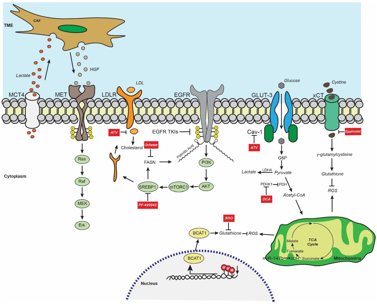
The use of epidermal growth factor receptor (EGFR) tyrosine kinase inhibitors (TKIs) as first-line treatment in patients with lung adenocarcinoma (LUAD) harboring EGFR-activating mutations has resulted in a dramatic improvement in the management of the disease. Lung cancer remains one of the most frequent and deadly types of cancer for both genders, while predictions about the future do not suggest dramatic improvements in incidence and mortality rates. There are different histological types of primary lung cancer, classified mainly into non-small-cell lung cancer (NSCLC) and small-cell lung cancer, with an annual incidence of approximately 85% and 15%, respectively. Lung adenocarcinoma (LUAD) is the most common subtype of NSCLC, accounting for around 40% of NSCLC cases.

  • lung adenocarcinoma
  • epidermal growth factor receptor
  • tyrosine kinase inhibitor
  • resistance
  • metabolism

1. Introduction

Lung cancer remains one of the most frequent and deadly types of cancer for both genders, while predictions about the future do not suggest dramatic improvements in incidence and mortality rates [1,2]. There are different histological types of primary lung cancer, classified mainly into non-small-cell lung cancer (NSCLC) and small-cell lung cancer, with an annual incidence of approximately 85% and 15%, respectively. Lung adenocarcinoma (LUAD) is the most common subtype of NSCLC, accounting for around 40% of NSCLC cases [3]. In 2004, the discovery of activating mutations in the gene encoding for epidermal growth factor receptor (EGFR) opened a new era for Lung adenocarcinoma (LUAD) treatment. EGFR mutations occur in nearly 50% of Asian and approximately 15% of Caucasian patients with LUAD [4][1]. Specifically, exon 19 deletions at codons 746–750 and L858R mutation in exon 21 account for approximately 90% of EGFR-sensitizing mutations. Several phase 3 clinical trials have consistently shown the superior efficacy of first-generation (e.g., gefitinib, erlotinib, icotinib) and second-generation (e.g., afatinib) EGFR tyrosine kinase inhibitors (TKIs) in comparison with standard first-line platinum-based chemotherapy in patients with LUAD harboring these EGFR-activating mutations [5,6,7,8,9][2][3][4][5][6]. Despite the robust clinical activity exerted by EGFR TKIs, the majority of patients develop resistance after an average period of 9–15 months. Acquired EGFR-dependent and EGFR-independent genetic alterations are known to be associated with resistance to EGFR TKIs. Among them, the secondary “gatekeeper” mutation EGFR T790M in exon 20 is the dominant resistance mechanism occurring in around 50% of patients treated with first- or second-generation TKIs [10,11][7][8]. Osimertinib is an irreversible third-generation EGFR TKI that is highly selective for both EGFR-activating mutations and the EGFR T790 mutation. In the FLAURA trial, osimertinib resulted in improved survival outcomes compared to first-generation EGFR TKIs, and consequently entered in clinical practice as standard therapy for treatment-naïve patients [12,13][9][10]. Unfortunately, similarly to patients treated with previous-generation EGFR TKIs, patients inevitably develop secondary resistance to osimertinib. In this setting, tertiary acquired mutations such as the EGFR C797S mutation in exon 20 are reported in only 7% of patients treated with first-line osimertinib [14,15][11][12]. In contrast, multiple co-existing molecular alterations such as KRAS mutations, MET amplification, small-cell transformation, and gene fusions have been observed in a considerable percentage of patients [16,17][13][14]. The heterogeneity and coexistence of multiple molecular alterations pose a significant challenge due to the paucity of post-osimertinib pharmacological options and underline the need of new alternative approaches in order to overcome resistance.

2. EGFR-Mutant LUAD Global Metabolic Profile

Physiologically, EGFR activation stimulates different signaling pathways such as PI3K/Akt and MEK/Erk that are tightly linked downstream with the regulation of a highly diverse set of metabolic pathways [22,23,24][15][16][17]. In the context of EGFR-mutant LUAD, the aberrant overactivation of EGFR signaling axis leads to increased glucose uptake, excessive lactate formation, upregulation of the pentose phosphate pathway (PPP), and de novo pyrimidine biosynthesis in EGFR-mutant in vitro models [25][18]. Moreover, the decisive role of EGFR signaling in reprogramming the metabolic landscape of EGFR-mutant LUAD has been demonstrated by treating gefitinib/erlotinib-sensitive (HCC827, PC9) and -resistant (H1975) EGFR-mutant LUAD cells. In particular, the treatment abrogated the activity of crucial metabolic pathways including glycolysis and PPP, downregulating effectively intermediate metabolites such as fructose 1,6-bisphosphate, lactate, and dihydroxyacetone phosphate in EGFR TKI-sensitive cells compared to resistant cells. Interestingly, quantification of these oncometabolites and establishment of cut-offs in the clinic might serve as auxiliary prediction biomarkers to EGFR TKI efficacy in patients with EGFR-mutant LUAD [25][18].

3. EGFR TKI Resistance and Glycolysis/Lactate Metabolism

Glycolysis is a multi-level metabolic process assigned to numerous enzymes. Pyruvate dehydrogenase complex (PDH) is a multienzyme complex that catalyzes the oxidative phosphorylation of pyruvate towards acetyl coenzyme A (Acetyl-CoA). However, phosphorylation of PDH by pyruvate dehydrogenase kinase 1 (PDHK1) inactivates it, resulting in the overproduction of lactate. Interestingly, mRNA levels of PDHK1 along with genes of multiple glycolytic enzymes have been found to be significantly higher in NSCLC compared to normal tissue [26][19]. Today, numerous metabolism-modulating agents are available, and some of them seem to exert an anti-cancer effect.
The tumor microenvironment (TME) is a mixture of active and dynamic components, including a plethora of metabolites that all interact to promote cancer cell survival and proliferation. In particular, the lactate secreted by EGFR TKI-resistant cells is engulfed by cancer-associated fibroblasts (CAFs), triggering the overproduction of hepatocyte growth factor (HGF) and the subsequent activation of MET signaling, suggesting the presence of a non-cell-autonomous metabolism-based mechanism of resistance to EGFR TKIs [28][20]. Erlotinib-resistant in vivo models were generated by subcutaneously injecting the EGFR exon 19 deletion-containing LUAD HCC827 cell line sensitive to EGFR TKIs. Interestingly, derived cell lines from these models were not resistant to erlotinib in vitro. The resistant phenotype was rescued when CAF-enriched conditioned media was cocultured. Importantly, monocarboxylate transporter 4 (MCT4), a lactate transporter and well-known readout of the Warburg effect, was found to be increased in resistant HCC827 cells compared to parental cells, confirming the metabolic rewiring towards aerobic glycolysis. Moreover, stromal HGF and MCT4 were upregulated in NSCLC samples progressed after gefitinib/erlotinib treatment, suggesting potential clinical relevance of the findings. Collectively, the implication of lactate metabolism and MET signaling in EGFR TKI resistance warrants additional research exploring the efficacy of combinatorial treatments with lactate/MET inhibitors and EGFR TKIs [28][20].

4. EGFR TKI Resistance and Fatty Acid Metabolism

In the clinical setting, the presence of EGFR-sensitizing mutations does not guarantee an equal survival benefit in all patients with EGFR-mutant LUAD. Therefore, and besides the inevitable resistance mechanisms, the high variability of response rate must be taken into consideration, demonstrating the urgent need for identifying the factors responsible for this reality. To that end, oneit studywas analyzed the serum metabolic profile of 44 patients with EGFR-mutant LUAD treated with icotinib and divided them into equal groups of good and poor responders, considering progression-free survival longer or shorter than 11 months [37][21]. Among 80 metabolites, a set of seven lipid factors based on their differential expression was able to distinguish good from poor responders. Specifically, lysophosphatidylcholine 16:1, lysophosphatidylcholine 22:5-1, and phosphatidylethanolamine 18:2 were upregulated in good responders, while ceramide 36:1-3, ceramide 38:1-3, sphingomyelin 36:1-2, and sphingomyelin 42:2 were elevated in poor responders [37][21].
Many non-oncological drugs have been repurposed for anti-cancer effects [38][22]. For example, statins are a class of cholesterol-lowering drugs widely prescribed to patients with hyperlipidemia. Intriguingly, there is growing evidence that statins augment the efficacy of EGFR TKIs in a synergistic manner [39,40][23][24]. Interestingly, exposure to gefitinib or erlotinib has been found to significantly downregulate and elevate cholesterol levels in EGFR-mutant sensitive (HCC827, PC9) and resistant (H1975) LUAD cell lines, respectively. The use of statins such as atorvastatin (ATV) has been demonstrated to abrogate the function of caveolin-1 (CAV-1), a factor that regulates cellular cholesterol homeostasis. Indeed, ATV abrogated the expression of CAV-1 and decreased the survival of EGFR-mutant LUAD cells by upregulating apoptosis. Interestingly, a combination of ATV with gefitinib significantly enhanced cell death compared to ATV alone. However, the addition of mevalonate, an intermediate metabolite of cholesterol formation, restricted the tumor cell suppression and reinduced the CAV-1 expression in ATV-treated cells, demonstrating the tight link between cholesterol and CAV-1 [30][25]. Glucose transporters 1 (GLUT-1) and 3 (GLUT-3) are the most upregulated transporters in cancer [41][26], and GLUT-3 is activated through EMT in NSCLC [42][27]. CAV-1 upregulated the expression of GLUT-3, then formed a heterodimer only in PC9-resistant cells increasing glucose uptake, while the EGFR TKI-resistant cells presented a higher dependency on CAV-1/GLUT-3-mediated glucose uptake for their survival compared to sensitive cells. Similarly, in vivo analysis has demonstrated that ATV-treated resistant xenograft models had decreased cholesterol levels inducing downregulation of the CAV-1/GLUT-3 axis, abrogating the glucose uptake, and resulting in lower tumor volumes [30][25].

5. EGFR TKI Resistance and Redox Homeostasis

The cells during physiological processes such as oxidative metabolism or response to cytokines or pathogen infection produce by-products of so-called reactive oxygen species (ROS). When ROS are accumulated without clearance, they induce cell death [48][28]. Moreover, EGFR-mutant LUAD enhances glycolysis to support cell survival. At a specific stage of glycolysis, LUAD cells exert an activating transcription factor 4-mediated metabolic rewiring, shifting glycolysis towards serine synthesis as demonstrated by upregulation of serine synthesis pathway metabolic enzymes such as phosphoserine phosphatase and phosphoglycerate dehydrogenase. Serine is a precursor of cystine which leads to glutathione (GSH) formation, which acts as an antioxidant and participates in ROS scavenging, inhibiting cell death [49][29]. Another study performed RNA sequencing analysis and immunoblotting in EGFR TKI-resistant HCC827 and H1975 models, identifying the upregulation of aldo-keto reductase family 1 member B1 (AKR1B1). Downregulation of AKR1B1 restored the sensitivity to osimertinib both in vitro and in vivo, while its ectopic overexpression rescued the resistant phenotype, revealing a crucial role of AKR1B1 in the occurrence of resistance [33][30]. Transcriptomic and metabolic comparisons of the models identified GSH metabolism as one of the most altered pathways. Specifically, resistant cells demonstrated an upregulation of major metabolites in the GSH de novo synthesis pathway, a finding which was confirmed both in vivo and in relapsed patients who had higher levels of GSH and oxidative GSH in blood compared to EGFR TKI-sensitive patients. Mechanistical studies demonstrated that AKR1B1 promotes the cystine uptake and consequent GSH de novo synthesis by interacting with and activating p-STAT3, which in turn activates cystine transporter SLC7A11/xCT. The elevated levels of GSH act as a major antioxidant, scavenging ROS and protecting tumor cells from treatment-induced stress. Disruption of AKR1B1, either genetically or due to epalrestat, an approved antidiabetic agent, inhibited the increase of GSH. The combination of epalrestat and either gefitinib or osimertinib therefore resulted in the restriction of tumor growth and delayed relapse by abrogating the axis of AKR1B1/p-STAT3/SLC7A11 in resistant models [33][30].
Figure 1. Overview of the metabolic remodeling in EGFR-mutant LUAD and the proposed treatment strategies for circumventing the EGFR TKI resistance. EGFR TKI-resistant LUAD cells secrete lactate, which is incorporated by CAFs located in TME, leading to overproduction of HGF ligands and subsequent activation of MET signaling and its downstream oncogenic effectors such as Ras/Raf/MEK/Erk signaling pathway. Moreover, the FASN-mediated EGFR palmitoylation allows the maintenance of EGFR constitutive signaling in EGFR TKI-resistant cells. Abrogation of palmitic acid synthesis using the anti-obesity drug orlistat (FASN inhibitor) promotes EGFR degradation. The use of statins such as ATV significantly downregulates the cholesterol levels, decreasing glucose uptake and lipogenesis simultaneously in EGFR-mutant LUAD. Further restriction of fatty acid synthesis by inhibiting SREBP1 using PF-429242 impairs both the levels of FASN and LDLR. Interestingly, blocking the PDHK1-mediated phosphorylation of PDH by treating EGFR-mutant LUAD cells with DCA shifts the glucose metabolism towards oxidative phosphorylation, preventing the accumulation of lactate, which has been correlated with EGFR TKI resistance. Furthermore, EGFR TKI-resistant cells have elevated levels of glutathione, the master antioxidant, thus efficiently scavenging ROS, escaping oxidative stress and cell death. Treatment with drugs such as epalrestat and BSO reduces glutathione synthesis, allowing ROS accumulation which eventually leads to ROS-mediated cell death in EGFR TKI-resistant models. Finally, epigenetic downregulation of SDH activity by miR-147b induces TCA cycle arrest and increased levels of succinate in EGFR TKI-tolerant cells. Abbreviations: CAF, cancer-associated fibroblast; TME, tumor microenvironment; MCT4, monocarboxylate 4; HGF, hepatocyte growth factor; LDLR, low-density lipoprotein receptor; LDL, low-density lipoprotein; EGFR, epidermal growth factor receptor; GLUT-3, glucose transporter 3; Cav-1, caveolin-1; G6P, glucose 6-phosphate; TCA cycle, tricarboxylic acid cycle; SDH, succinate dehydrogenase; ATV, atorvastatin; FASN, fatty acid synthase; PI3K, phosphoinositide 3-kinase; mTORC1, mTOR complex 1; ROS, reactive oxygen species; DCA; dichloroacetate; PDH, pyruvate dehydrogenase; PDHK1, pyruvate dehydrogenase kinase 1; Acetyl-CoA, acetyl coenzyme A; LDHA, lactate dehydrogenase A; SRBP1, sterol regulatory-element-binding protein 1; BSO, buthionine sulfoximine; BCAT1, branched-chain amino acid aminotransferase 1.

6. EGFR TKI Resistance and Metabolism-Mediated Stemness

The acquisition of a stem-cell-like phenotype is among the resistance mechanisms that EGFR-mutant LUAD cells develop to tolerate EGFR TKI treatment [54][31]. Indeed, Iroquois-class homeodomain protein 4 (IRX4), a factor that positively regulates pluripotency transcription factors such as NANOG, Sox2, and CD133, providing cells with stemness characteristics, was found to be significantly higher in PC9 gefitinib-resistant cells than PC9 parental cells [35][32]. Of note is the use of 1,25-dihydroxyvitamin D3 (1,25D), the biologically active form of vitamin D which activates the vitamin D receptor (VDR), which has been reported to abrogate a stemness phenotype in ovarian cancer cells [55][33]. Similarly, treating PC9-resistant cells with 1,25D increased VDR expression while significantly diminishing the levels of IRX4 and NANOG by negatively regulating the TGF-β1/SMAD3/IRX4/NANOG axis. Interestingly, a combination of 1,25D with gefitinib resulted in significantly higher efficacy of treatment both in in vitro and in vivo gefitinib-resistant models compared to single treatment. Collectively, impairing stem-like characteristics in EGFR TKI-resistant cells, as in this case by activating the 1,25D/VDR axis, led to resistance mitigation [35][32]. Moreover, preliminary findings suggested that a combination of osimertinib with a novel FASN inhibitor, AZ12756122, presented a synergistic effect overcoming osimertinib resistance in PC9 cells, likely by attenuating cancer stem cell features [36][34].

References

  1. Cancer Genome Atlas Research, N. Comprehensive molecular profiling of lung adenocarcinoma. Nature 2014, 511, 543–550.
  2. Mok, T.S.; Wu, Y.L.; Thongprasert, S.; Yang, C.H.; Chu, D.T.; Saijo, N.; Sunpaweravong, P.; Han, B.; Margono, B.; Ichinose, Y.; et al. Gefitinib or carboplatin-paclitaxel in pulmonary adenocarcinoma. N. Engl. J. Med. 2009, 361, 947–957.
  3. Douillard, J.Y.; Ostoros, G.; Cobo, M.; Ciuleanu, T.; McCormack, R.; Webster, A.; Milenkova, T. First-line gefitinib in Caucasian EGFR mutation-positive NSCLC patients: A phase-IV, open-label, single-arm study. Br. J. Cancer 2014, 110, 55–62.
  4. Rosell, R.; Carcereny, E.; Gervais, R.; Vergnenegre, A.; Massuti, B.; Felip, E.; Palmero, R.; Garcia-Gomez, R.; Pallares, C.; Sanchez, J.M.; et al. Erlotinib versus standard chemotherapy as first-line treatment for European patients with advanced EGFR mutation-positive non-small-cell lung cancer (EURTAC): A multicentre, open-label, randomised phase 3 trial. Lancet Oncol. 2012, 13, 239–246.
  5. Zhou, C.; Wu, Y.L.; Chen, G.; Feng, J.; Liu, X.Q.; Wang, C.; Zhang, S.; Wang, J.; Zhou, S.; Ren, S.; et al. Erlotinib versus chemotherapy as first-line treatment for patients with advanced EGFR mutation-positive non-small-cell lung cancer (OPTIMAL, CTONG-0802): A multicentre, open-label, randomised, phase 3 study. Lancet Oncol. 2011, 12, 735–742.
  6. Yang, J.C.; Wu, Y.L.; Schuler, M.; Sebastian, M.; Popat, S.; Yamamoto, N.; Zhou, C.; Hu, C.P.; O’Byrne, K.; Feng, J.; et al. Afatinib versus cisplatin-based chemotherapy for EGFR mutation-positive lung adenocarcinoma (LUX-Lung 3 and LUX-Lung 6): Analysis of overall survival data from two randomised, phase 3 trials. Lancet Oncol. 2015, 16, 141–151.
  7. Wu, S.G.; Shih, J.Y. Management of acquired resistance to EGFR TKI-targeted therapy in advanced non-small cell lung cancer. Mol. Cancer 2018, 17, 38.
  8. Yu, H.A.; Arcila, M.E.; Rekhtman, N.; Sima, C.S.; Zakowski, M.F.; Pao, W.; Kris, M.G.; Miller, V.A.; Ladanyi, M.; Riely, G.J. Analysis of tumor specimens at the time of acquired resistance to EGFR-TKI therapy in 155 patients with EGFR-mutant lung cancers. Clin. Cancer Res. 2013, 19, 2240–2247.
  9. Mok, T.S.; Wu, Y.L.; Ahn, M.J.; Garassino, M.C.; Kim, H.R.; Ramalingam, S.S.; Shepherd, F.A.; He, Y.; Akamatsu, H.; Theelen, W.S.; et al. Osimertinib or Platinum-Pemetrexed in EGFR T790M-Positive Lung Cancer. N. Engl. J. Med. 2017, 376, 629–640.
  10. Ramalingam, S.S.; Vansteenkiste, J.; Planchard, D.; Cho, B.C.; Gray, J.E.; Ohe, Y.; Zhou, C.; Reungwetwattana, T.; Cheng, Y.; Chewaskulyong, B.; et al. Overall Survival with Osimertinib in Untreated, EGFR-Mutated Advanced NSCLC. N. Engl. J. Med. 2020, 382, 41–50.
  11. Papadimitrakopoulou, V.; Wu, Y.L.; Han, J.Y.; Ahn, M.J.; Ramalingam, S.; John, T.; Okamoto, I.; Yang, J.C.H.; Bulusu, K.; Laus, G.; et al. LBA51Analysis of resistance mechanisms to osimertinib in patients with EGFR T790M advanced NSCLC from the AURA3 study. Ann. Oncol. 2018, 29, viii741.
  12. Oxnard, G.R.; Hu, Y.; Mileham, K.F.; Husain, H.; Costa, D.B.; Tracy, P.; Feeney, N.; Sholl, L.M.; Dahlberg, S.E.; Redig, A.J.; et al. Assessment of Resistance Mechanisms and Clinical Implications in Patients With EGFR T790M-Positive Lung Cancer and Acquired Resistance to Osimertinib. JAMA Oncol. 2018, 4, 1527–1534.
  13. Schmid, S.; Li, J.J.N.; Leighl, N.B. Mechanisms of osimertinib resistance and emerging treatment options. Lung Cancer 2020, 147, 123–129.
  14. Du, X.; Yang, B.; An, Q.; Assaraf, Y.G.; Cao, X.; Xia, J. Acquired resistance to third-generation EGFR-TKIs and emerging next-generation EGFR inhibitors. Innovation 2021, 2, 100103.
  15. Wee, P.; Wang, Z. Epidermal Growth Factor Receptor Cell Proliferation Signaling Pathways. Cancers 2017, 9, 52.
  16. Hoxhaj, G.; Manning, B.D. The PI3K-AKT network at the interface of oncogenic signalling and cancer metabolism. Nat. Rev. Cancer 2020, 20, 74–88.
  17. Papa, S.; Choy, P.M.; Bubici, C. The ERK and JNK pathways in the regulation of metabolic reprogramming. Oncogene 2019, 38, 2223–2240.
  18. Makinoshima, H.; Takita, M.; Matsumoto, S.; Yagishita, A.; Owada, S.; Esumi, H.; Tsuchihara, K. Epidermal growth factor receptor (EGFR) signaling regulates global metabolic pathways in EGFR-mutated lung adenocarcinoma. J. Biol. Chem. 2014, 289, 20813–20823.
  19. Dyrstad, S.E.; Lotsberg, M.L.; Tan, T.Z.; Pettersen, I.K.N.; Hjellbrekke, S.; Tusubira, D.; Engelsen, A.S.T.; Daubon, T.; Mourier, A.; Thiery, J.P.; et al. Blocking Aerobic Glycolysis by Targeting Pyruvate Dehydrogenase Kinase in Combination with EGFR TKI and Ionizing Radiation Increases Therapeutic Effect in Non-Small Cell Lung Cancer Cells. Cancers 2021, 13, 941.
  20. Apicella, M.; Giannoni, E.; Fiore, S.; Ferrari, K.J.; Fernandez-Perez, D.; Isella, C.; Granchi, C.; Minutolo, F.; Sottile, A.; Comoglio, P.M.; et al. Increased Lactate Secretion by Cancer Cells Sustains Non-cell-autonomous Adaptive Resistance to MET and EGFR Targeted Therapies. Cell Metab. 2018, 28, 848–865.e6.
  21. Han, X.; Luo, R.; Wang, L.; Zhang, L.; Wang, T.; Zhao, Y.; Xiao, S.; Qiao, N.; Xu, C.; Ding, L.; et al. Potential predictive value of serum targeted metabolites and concurrently mutated genes for EGFR-TKI therapeutic efficacy in lung adenocarcinoma patients with EGFR sensitizing mutations. Am. J. Cancer Res. 2020, 10, 4266–4286.
  22. Corsello, S.M.; Nagari, R.T.; Spangler, R.D.; Rossen, J.; Kocak, M.; Bryan, J.G.; Humeidi, R.; Peck, D.; Wu, X.; Tang, A.A.; et al. Discovering the anti-cancer potential of non-oncology drugs by systematic viability profiling. Nat. Cancer 2020, 1, 235–248.
  23. Hung, M.S.; Chen, I.C.; Lee, C.P.; Huang, R.J.; Chen, P.C.; Tsai, Y.H.; Yang, Y.H. Statin improves survival in patients with EGFR-TKI lung cancer: A nationwide population-based study. PLoS ONE 2017, 12, e0171137.
  24. Nguyen, P.A.; Chang, C.C.; Galvin, C.J.; Wang, Y.C.; An, S.Y.; Huang, C.W.; Wang, Y.H.; Hsu, M.H.; Li, Y.J.; Yang, H.C. Statins use and its impact in EGFR-TKIs resistance to prolong the survival of lung cancer patients: A Cancer registry cohort study in Taiwan. Cancer Sci. 2020, 111, 2965–2973.
  25. Ali, A.; Levantini, E.; Fhu, C.W.; Teo, J.T.; Clohessy, J.G.; Goggi, J.L.; Wu, C.S.; Chen, L.; Chin, T.M.; Tenen, D.G. CAV—GLUT3 signaling is important for cellular energy and can be targeted by Atorvastatin in Non-Small Cell Lung Cancer. Theranostics 2019, 9, 6157–6174.
  26. Martinez, C.A.; Scafoglio, C. Heterogeneity of Glucose Transport in Lung Cancer. Biomolecules 2020, 10, 868.
  27. Masin, M.; Vazquez, J.; Rossi, S.; Groeneveld, S.; Samson, N.; Schwalie, P.C.; Deplancke, B.; Frawley, L.E.; Gouttenoire, J.; Moradpour, D.; et al. GLUT3 is induced during epithelial-mesenchymal transition and promotes tumor cell proliferation in non-small cell lung cancer. Cancer Metab. 2014, 2, 11.
  28. Schumacker, P.T. Reactive oxygen species in cancer cells: Live by the sword, die by the sword. Cancer Cell 2006, 10, 175–176.
  29. Jin, N.; Bi, A.; Lan, X.; Xu, J.; Wang, X.; Liu, Y.; Wang, T.; Tang, S.; Zeng, H.; Chen, Z.; et al. Identification of metabolic vulnerabilities of receptor tyrosine kinases-driven cancer. Nat. Commun. 2019, 10, 2701.
  30. Zhang, K.R.; Zhang, Y.F.; Lei, H.M.; Tang, Y.B.; Ma, C.S.; Lv, Q.M.; Wang, S.Y.; Lu, L.M.; Shen, Y.; Chen, H.Z.; et al. Targeting AKR1B1 inhibits glutathione de novo synthesis to overcome acquired resistance to EGFR-targeted therapy in lung cancer. Sci. Transl. Med. 2021, 13, eabg6428.
  31. Hashida, S.; Yamamoto, H.; Shien, K.; Miyoshi, Y.; Ohtsuka, T.; Suzawa, K.; Watanabe, M.; Maki, Y.; Soh, J.; Asano, H.; et al. Acquisition of cancer stem cell-like properties in non-small cell lung cancer with acquired resistance to afatinib. Cancer Sci. 2015, 106, 1377–1384.
  32. Jia, Z.; Zhang, Y.; Yan, A.; Wang, M.; Han, Q.; Wang, K.; Wang, J.; Qiao, C.; Pan, Z.; Chen, C.; et al. 1,25-dihydroxyvitamin D3 signaling-induced decreases in IRX4 inhibits NANOG-mediated cancer stem-like properties and gefitinib resistance in NSCLC cells. Cell Death Dis. 2020, 11, 670.
  33. Ji, M.; Liu, L.; Hou, Y.; Li, B. 1alpha,25Dihydroxyvitamin D3 restrains stem celllike properties of ovarian cancer cells by enhancing vitamin D receptor and suppressing CD44. Oncol. Rep. 2019, 41, 3393–3403.
  34. Polonio, E.; Palomeras, S.; Porta-Balanya, R.; Bosch-Barrera, J.; Vásquez, C.A.; Ciurana, J.; Ruiz-Martínez, S.; Puig, T. 3P AZ12756122, a novel fatty acid synthase (FASN) inhibitor, reduces resistance properties in gefitinib- and osimertinib-resistant EGFR-mutated non-small cell lung cancer models. J. Thorac. Oncol. 2021, 16, S700.
More
ScholarVision Creations