DOX Delivery Systems: Comparison
Please note this is a comparison between Version 2 by Rita Xu and Version 1 by Waad Hussein Abuwatfa.

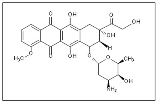
Doxorubicin (DOX) is one of the most widely used anthracycline anticancer drugs due to its high efficacy and evident antitumoral activity on several cancer types. However, its effective utilization is hindered by the adverse side effects associated with its administration, the detriment to the patients’ quality of life, and general toxicity to healthy fast-dividing cells. Thus, delivering DOX to the tumor site encapsulated inside nanocarrier-based systems (like liposomes, micelles, metal-organic frameworks (MOFs)) is an area of research that has garnered colossal interest in targeted medicine. Nanoparticles can be used as vehicles for the localized delivery and release of DOX, decreasing the effects on neighboring healthy cells and providing more control over the drug’s release and distribution.

  • doxorubicin
  • liposomes
  • micelles
  • metal-organic frameworks (MOFs)

1. Introduction

Cancer refers to uncontrolled cell division due to DNA mutations. It occurs when cells are induced to over-proliferate, caused by the permanent activation of proto-oncogenes in upregulated oncogenes [1]. Throughout their normal life cycle, cells divide controllably, differentiate, and eventually die through the programmed cell death mechanism known as apoptosis. However, mutations perturbing the ordinary growth pathway of the cells can introduce behavioral changes on genetic and epigenetic levels, where these transformed cells exhibit different growth characteristics, including enhanced mobility and the decreased contact inhibition of growth mechanisms [2]. Some cancer cells incur tumor-initiating abilities and can travel to other parts of the body through the bloodstream or lymph vessels (metastasis) [3].
Anticancer drugs are classified into three categories: chemotherapy, immunotherapy, and hormonal therapy. These therapies can target tumor cells at the DNA, RNA, or protein level [4,5,6][4][5][6]. Chemotherapy is the invasive anticancer therapeutic regimen used worldwide. It was introduced by the German chemist Paul Ehrlich and has been used to treat cancer since the beginning of the 20th century [7,8,9][7][8][9].
The formulations of antineoplastic agents were divided, according to their chemical structure and mechanism of action, into alkylating agents, antibiotics, antimetabolites, topoisomerase inhibitors, and others [5,6,7,10,11,12][5][6][7][10][11][12]. The last decades have witnessed colossal efforts to discover novel chemotherapeutic drugs including, but not limited to, doxorubicin, epirubicin, pirarubicin, methotrexate, pemetrexed, 5-fluorouracil, paclitaxel, cisplatin, gemcitabine, and others [3,10,13][3][10][13]. Chemotherapeutics are usually administered either orally or intravenously to achieve systemic distribution, thus maximizing their effect. Unfortunately, the practicality of chemotherapy drugs is counteracted by their lack of selectivity, causing grave damage to healthy cells with high division rates like bone marrow, hair follicles, and gastrointestinal epithelia. In addition, both spermatogenesis and oogenesis processes are highly susceptible to the cytotoxic side effects of chemotherapeutics.
Although surgery and radiotherapy are ideal for treating localized solid cancers, their successful use is hampered in many cases where cancer has metastasized to other organs in the body. Thus, chemotherapy is commonly offered to patients at advanced cancer stages. It is a systemic therapy that targets rapidly dividing cells by employing agents that interfere with their life cycle and replication mechanisms. It is advantageous over conventional therapies because it can act throughout the whole body, thus targeting cancer cells that metastatically spread far from the primary tumor site and helping to shrink bulky tumors that might otherwise be unresectable [14]. From the 1960s onwards, the success of chemotherapeutic drugs in treating various advanced cancers led to the development of the so-called adjuvant therapy, in which chemotherapeutics are combined with surgery to eradicate potential cancer dissemination or recurrence [15,16][15][16]. Sometimes, chemotherapy combined with radiotherapy is more effective than either modality alone.
Among the most widely used class of chemotherapeutics are anthracycline agents, which are prevalently successful in achieving short-time cancer progression inhibition, high response, and improved survival rates [17]. Anthracyclines are the first anti-tumor antibiotics approved by the Food and Drug Administration (FDA), among which is Doxorubicin (DOX), one of the first effective anthracycline antibiotic cytotoxic drugs discovered. It was isolated from the red pigment-producing soil bacterium; Streptomyces peucetius via mutagenic treatment [18]. DOX (Figure 1) is extensively used to treat lymphomas, leukemia, as well as ovarian, breast, small cell lung, stomach, and liver cancers [4,7,19,20][4][7][19][20].
Figure 1. The chemical structure of DOX [7].
Like most anthracycline antibiotics, DOX oxidizes and inhibits the activity of topoisomerase II, an enzyme responsible for DNA transcription and replication, generating unstable, highly active free radicals that damage the DNA and cause cell death [4,7,19][4][7][19]. Although it is an effective anti-tumor agent, its side effects are most evident on cells exhibiting high division rates, such as hair follicles and the gastrointestinal tract lining; thus, hair loss, digestive tract ulcerations, vomiting, nausea, and diarrhea are all common complications/side effects [21]. Also, it has been known to stimulate myelosuppression and induce cardiotoxicity by the upregulation of apoptosis receptors in cardiomyocytes [22,23][22][23]. Clinical investigations by Von Hoff et al. [24] showed 7.5% cardiomyopathy occurrences associated with cumulative DOX doses of 550 mg/m2. Thus, cumulative doses of 400–500 mg/m2 are generally employed for free administration once every three weeks to prevent cardiotoxic effects. In addition, DOX is a vesicant and can cause blistering and necrosis if extravasation occurs at the time of drug administration [14]. Moreover, drug resistance in the tumor cells is another problem that limits the clinical use of DOX [19,25,26][19][25][26]. In general, the toxicity of chemotherapeutic drugs limits their effective dosage threshold. In most cases, a therapeutic dose of the chemotherapy drugs capable of efficiently killing the tumor cells is highly toxic to the fast-growing normal cell and, therefore, cannot be given to patients (Figure 2). Instead, a lower dosage capable of destroying considerable numbers of tumor cells with lower toxic effects on normal cells is utilized [14].
Figure 2. Dose responsive curve for chemotherapy drugs kills both tumor cells and healthy cells. The therapeutic dose (A) is close to the toxic dose (B). It is not safe to give this drug at a therapeutic dose. A safe dose (C) is chosen for administration to patients. Reproduced with permission from [14], Elsevier, 2008.

2. Nanoparticles as Drug Delivery Systems (DDS)

Due to the detriment to the patients’ quality of life and the potential lethality of some of the side effects associated with conventional chemotherapy, novel drug delivery systems (DDS) aim to reduce the adverse side effects and enhance the specificity of chemotherapeutic drugs. In 1964, Cheng initiated the use of encapsulation of enzymes and other biologically active materials in semipermeable vesicles and tested their use to suppress the growth of lymphosarcoma in a mice model [28,29][27][28]. This approach has evolved and been extensively extended into nanomedicine, biotherapeutics, blood substitutes, drug delivery, enzyme/gene therapy, cancer therapy, nanoparticles, liposomes, bioencapsulation, regenerative medicine, nanobiotechnology, and nanotechnology [30][29]. Earlier studies on the transformed cancer cells showed that they had adopted many biochemical strategies to support their uncontrolled growth [31,32][30][31]. A deep understanding of the key enzymes and antagonistic pathways of synthesis and catabolism involved in tumor progression resulted in the development of “targeted therapy”. Targeted DDS are nanoplatforms that incorporate nanoparticles (NPs) as drug delivery vehicles, ranging in size from 1 nm to 1000 nm [33,34][32][33]. These include, but are not limited to, gold NPs, dendrimers, polymeric nanogels, micelles, metal-organic frameworks (MOFs), liposomes, and quantum dots. The synthesis routes of these NPs vary and are generally divided into chemical and biological ways, where the latter are preferred as they are safer and innocuous [35][34].
Utilizing these nanocarriers for the remote delivery of appropriate dosages to targeted anatomical sites under controlled release conditions can overcome the shortcomings of traditional treatment approaches. The localized accumulation of the nanoparticles at the neoplastic site is mainly achieved by passive and/or active targeting routes. In passive targeting, the intrinsic features of the tumor neovasculature beneficially provide fenestrations where the NPs accumulate. As a tumor undergoes rapid, chaotic growth, it exhibits a disorganized vascular network and becomes hypoxic due to insufficient oxygen supply. Tumor cells can secrete growth factors to induce vascularization to resolve this hypoxia and get nutrients from neighboring healthy cells by a process referred to as angiogenesis [35,36][34][35]. The newly formed capillitial endothelium in tumor tissues is disorganized and leaky with improper lymphatic drainage and inadequate transport phenomena mechanisms (Figure 3) [37][36]. Consequently, the tumor site suffers from abnormal molecular and fluid dynamics. These features allow the NPs to extravasate into the tumor’s interstitium [38][37] and accumulate inside the diseased tissues, in a phenomenon referred to as the enhanced permeability and retention (EPR) effect.
Figure 3. (A) a normal vascular network where the vessels are parallel-aligned next to each other; (B) a tumoral vasculature with chaotic defective arrangement. Adapted with permission from [37][36], Oxford University Press, 2006.
Yet, designing DDS with complete dependence on passive targeting has significant limitations, such as the possible accumulation of the NPs in the spleen and liver as these organs have fenestrated vasculature as well as the inability of the NPs to sufficiently penetrate deep enough through the complex tumoral network due to heterogeneities in structure [39][38]. Thus, the development of systems that incorporate both passive and active targeting mechanisms became imperative. Since tumor cells overexpress receptors that participate in growth and survival pathways, such receptors make promising active targets. To this end, nanocarriers could be conjugated to natural ligands to target these receptors leading to their accumulation and internalization by the cancer cells [40][39].
This review will discuss three types of nanocarriers: liposomes, micelles, and metal-organic frameworks (MOFs) delivering the antineoplastic agent DOX to treat different types of solid cancers, including their DOX loading techniques, encapsulation efficiency, and their ability to eradicate tumors.

3. DOX Delivery Systems Based on Liposomes

Bangham and co-workers used multilamellar bilayer lipid microspheres and liposomes as models of biological membranes for basic research in the 1960s [41,42][40][41]. It was Gregoriadis’ group that first used liposomes as drug carriers in the 1970s [43][42]. Later, the multilamellar onion-like microspheres were developed into submicron dimension bilayer lipid vesicles (Figure 4). The hydrophilic heads of the phospholipids are directed outwards, whereas the hydrophobic tails are directed inwards. Cholesterol is usually added to the phospholipids to increase the mechanical rigidity of liposomes [44,45][43][44]. Liposomes have gained immense interest as drug carriers because of their biocompatible, biodegradable, non-toxic, and targetable nature. Moreover, they deliver a high drug-to-lipid ratio and can also be functionalized to avoid rapid elimination from the body [46][45].
Figure 4. Structure of liposomes. Reproduced from [46][45], IntechOpen, 2014.
For the encapsulation of lipophilic DOX into liposomes, two loading routes (i.e., passive and active) are generally used (Figure 5). One of the most common passive loading methods is based on the thin-film hydration method, where the drug solution is added during the liposome formation process, either while the film is being formed or during the hydration step. To enhance trapping efficiency, the drug is typically added during the film formation of liposomes containing negatively charged (acidic) lipids [44,45][43][44]. The dried lipids are initially randomly orientated, but upon exposure to an aqueous solution, the water causes the molecules to self-organize into a bilayer configuration to minimize entropic interactions. In the presence of hydrophilic and/or hydrophobic drugs in solution, some of the drug molecules would passively partition and accumulate in the cores and/or the shells of the liposomes. Although this method is simple, it yields very small drug encapsulation percentages. In 1983, DOX was passively entrapped in the bi-layer of positively (phosphatidylcholine-cholesterol-stearyl amine) and negatively (phosphatidylcholine-cholesterol-phosphatidylserine) charged liposomes. The drug was added to the phospholipids before film formation [47][46]. One day after DOX loading, the mean diameters of positive and negative non-filtered multilamellar liposomes were between 500-600 nm and 1 μm, respectively. The maximum loading capacities ranged between 60-75 mmol DOX/mol phospholipid for the negative and approximately 55 mmol DOX/mol phospholipid for the positive, non-filtered, non-sonicated liposomes [47][46].
Figure 5. Two approaches for DOX loading into liposomes: passive and active. (A) before “pasive” and (B) before “active”.
On the other hand, the size stability and release profiles were determined over an extended period. The filtered and sonicated/centrifuged negatively-charged liposomes were stable during the entire storage period and maintained average diameters of 270 and 120 nm, respectively. However, the sonicated/centrifuged positively-charged liposomes were not stable and had a size of 120 nm with a standard deviation of 40 nm over 2 weeks of storage. The negative filtered liposomes released only 10% of DOX during 75 days, whereas 40% was released over 20 days from the negative sonicated/centrifuged liposomes. For the release from positive sonicated/centrifuged liposomes, the baseline was set after 12 days, and the cumulative release was ~30% after 48 days [47][46].
Other passive loading methods include the freezing-and-thawing technique (FAT). The liposomal solution undergoes a series of freezing and thawing processes, causing the interlayer distance within the liposomes to increase [48][47]. As a result, ice crystals cause transient holes and pores to form within the liposomal structures, allowing for passive drug entrapment. However, this method yields 5–20% loading efficiency and requires heavy post-processing to remove the excess drug. Likewise, the reverse-evaporation method can load drugs with efficiencies up to 50%. Still, the formulations lack reliable in vivo drug retention and often suffer from rapid cargo release under physiological conditions. Thus, occurrences of “burst release”, where a large fraction of the entrapped drug is abruptly released due to poor association and retention in the vesicles, combined with the relatively low drug-to-lipid entrapment ratio, typically do not exceed 0.05% (w/w), making it imperative to divert to other loading techniques [48][47]. Moreover, the high variability and dependence of the encapsulation efficiency on operational factors like the drug’s solubility, nanocarriers’ chemistry, size, and preparation method deemed active loading methods more preferable and more commonly used for the stable entrapment of DOX into liposomes.
On the other hand, active methods, also known as remote loading methods, employ pH gradients between the internal acidic core buffer of blank liposomes and the external buffer containing DOX at neutral physiological conditions. Under such conditions, the drug molecules incubated with the formed liposomes can permeate selectively through the lipids transmembrane, where they become protonated intra-vesicularly [45][44]. Work by Bally and colleagues [49][48] investigated the effects of creating a membrane potential by altering the pH of the intraliposomal solution from a 300 mM citrate buffer at pH 4.0 to a HEPES buffer at pH 7.4 on the entrapment of DOX and biogenic amines in liposomes. High encapsulation efficiencies of up to 98% were achieved, with a drug-to-lipid ratio of 0.3 (w/w%) corresponding to 400 nmol DOX/µmol phospholipid (~260 mM internal concentration). Also, retention times (T50) significantly increased from 1 h to 30 h in passively versus actively DOX-loaded formulations, respectively. The proposed “citrate” method resulted in highly stable liposomes with remarkably enhanced drug retention and release characteristics, with the added benefit of cost-effectiveness. A commercially available formulation of liposomal DOX, Myocet®, is loaded using the same approach (Figure 6). The Myocet® liposomes (~150 nm) are made from egg phosphatidylcholine and cholesterol at a ratio of 55/45 (mole/mole %). As a result of the pH gradient, DOX molecules diffuse and accumulate in the liposomal cores and form a complex with the citrate anions. The drug entrapment efficiencies (>95%) and drug-to-lipid ratio (~0.27) exceeded the theoretical predictions as the bundled fibers complexes allowed higher DOX loading beyond its solution solubility limits. The DOX-citrate complexes resembled the appearance of coffee-bean liposomes, where the linear bundles prevented drug leakage and premature release. The formulation also exhibited prolonged circulation times and improved in vivo tissue distribution compared to free DOX [50][49]. According to Li et al. [51][50], 99% of DOX loaded into citrate buffered liposomes appears in the form of fiber aggregates. The threshold for forming these fibrous-like DOX-citrate structures can be as low as ~20 mM internal DOX concentration. As the DOX concentration increases, the fibers begin cross-linking via citrates and then pack into bundles. Kanter and co-workers [52][51] demonstrated that the Myocet liposomal formulation effectively reduced the effects of myocardial degeneration, commonly associated with anthracycline-based treatments. Histological analysis on beagle dogs treated with conventional DOX versus those treated with Myocet (single i.v. administration every 3 weeks; 8 cycles; cumulative dose = 12 mg/kg) showed that the latter group did not suffer from any anthracycline-induced cardiotoxicity, while the first group had lesions of myocardial degeneration.
Figure 6. (A) DOX loading via citrate transmembrane gradient method, (B,C) confocal images of DOX-loaded liposomes showing the coffee-beans liposomes appearance where DOX-citrate complexes appear in rod, circular and U-shaped structures. A,B are adapted with permission from [50][49], Elsevier, 2001. C is adapted with permission from [51][50], Elsevier, 1998.
Along the same lines, Haran et al. [53][52] generated a pH gradient across the liposomes using ammonium sulfate salt. The liposomes initially encapsulated a 300 mM ammonium sulfate solution at pH 5.5 in a pH 7.4 external buffer. The higher concentration of ammonium ions on the inside led to the diffusion of the neutral ammonia molecules, and with every molecule diffusing from the core, a proton was left behind. Due to salting-out effects and the acidification of the intraliposomal compartment, high fractions of DOX can accumulate in the liposomes in an aggregated form. Liposomes loaded using this technique exhibit a prolonged stable storage period beyond 6 months because of the gelation effects of DOX with the sulfate salt, which inhibit membrane re-permeation. Alyane et al. [54][53] examined the stability and release behavior of liposomes loaded with DOX via the ammonium sulfate transmembrane method. Liposomes comprised of hydrogenated egg yolk phosphatidylcholine (HEPS), cholesterol, and DSPE-PEG2000 at a molar ratio of 185:1:15 showed an encapsulation efficiency exceeding 90%, with a drug-to-lipid ratio of 1:20 (w/w). Upon incubation at 37 °C for 24 h in culture media and PBS, the liposomes retained 98% and 90% of the encapsulated drug, respectively, showing minimal leakage under the stated conditions. When the incubation medium was changed to phosphate buffer at pH 5.3, the maximum release achieved was 37%. The pH responsiveness of the liposomes suggested their suitability for controlled release in acidic compartments (i.e., tumor tissues) as they would keep their cargo intact under physiological conditions. The study also revealed that encapsulating DOX in the liposomes decreased the agent’s uptake by rat myocardial H9C2 cells, suggesting the decreased cardiac toxicity of the formulation. Flow cytometry and MTT assay analysis showed that the toxic effects of DOX were reduced when compared against that of free DOX, up to 20 h of incubation time. After 20 h, the liposomal formulation became more toxic to the cells than the free one [54][53].
DOXIL® is a commercially available PEGylated liposomal DOX formulation that uses the ammonium sulfate gradient method to encapsulate the drug. An in vivo study by Sakakibara et al. [55][54] investigated the performance of DOXIL®, conventional DOX-liposomes, and free DOX on the treatment of human lung tumors. The DOXIL® liposomes were composed of hydrogenated soy phosphatidylcholine, cholesterol, PEG-DSPE, and dl-a-tocopherol in a molar ratio of 56.1:38.2:5.5:0.2. In contrast, the non-PEGylated liposomes had phosphatidylglycerol, phosphatidylcholine, cholesterol, and DL- α -tocopherol in a molar ratio of 1:4:3:0.02. To assess and compare the different treatment groups (dosage: 1.5 mg/kg) of anti-tumor activity, human lung tumor xenografts were engrafted into the gonadal fat pad of severe combined immunodeficient (SCID) mice. The tumor eventually metastasized from the primary site into the mice’s peritoneal cavity (i.e., liver, lung). It was concluded that the PEGylated formulation successfully suppressed the primary tumor growth and arrested metastasis in the peritoneal cavity, while the free DOX was only able to stop the growth of the primary tumor without significant effects on preventing the spread. The PEGylated liposomes showed 5-folds higher circulation times than conventional liposomes, while the uptake by vital organs like the spleen was reduced. Also, a significant increase in extravasation and accumulation of the PEGylated formulation was observed, as considerable amounts were detectable at the tumors after 1 week following administration [55][54]. Another study [56][55] compared the in vivo pharmacokinetic performance of free DOX with PEGylated and non-PEGylated liposomal formulations. Interestingly, the PEGylated liposomal DOX clearance rate decreased by 100-folds (Cl = 0.023 L/h), and its half-life (t1/2 = 83.7 h) was prolonged by 8-folds, compared to free DOX (Cl = 25.3 L/h, t1/2 = 10.4 h). Moreover, the distribution volume decreased significantly from 364 L to 139 L to 3.0 L in the free DOX, non-PEGylated, and PEGylated liposomal DOX, respectively. This conclusion demonstrated that PEGylation prevents premature drug release and that most of it remains entrapped without leakage.
Fritze et al. [57][56] introduced another DOX remote loading method based on a phosphate ((NH4)2HPO4) transmembrane gradient, using different ammonium and sodium salts. The liposomes were composed of egg phosphatidylcholine (EPC) and cholesterol in the molar ratio 7:3. After hydrating the liposomes with 300 mM salt solutions at neutral pH, they were incubated with DOX.HCl for 12 h at 7 °C to achieve a drug-to-lipids ratio of 1:3 (mol/mol). The sizes of liposomes containing DOX dissolved in different ammonium and sodium salts, as well as their encapsulation efficiencies (EE%), are summarized in Table 1. Results showed that loading did not significantly alter the size of the liposomes, and those loaded with the ammonium salts gradients showed higher EE than the sodium salts. Synergistic effects are suggested when using ammonium salts as they act as a reservoir for donating free protons when DOX is internalized and protonated in the acidic interior of the liposomes. Thus, intraliposomal protonation and DOX precipitation led to increased encapsulation efficiencies. Further analysis was conducted to assess the impact of varying the intravesicular ammonium concentration on liposome size and EE. DOX was dissolved in 10 mM isotonic HEPES buffered saline (HBS), 50, 100, 200, and 300 mM ammonium phosphate at pH 7.2. The sizes of the DOX-loaded liposomes were found to be 84 ± 0.4, 102 ± 1.4, 114 ± 3.6, 95 ± 1.5, and 92 ± 1.6 nm, respectively, with EE of 2.81, 23.52, 61.03, 83.35, 97.98%. Increasing the ammonium ion concentration augmented the effects of drug protonation and intravesicular acidification, which led to increased DOX diffusion across the transmembrane gradient. The least efficiency (<5%) was observed when incubating in HBS, which contains no phosphate ions (no decrease in the intraliposomal pH level), while the highest efficiency approaching 100% was observed at the highest concentration of the ammonium phosphate (300 mM).
Table 1. Summary of the size and encapsulation efficiency of loaded liposomes achieved via different salts.
Salt Gradient Size ± SD (nm) EE (%)
Ammonium Phosphate 129.3 ± 3.7 98
Ammonium Sulfate 129.2 ± 2.9 95
]. Benefiting from the tumor cells’ overexpression of receptors to aid in augmenting their growth and survival pathways, such receptors make promising active targets. To this end, nanocarriers can be conjugated to the ligands complementing these receptors to enhance DOX accumulation and internalization by the cancerous cells. Xing and co-workers [63][62] examined the in vitro effects of functionalizing DOX-loaded PEGylated liposomes with a DNA aptamer (Apt-Urn liposomes) on MCF-7 cells. Flow cytometry results showed a 6.6-fold increase in drug uptake and efficacy of the functionalized loaded liposomes as opposed to the conventional ones, as the fluorescence response upon 4 h of incubation with the treatments corresponded to 93.6% and 57.0%, in the cells treated with Apt-Urn and control liposomes, respectively. Moreover, MTT assay results analyzed the cytotoxic effects of the Apt-Urn liposomes on cell viability. The cells were treated with different liposomal concentrations followed by an incubation period of 6 h, then re-cultured in fresh media, and the MTT test was done after 72 h. At a liposomal DOX concentration of 500 nM, cells treated with the functionalized liposomes exhibited a viability of 57.0 ± 6%, whereas cells treated with the control liposomes exhibited a viability of 92.4 ± 9%, evidencing the superior localization and internalization of the nanocarriers as a function of surface modification.
Yang [64][63] conducted a recent preclinical investigation on DOX-loaded liposomes composed of EPC and cholesterol for treating head and neck cancer. The study compared three treatment groups: free DOX, DOX-loaded liposomes, and DOX-loaded peptide-conjugated liposomes. Results showed the dependence of the encapsulation efficiency on the cholesterol content in the formulations, as it increased from 20% to 79% upon the incorporation of cholesterol with EPC. Varying the EPC and cholesterol ratio yielded different entrapment efficiencies ranging from ~48% to ~82%. Both liposomal formulations, the functionalized and the conventional ones, showed sustained release profiles over a period of 40 h, but the DOX-loaded peptide-conjugated liposomes had a faster in vitro release behavior. In vivo analysis of the formulations’ efficacy was carried out on nude mice bearing HSCC (human squamous cell carcinomas) xenograft. The tumor progression was suppressed when treated with both liposomal formulations; however, the functionalized liposomes arrested metastasis and increased the median survival time of the animals in that group by 100%. The enhanced performance was due to the increased accumulation at the tumor site, resorted to localized targeting by binding to the overexpressed surface-specific receptors (Hsp47/CBP2) abundant on the head and neck cancer cells.
Another study [65][64] examined the performance of DOX-loaded liposomes, functionalized with the monoclonal antibody Trastuzumab (TRA) for the enhanced targeting of breast cancer cells overexpressing the Human Epidermal growth factor Receptor 2 (HER2). The liposomes consisted of cholesterol, DPPC, and DSPE-PEG(2000)-NH2 at a molar ratio of 30:65:5, respectively. DOX was loaded via the ammonium sulfate transmembrane gradient method, yielding a drug-to-lipids ratio of 1:6. The cargo release from the liposomal formulations, conventional and TRA-conjugated, was triggered using pulsed ultrasound. In vitro results showed synergistic effects on the DOX uptake and internalization upon sonication and surface modification with TRA. Similarly, Chowdhury and co-workers [66][65] designed an in vitro study where they tested different targeted (functionalized with Aptamer-A6) and untargeted liposomal-DOX formulations for the localized treatment of HER2-overexpressing breast cancers. Twelve liposomal formulations (F1-F12) were prepared using the thin-film hydration method by varying the compositions of the phospholipid mixtures. The liposome sizes slightly increased upon DOX loading and ranged from 98.7 to 181.2 nm, with 74.9 to 94.1% entrapment efficiencies. The optimized formulations for further complexation with Aptamer-A6 were determined based on the smallest size to benefit from the EPR effect and the highest encapsulation efficiency. Formulations 5 and 8 (10 mg, 150 mg, 40 mg, 40 mg, 20 mg, and 0.25 mg of DOX, POPC, DOTAP, DOPE, DSPE-mPEG2000, and Mal-PEG, respectively) had sizes of 101.70 ± 14.04 nm and 98.7 ± 13.25 nm, respectively, and DOX entrapment efficiency of 92.8% and 94.1%, respectively. These results suggest the statistical insignificance of the hydrophobicity and charge (i.e., cationic) of the used phospholipids on the encapsulation efficiencies. However, the presence of PEG chains and the charge of the liposomal complexes directly affect their stability and integrity in long-term storage. Formulations with cationic lipids exhibited a relatively smaller size and extended shelf-life lasting up to 10 weeks, and PEG prevented liposomes from cross-linking and aggregating. Integrating Mal-PEG into the formulations aided in linking the Aptamer-A6 at its amino-terminal. In vitro analysis was done on SKBR3 and MCF7 cells, which overexpress HER2 on their surfaces. It was observed that F5 was internalized the fastest and the most by both cell lines, as flow cytometry uptake results showed the fluorescence intensities upon 2-h incubation to be 98.6% and 66.5%, respectively. SKBR3 cells uptake of F5 compared to MDA-MB-231 cells (HER2 negative cell line) was more by 1.79 times, substantiating the merit of targeted therapy. Table 2 presents some studies involving liposomal DOX.
Table 2. Different liposomal-based DDS encapsulating DOX via the (NH4)2SO4 transmembrane gradient method.
Preparation Method Target Cancer Functionalization Study Model Triggering Modality Findings Ref.
Ethanol injection osteosarcoma Estrogen In vitro flow cytometry and MTT analysis on MG63 (estrogen overexpressing) cells and LO2 (negative liver cells).

Ex vivo imaging of MG63 tumors extracted from Male BALB/c nude mice.
Redox-sensitivity and glutathione responsiveness Loaded decorated liposomes size~110 nm.

Exhibited high encapsulation efficiency.

Ex vivo analysis of the functionalized liposomes showed more selective accumulation in tumor tissues compared to other vital organs, and in vitro results showed higher cytotoxicity towards overexpressing cells. [67][66]
Thin-film hydration Lymphoma anti-CD19 moiety; PEG grafted by disulfide links (mPEG-S-S-DSPE)
Ammonium Acetate 115.9 ± 1.0 77
Ammonium Citrate 114.9 ± 1.2 100
Sodium Phosphate 113.4 ± 1.6 52
Sodium Sulfate 111.8 ± 1.9 44
Sodium Acetate 113.4 ± 1.6 16
Sodium Citrate 151.7 ± 3.8 54
Another study [58][57] investigated the effect of varying the bilayer composition of DOX entrapment and release efficiency. Three liposomal formulations were tested, which included non-thermosensitive (NTS) liposomes composed of L-α-phosphatidylcholine (PC), thermosensitive (TS) liposomes composed of dipalmitoylphosphatidylcholine (DPPC) and distearoylphosphatidylcholine (DSPC), lyso-thermosensitive (LTS) liposomes composed of DPPC, DSPC, and 1-palmitoyl-2-lyso-glycero-3-phosphocholine (P-lyso-PC). All three formulations contained small amounts of cholesterol. The encapsulation efficiency of DOX dissolved in 0.9% NaCl solution was very low for TS and LTS liposomes (5.8 and 5.6%), while it was slightly higher (17.29%) for NTS liposomes because of the possibility of controlling the temperature effectively during the loading (above the phase transition temperature). In vitro drug release was modeled at controlled hyperthermia conditions (41–42 °C), and results showed that for a period of 5 h, 2%, 36%, and 54% DOX diffused from NTS, TS, and LTS liposomes. The relatively lower encapsulation efficiency and higher release performance of LTSL resort to the temperature-induced membrane instability when operating at temperatures beyond the lipids transition temperature [58][57]. Generally, drug loading via active loading depends on several factors besides the drug’s physiochemical properties, like the extraliposomal medium condition, pH, conductivity, electrolytic activity, loading duration, as well as operating temperatures [44][43].
Research efforts have also focused on the synergistic effects of using release triggering modalities, like ultrasound and pH, on the internalization and efficacy of DOX. Pitt et al. [59][58] studied the in vivo performance of ultrasonically-triggered DOX-loaded liposomes on BDIX rats bearing colonic carcinoma. The liposomes were comprised of soy phosphatidyl choline, cholesterol, DSPE-PEG, and alpha-tocopherol combined in the molar ratio 3:1:1:0.004, and DOX was entrapped via the ammonium sulfate gradient method. Using sonication remarkably enhanced the DOX release kinetics and effects on mice. The group treated with liposomes followed by ultrasound exposure (20 kHz for 15 min, once for 4 weeks) showed significant regression in tumors growth to an immeasurable size by the end of the treatment period. Likewise, another in vivo study [60][59] on mice bearing SCC7 murine squamous carcinoma cells showed that sonication using pulsed high-frequency US (HFUS) enhanced the performance of the proposed liposomal drug delivery system. Fluorescent spectrophotometry results proved that the mean DOX concentration in the sonicated tumors was 124% more than in the control, which received the liposomal treatment but without sonication. Work by Zhang et al. [61][60] synthesized pH-sensitive liposomes by modifying their surfaces via the insertion of poly(2-ethyl-2-oxazoline)-cholesteryl methyl carbonate (PEOZ-CHMC) copolymer. The performance of these liposomes was compared to others grafted with PEG-DSPE chains. DOX hydrochloride was loaded into both types of liposomes by the ammonium sulfate transmembrane gradient method. It was revealed that functionalization with PEOZ-CHMC and PEG-DSPE had no effects either on the drug’s encapsulation efficiency, which was 97.3 ± 1.4, or on the size, which was ~120 nm. In vitro release done via dialysis showed that the release profile from PEOZ-CHMC-DOX liposomes in PBS at pH 5.0 significantly surpassed that observed at pH 7.4, suggesting a strong relationship between medium acidity and cargo release from the modified liposomes. Moreover, MTT assay results proved a direct relationship between the antiproliferative effects of the PEOZ-CHMC-DOX liposomes and pH conditions. The pH-sensitive formulation showed higher activity inhibition to the MCF-7 cells at lower pH; herein, pH can be used as a triggering mechanism for drug release.
As mentioned, scheming highly efficient liposomal drug delivery systems with complete dependence on passive targeting is hampered by limitations like the possible accumulation of the nanocarriers in the spleen and liver as these organs have fenestrated vasculature and their incapability to sufficiently penetrate deep enough through the complex tumoral network due to heterogeneities in structure [62][61
In vitro MTT assay


In vivo model: Female BALB/c Cr Alt B/M mice bearing Namalwa cells pH sensitivity Liposomes decorated with cleavable PEG chains rapidly dissociated in the plasma. The pH-sensitive liposomes, targeting the CD19 epitope excessively abundant on B-lymphoma cells,

showed increased selective cytotoxicity towards these cells, and enhanced release kinetics at lower pH levels. [68][67]
Post-insertion; mixing with preformed DOXIL Cancer Stem Cells (CSCs) anti-CD44 monoclonal antibody (mAb) In vitro flow cytometry and MTT assay on C-26 and NIH-3T3 (non-tumor) cells.

In vivo model: female BALB/c mice bearing C-26 colon carcinoma.
N/A Functionalization of DOXIL liposomes significantly increased their size. The IC50 values were lower on the C-26 cell line overexpressing CD44, while higher values were reported for the negative cell line (NIH-3T3). [69][68]
Solvent evaporation Various cancers Cationic Polymethacrylate Eudragit RL100 In vitro flow cytometry and MTT assay on MCF7/adr and H22 cells.

In vivo model: ICR mice bearing aggressive liver cancer H22 cells.
N/A Functionalization of liposomes with Polymethacrylate derivatives increases their cellular internalization and antitumoral activity. The in vivo results showed that four injections of the functionalized formulation led to tumor size reduction by 60%. [70][69]
Thin-film hydration Metastatic lung cancer CXCR4-antagonist cyclic peptide (peptide R) In vitro cytotoxicity assay.

In vivo model: C57BL/6 mice bearing B16 human melanoma cells
N/A In vitro results showed that targeting significantly decreased the IC50 while reducing metastasis and regression in tumor size growth. [71][70]
Film dispersion hepatocellular carcinoma (HCC) glycyrrhetinic acid (GA) and peanut agglutinin (PNA) In vitro specific uptake of HepG2, MCF-7, and SMMC-7721 cells

In vivo model: male BALB/C-nu mice bearing SMMC-7721 xenografts.
N/A HepG2 cells showed the highest uptake towards the liposomes functionalized with GA alone, while MCF-7 showed the highest affinity towards the PNA functionalized liposomes. The dual-targeted liposomal formulation was most internalized by the SMMC-7721 [72][71]
As many liposomal DOX formulations have successfully translated to clinical applications, and others are still in the clinical testing phase, it is essential to compare their performance and overall pharmacovigilance against the free drug. Table 3 presents a comparison between phase III clinical trials results of two current commercially available liposomal DOX formulations, namely Myocet® and DOXIL®, against conventional DOX. Clinical studies have shown that DOXIL® reduced the constraints on the dose limits of free DOX administration, in addition to reducing the cardiotoxic and myocardial damage incidences even at high doses exceeding the 500 mg/m2 threshold [73,74][72][73]. Fukuda et al. [75][74] presented the first large-scale study that compared the adverse effects (AEs) associated with treatments of conventional, PEGylated (DOXIL®, Caelyx®, and LipoDox®), and non-PEGylated (Myocet®) liposomal DOX formulations, based on data collected from 7,561,254 patient reports (from 2004 to 2015) retrieved from the Food and Drug Administration Adverse Event Reporting System (FAERS). The analysis was based on the top 30 AEs correlated with conventional DOX treatments, including nausea, diarrhea, vomiting, anemia, cardiomyopathy, and cardiotoxicity. Figure 7 summarizes the findings, which show the reporting odds ratios (RORs) of each treatment for each AE. ROR is a pharmacovigilance index that reflects the chances of occurrence, detection, and prevention of AEs of drug formulations. The lower the ROR, the more patient-friendly the formulation is. As reported, all DOX treatments have been correlated with severities of myelosuppression, cardiotoxicity, alopecia, nausea, and vomiting; however, both liposomal DOX formulations show relatively better safety profiles than conventional DOX.
Figure 7. Summary of ROR of conventional, PEGylated, and non-PEGylated liposomal DOX in the FAERS database. (High resolution image at https://doi.org/10.1371/journal.pone.0185654.g002 accessed on 3 January 2022). Adapted from [75][74], PLOS, 2017.

References

  1. Husseini, G.A.; Jabbar, N.A.; Mjalli, F.S.; Pitt, W.G. Modeling and sensitivity analysis of acoustic release of Doxorubicin from unstabilized pluronic P105 using an artificial neural network model. Technol. Cancer Res. Treat. 2007, 6, 49–56.
  2. Hagos, E.G.; Ghaleb, A.M.; Dalton, W.B.; Bialkowska, A.B.; Yang, V.W. Mouse embryonic fibroblasts null for the Krüppel-like factor 4 gene are genetically unstable. Oncogene 2009, 28, 1197–1205.
  3. Sudhakar, A. History of Cancer, Ancient and Modern Treatment Methods. J. Cancer Sci. Ther. 2009, 1, 1–4.
  4. Espinosa, E.; Zamora, P.; Feliu, J.; Barón, M.G. Classification of anticancer drugs—a new system based on therapeutic targets. Cancer Treat. Rev. 2003, 29, 515–523.
  5. Espinosa, E.; Raposo, C.G. Classification of Anticancer Drugs Based on Therapeutic Targets. In Macromolecular Anticancer Therapeutics; Springer: New York, NY, USA, 2009; pp. 3–35.
  6. Wu, X.-Z. A new classification system of anticancer drugs—Based on cell biological mechanisms. Med. Hypotheses 2006, 66, 883–887.
  7. Bhattacharya, B.; Mukherjee, S. Cancer Therapy Using Antibiotics. J. Cancer Ther. 2015, 6, 849–858.
  8. Mier, W.; Hoffend, J.; Haberkorn, U.; Eisenhut, M. Current Strategies in Tumor-Targeting. In Apoptotic Pathways as Targets for Novel Therapies in Cancer and Other Diseases; Springer: Boston, MA, USA, 2005; pp. 343–355.
  9. DeVita, V.T., Jr.; Chu, E. A History of Cancer Chemotherapy. Cancer Res. 2008, 68, 8643–8653.
  10. Guimarães, I.; dos Santos Guimarães, I.; Daltoé, R.D.; Herlinger, A.L.; Madeira, K.P.; Ladislau, T.; Valadão, I.C.; Lyra, P.C.M., Jr.; Teixeira, S.F.; Amorim, G.M.; et al. Conventional cancer treatment. In Cancer Treatment—Conventional and Innovative Approaches; Rangel, L., Ed.; Intech: Rijeka, Croatia, 2013; pp. 3–35.
  11. Kakde, D.; Kakde, R.; Patil, A.T.; Shrivastava, V.; Jain, D. Cancer Therapeutics-Opportunities, Challenges and Advances in Drug Delivery. J. Appl. Pharm. Sci. 2011, 1, 1–10.
  12. Nussbaumer, S.; Bonnabry, P.; Veuthey, J.-L.; Fleury-Souverain, S. Analysis of anticancer drugs: A review. Talanta 2011, 85, 2265–2289.
  13. Akhdar, H.; Legendre, C.; Aninat, C.; More, F. Anticancer Drug Metabolism: Chemotherapy Resistance and New Therapeutic Approaches. In Topics on Drug Metabolism; Paxton, J., Ed.; InTech: Rijeka, Croatia, 2012; pp. 138–170.
  14. Corrie, P.G. Cytotoxic chemotherapy: Clinical aspects. Medicine 2011, 39, 717–722.
  15. DeVita, V.T. The Evolution of Therapeutic Research in Cancer. N. Engl. J. Med. 1978, 298, 907–910.
  16. Osler, W. Principles Practice Medicine; D. Appleton and Company: New York, NY, USA, 1893; Volume 708.
  17. Fujiwara, A.; Hoshino, T.; Westley, J.W. Anthracycline Antibiotics. Crit. Rev. Biotechnol. 1985, 3, 133–157.
  18. Ajaykumar, C. Overview on the Side Effects of Doxorubicin. In Advances in Precision Medicine Oncology; IntechOpen: London, UK, 2020.
  19. Tyleckova, J.; Hrabakova, R.; Mairychova, K.; Halada, P.; Radova, L.; Džubák, P.; Hajduch, M.; Gadher, S.J.; Kovarova, H. Cancer Cell Response to Anthracyclines Effects: Mysteries of the Hidden Proteins Associated with These Drugs. Int. J. Mol. Sci. 2012, 13, 15536–15564.
  20. Maksimenko, A.; Dosio, F.; Mougin, J.; Ferrero, A.; Wack, S.; Reddy, L.H.; Weyn, A.A.; Lepeltier, E.; Bourgaux, C.; Stella, B.; et al. A Unique Squalenoylated and Nonpegylateddoxorubicin Nanomedicine with Systemiclong-Circulating Properties and Anticancer Activity. Proc. Natl. Acad. Sci. 2014, 111, E217–E226.
  21. Volkova, M. Anthracycline Cardiotoxicity: Prevalence, Pathogenesis and Treatment. Curr. Cardiol. Rev. 2012, 7, 214–220.
  22. Fernando, A.; Gatot, D.; Sitepu, Y.I.F. The Differences of Myelosuppression before and after Doxorubicin Chemotherapy in Breast Cancer Patients in Rsup. H. Adam Malik Medan. Int. J. Res. Rev. 2021, 8, 18–24.
  23. Zhao, L.; Zhang, B. Doxorubicin induces cardiotoxicity through upregulation of death receptors mediated apoptosis in cardiomyocytes. Sci. Rep. 2017, 7, 44735.
  24. Von Hoff, D.D.; Layard, M.W.; Basa, P.; Davis, H.L.; Von Hoff, A.L.; Rozencweig, M.; Muggia, F.M. Risk Factors for Doxorubicin-lnduced Congestive Heart Failure. Ann. Intern. Med. 1979, 91, 710–717.
  25. Thorn, C.F.; Oshiro, C.; Marsh, S.; Hernandez-Boussard, T.; McLeod, H.; Klein, T.E.; Altman, R.B. Doxorubicin pathways: Pharmacodynamics and adverse effects. Pharmacogenet. Genom. 2011, 21, 440–446.
  26. Tacar, O.; Sriamornsak, P.; Dass, C.R. Doxorubicin: An update on anticancer molecular action, toxicity and novel drug delivery systems. J. Pharm. Pharmacol. 2013, 65, 157–170.
  27. Chang, T.M.S. Semipermeable Microcapsules. Science 1964, 146, 524–525.
  28. Chang, T.M.S. The in vivo Effects of Semipermeable Microcapsules containing L-Asparaginase on 6C3HED Lymphosarcoma. Nature 1971, 229, 117–118.
  29. Chang, T.M.S. ARTIFICIAL CELL evolves into nanomedicine, biotherapeutics, blood substitutes, drug delivery, enzyme/gene therapy, cancer therapy, cell/stem cell therapy, nanoparticles, liposomes, bioencapsulation, replicating synthetic cells, cell encapsulation/scaffold, biosorbent/immunosorbent haemoperfusion/plasmapheresis, regenerative medicine, encapsulated microbe, nanobiotechnology, nanotechnology. Artif. Cells Nanomed. Biotechnol. 2019, 47, 997–1013.
  30. Skipper, H.E.; Thomson, J.R.; Bell, M. Attempts at dual blocking of biochemical events in cancer chemotherapy. Cancer Res. 1954, 14, 503–507.
  31. Weber, G. Biochemical Strategy of Cancer Cells and the Design of Chemotherapy: G.H.A. Clowes Memorial Lecture. Cancer Res. 1983, 43, 3466–3492.
  32. Prabha, S.; Arya, G.; Chandra, R.; Ahmed, B.; Nimesh, S. Effect of size on biological properties of nanoparticles employed in gene delivery. Artif. Cells Nanomed. Biotechnol. 2014, 44, 83–91.
  33. Awad, N.S.; Paul, V.; AlSawaftah, N.M.; ter Haar, G.; Allen, T.M.; Pitt, W.G.; Husseini, G.A. Ultrasound-Responsive Nanocarriers in Cancer Treatment: A Review. ACS Pharmacol. Transl. Sci. 2021, 4, 589–612.
  34. Liu, D.; Yang, F.; Xiong, F.; Gu, N. The Smart Drug Delivery System and Its Clinical Potential. Theranostics 2016, 6, 1306–1323.
  35. Tomao, S.; Tomao, F.; Rossi, L.; Zaccarelli, E.; Caruso, D.; Zoratto, F.; Panici, P.B.; Papa, A. Angiogenesis and antiangiogenic agents in cervical cancer. OncoTargets Ther. 2014, 7, 2237–2248.
  36. Dreher, M.; Liu, W.; Michelich, C.; Dewhirst, M.; Yuan, F.; Chilkoti, A. Tumor Vascular Permeability, Accumulation, and Penetration of Macromolecular Drug Carriers. J. Natl. Cancer Inst. 2006, 5, 335–344.
  37. Maeda, H.; Tsukigawa, K.; Fang, J. A Retrospective 30 Years After Discovery of the Enhanced Permeability and Retention Effect of Solid Tumors: Next-Generation Chemotherapeutics and Photodynamic Therapy-Problems, Solutions, and Prospects. Microcirculation 2015, 23, 173–182.
  38. Wang, M.; Thanou, M. Targeting nanoparticles to cancer. Pharmacol. Res. 2010, 62, 90–99.
  39. Al Basha, S.; Salkho, N.; Dalibalta, S.; Husseini, G.A.; Sameer, A.S.; Banday, M.Z.; Nissar, S.; Saeed, S.A. Liposomes in Active, Passive and Acoustically-Triggered Drug Delivery. Mini-Rev. Med. Chem. 2019, 19, 961–969.
  40. Bangham, A.; Horne, R. Negative staining of phospholipids and their structural modification by surface-active agents as observed in the electron microscope. J. Mol. Biol. 1964, 8, 660–668.
  41. Bangham, A.D.; Hill, M.W.; Miller, N.G.A. Preparation and Use of Liposomes as Models of Biological Membranes. In Methods in Membrane Biology; Korn, E., Ed.; Springer: Boston, MA, USA, 1974; pp. 1–68.
  42. Gregoriadis, G.; Buckland, R.A. Enzyme-containing Liposomes alleviate a Model for Storage Disease. Nature 1973, 244, 170–172.
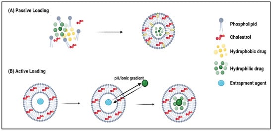
  43. Cullis, P.; Mayer, L.; Bally, M.; Madden, T.; Hope, M. Generating and loading of liposomal systems for drug-delivery applications. Adv. Drug Deliv. Rev. 1989, 3, 267–282.
  44. Chonn, A.; Cullis, P.R. Recent advances in liposomal drug-delivery systems. Curr. Opin. Biotechnol. 1995, 6, 698–708.
  45. Çağdaş, M.; Sezer, A.D.; Bucak, S. Liposomes as Potential Drug Carrier Systems for Drug Delivery. In Application of Nanotechnology in Drug Delivery; Sezer, A.D., Ed.; Intech: Rijeka, Croatia, 2014.
  46. Crommelin, D.; van Bloois, L. Preparation and characterization of doxorubicin-containing liposomes. II. Loading capacity, long-term stability and doxorubicin-bilayer interaction mechanism. Int. J. Pharm. 1983, 17, 135–144.
  47. Gubernator, J. Active methods of drug loading into liposomes: Recent strategies for stable drug entrapment and increasedin vivoactivity. Expert Opin. Drug Deliv. 2011, 8, 565–580.
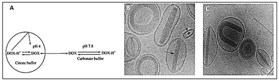
  48. Mayer, L.; Bally, M.; Cullis, P. Uptake of adriamycin into large unilamellar vesicles in response to a pH gradient. Biochim. Biophys. Acta (BBA) Biomembr. 1986, 857, 123–126.
  49. Swenson, C.E.; Perkins, W.R.; Roberts, P.; Janoff, A.S. Liposome Technology and the Development of Myocet TM (Liposomal Doxorubicin Citrate). Breast 2001, 2, 1–7.
  50. Li, X.; Hirsh, D.J.; Cabral-Lilly, D.; Zirkel, A.; Gruner, S.M.; Janoff, A.S.; Perkins, W.R. Doxorubicin physical state in solution and inside liposomes loaded via a pH gradient. Biochim. Biophys. Acta (BBA) Biomembr. 1998, 1415, 23–40.
  51. Kanter, P.M.; A Bullard, G.; A Ginsberg, R.; Pilkiewicz, F.G.; Mayer, L.D.; Cullis, P.R.; Pavelic, Z.P. Comparison of the cardiotoxic effects of liposomal doxorubicin (TLC D-99) versus free doxorubicin in beagle dogs. Vivo 1993, 7, 17–26.
  52. Haran, G.; Cohen, R.; Bar, L.K.; Barenholz, Y. Transmembrane ammonium sulfate gradients in liposomes produce efficient and stable entrapment of amphipathic weak bases. Biochim. Biophys. Acta (BBA) Biomembr. 1993, 1151, 201–215.
  53. Alyane, M.; Barratt, G.; Lahouel, M. Remote loading of doxorubicin into liposomes by transmembrane pH gradient to reduce toxicity toward H9c2 cells. Saudi Pharm. J. 2015, 24, 165–175.
  54. Sakakibara, T.; Chen, F.-A.; Kida, H.; Kunieda, K.; Cuenca, R.E.; Martin, F.J.; Bankert3, R.B. Doxorubicin Encapsulated in Sterically Stabilized Liposomes Is Superior to Free Drug or Drug-Containing Conventional Liposomes at Suppressing Growth and Métastases of Human Lung Tumor Xenografts1. Cancer Res. 1996, 56, 3743–3746.
  55. Dosio, F.; Cattel, L. PEGylation of Proteins and Liposomes: A Powerful and Flexible Strategy to Improve the Drug Delivery. Curr. Drug Metab. 2012, 13, 105–119.
  56. Fritze, A.; Hens, F.; Kimpfler, A.; Schubert, R.; Peschka-Süss, R. Remote loading of doxorubicin into liposomes driven by a transmembrane phosphate gradient. Biochim. Biophys. Acta (BBA) Biomembr. 2006, 1758, 1633–1640.
  57. Achim, M.; Precup, C.; Gonganău-Niţu, D.; Barbu-Tudoran, L.; Porfire, A.S.; Scurtu, R.; Ciuce, C. Thermosensitive Liposomes Containing Doxorubicin. Preparation and In Vitro Evaluation. Farmacia 2009, 57, 6.
  58. Pitt, W.G.; Husseini, G.A.; Roeder, B.L.; Dickinson, D.J.; Warden, D.R.; Hartley, J.M.; Jones, P.W. Preliminary Results of Combining Low Frequency Low Intensity Ultrasound and Liposomal Drug Delivery to Treat Tumors in Rats. J. Nanosci. Nanotechnol. 2011, 11, 1866–1870.
  59. Yuh, E.L.; Shulman, S.G.; Mehta, S.A.; Xie, J.; Chen, L.; Frenkel, V.; Bednarski, M.D.; Li, K. Delivery of Systemic Chemotherapeutic Agent to Tumors by Using Focused Ultrasound: Study in a Murine Model. Radiology 2005, 234, 431–437.
  60. Xu, H.; Zhang, W.; Li, Y.; Ye, F.F.; Yin, P.P.; Yu, X.; Hu, M.N.; Fu, Y.S.; Wang, C.; Shang, D.J. The Bifunctional Liposomes Constructed by Poly(2-ethyl-oxazoline)-cholesteryl Methyl Carbonate: An Effectual Approach to Enhance Liposomal Circulation Time, pH-Sensitivity and Endosomal Escape. Pharm. Res. 2014, 31, 3038–3050.
  61. Faraji, A.H.; Wipf, P. Nanoparticles in cellular drug delivery. Bioorganic Med. Chem. 2009, 17, 2950–2962.
  62. Xing, H.; Tang, L.; Yang, X.; Hwang, K.; Wang, W.; Yin, Q.; Wong, N.Y.; Dobrucki, W.; Yasui, N.; Katzenellenbogen, J.A.; et al. Selective delivery of an anticancer drug with aptamer-functionalized liposomes to breast cancer cells in vitro and in vivo. J. Mater. Chem. B 2013, 1, 5288–5297.
  63. Yang, B. Preclinical study of Doxorubicine-loaded liposomal drug delivery for the treatment of head and neck cancer: Optimization by Box-Behnken statistical design. Acta Biochim. Pol. 2020, 67, 149–155.
  64. Elamir, A.; Ajith, S.; Al Sawaftah, N.; Abuwatfa, W.; Mukhopadhyay, D.; Paul, V.; Al-Sayah, M.H.; Awad, N.; Husseini, G.A. Ultrasound-triggered herceptin liposomes for breast cancer therapy. Sci. Rep. 2021, 11, 1–13.
  65. Chowdhury, N.; Chaudhry, S.; Hall, N.; Olverson, G.; Zhang, Q.-J.; Mandal, T.; Dash, S.; Kundu, A. Targeted Delivery of Doxorubicin Liposomes for Her-2+ Breast Cancer Treatment. AAPS PharmSciTech 2020, 21, 202.
  66. Yin, X.; Feng, S.; Chi, Y.; Liu, J.; Sun, K.; Guo, C.; Wu, Z. Estrogen-functionalized liposomes grafted with glutathione-responsive sheddable chotooligosaccharides for the therapy of osteosarcoma. Drug Deliv. 2018, 25, 900–908.
  67. Ishida, T.; Kirchmeier, M.; Moase, E.; Zalipsky, S.; Allen, T. Targeted delivery and triggered release of liposomal doxorubicin enhances cytotoxicity against human B lymphoma cells. Biochim. Biophys. Acta (BBA) Biomembr. 2001, 1515, 144–158.
  68. Arabi, L.; Badiee, A.; Mosaffa, F.; Jaafari, M.R. Targeting CD44 expressing cancer cells with anti-CD44 monoclonal antibody improves cellular uptake and antitumor efficacy of liposomal doxorubicin. J. Control. Release 2015, 220, 275–286.
  69. Wang, W.; Shao, A.; Zhang, N.; Fang, J.; Ruan, J.J.; Ruan, B.H. Cationic Polymethacrylate-Modified Liposomes Significantly Enhanced Doxorubicin Delivery and Antitumor Activity. Sci. Rep. 2017, 7, 43036.
  70. Ieranò, C.; Portella, L.; Lusa, S.; Salzano, G.; D’Alterio, C.; Napolitano, M.; Buoncervello, M.; Macchia, D.; Spada, M.; Barbieri, A.; et al. CXCR4-antagonist Peptide R-liposomes for combined therapy against lung metastasis. Nanoscale 2016, 8, 7562–7571.
  71. Li, X.; Diao, W.; Xue, H.; Wu, F.; Wang, W.; Jiang, B.; Bai, J.; Lian, B.; Feng, W.; Sun, T.; et al. Improved efficacy of doxorubicin delivery by a novel dual-ligand-modified liposome in hepatocellular carcinoma. Cancer Lett. 2020, 489, 163–173.
  72. Safra, T.; Muggia, F.; Jeffers, S.; Tsao-Wei, D.D.; Groshen, S.; Lyass, O.; Henderson, R.; Berry, G.; Gabizon, A. Pegylated liposomal doxorubicin (doxil): Reduced clinical cardiotoxicity in patients reaching or exceeding cumulative doses of 500 mg/m2. Ann. Oncol. 2000, 11, 1029–1034.
  73. Kesterson, J.P.; Odunsi, K.; Lele, S. High cumulative doses of pegylated liposomal doxorubicin are not associated with cardiac toxicity in patients with gynecologic malignancies. Chemotherapy 2010, 56, 108–111.
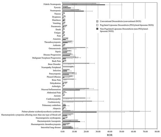
  74. Fukuda, A.; Tahara, K.; Hane, Y.; Matsui, T.; Sasaoka, S.; Hatahira, H.; Motooka, Y.; Hasegawa, S.; Naganuma, M.; Abe, J.; et al. Comparison of the adverse event profiles of conventional and liposomal formulations of doxorubicin using the FDA adverse event reporting system. PloS ONE 2017, 12, e0185654.
More
ScholarVision Creations