Golgi Metal Ion Homeostasis (Ca2+/Mn2+): Comparison
Please note this is a comparison between Version 1 by Yanzhuang Wang and Version 2 by Amina Yu.

The Golgi apparatus is an organelle found in most eukaryotic cells. Being part of the endomembrane system in the cytoplasm, it resides at the intersection of the exocytic and endocytic pathways, and works mainly in post-translational modifications and sorting of lipids and proteins. One unique characteristic of the Golgi is the multilayer stack that divides the Golgi membrane system into several sub-compartments known as cis-, medial, and trans-Golgi, each of which contains a set of glycosylation enzymes that sequentially remove or add various sugar monomers to proteins as they pass through the Golgi. To fulfill its function, the Golgi structure is highly dynamic, while Golgi structure and function are tightly regulated. Similarly, the microenvironment of each sub-compartment is also under strict regulation in response to intracellular environmental changes.

  • Golgi
  • homeostasis
  • transporter
  • channel
  • metal ion
  • calcium
  • manganese
  • zinc
  • copper

1. Ca2+/Mn2+ Transporters and Channels

As one of the most ubiquitous second messengers, Ca2+ plays a critical role in a variety of intracellular signaling events in the cell. The resting concentration of Ca2+ in the cytoplasm is normally maintained at around 100 nM, while the extracellular level is in low millimolar range. To maintain the low cytosolic concentration, most of the Ca2+ is stored in the lumen of the endoplasmic reticulum (ER) and the Golgi. Correlative laser scanning confocal fluorescence microscopy (LSCFM) and ion microscopy revealed that the Golgi is capable of storing up to 5% of total cellular Ca2+, depending on the cell type, and is more resistant to Ca2+ depletion than other cellular organelles [1][27]. The maintenance of high Ca2+ levels in the Golgi lumen is facilitated by Golgi-residing Ca2+ ATPases (transporters), Ca2+ release channels, and Ca2+ binding proteins.

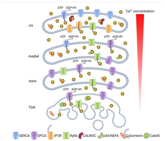
2. Ca2+/Mn2+ Transporters in the Golgi

The early Golgi compartments host many Ca2+ pumps that are also highly abundant in the ER, such as Sarcoplasmic/endoplasmic reticulum calcium ATPases (SERCAs), possibly because of the constant content exchange between the Golgi and ER, mediated by direct contact and vesicles [2][3][28,29]. On the other hand, the Golgi also contains unique Ca2+ transporters not found in the ER, including Secretory pathway Ca2+-transporting ATPases (SPCAs, also known as the Calcium-transporting ATPase type 2C members) and TMEM165 [3][29]. Different from ER Ca2+ pumps, they usually show high affinity to Mn2+ ion and therefore act as Mn2+ transporters as well.
It is widely accepted that SERCAs and SPCAs both contribute to the Ca2+ uptake into the Golgi since it is only partially inhibited by the SERCA inhibitor thapsigargin [4][5][38,39]. Isolated membranes show enrichment of SERCA in the ER and cis-Golgi while SPCA1 in the trans-Golgi membranes [6][40]. This conclusion was supported by immunoelectron microscopy (immuno-EM) [7][41]. Additionally, investigation using Ca2+ probes targeted to the cis- and trans-Golgi showed that the Ca2+ accumulation in these compartments is mainly facilitated by SERCA and SPCA1, respectively [5][39]. As Golgi-specific Ca2+ pumps, SPCAs have been shown to be the major Ca2+ pumps that facilitate the Ca2+ uptake in the trans-Golgi network (TGN).
TMEM165 was first identified in congenital disorders of glycosylation (CDG) [8][17] and localize to the Golgi [9][42]. Loss of TMEM165 function interrupts Ca2+, pH, and Mn2+ homeostasis and causes glycosylation abnormalities, which are restored by Mn2+ supplementation. Therefore, TMEM165 is proposed to be a Golgi-localized Ca2+/Mn2+ antiporter involved in glycosylation regulation [10][11][43,44]. Interestingly, TMEM165 is relocated to lysosomes and subjected to lysosomal degradation upon high Mn2+ exposure in a Rab7 and Rab5 independent manner, indicating a Mn2+-dependent TMEM165 quality control mechanism involving direct targeting of the protein to the lysosomal degradation pathway [12][45]. The Mn2+-induced translocation and lysosomal degradation of TMEM165 was later associated to the function of SPCA1, since the phenotype was rescued by a gain-of-function mutation of SPCA1. This suggests a possible functional link between the two proteins in response to the cytosolic Mn2+ change, however the underlying mechanism remains unclear [13][46]. One of the unanswered questions is why TMEM165 is degraded upon high Mn2+ exposure. One proposal is that TMEM165 works as a Mn2+ importer using the gradient of Golgi Ca2+. TMEM165 degradation would increase cytosolic Mn2+ and engage the major Mn2+ importer SPCA1 for detoxification. Another hypothesis is that TMEM165 imports Ca2+ into the Golgi using the Golgi Mn2+ gradient. Then TMEM165 degradation would restrain the Mn2+ in the Golgi to avoid further toxication. Therefore, TMEM165 is proposed to function in both directions depending on the combined effects of Ca2+ and Mn2+ [12][45].

3. Ca2+-Release Channels in the Golgi

The inositol 1,4,5-trisphosphate (IP3) receptors (IP3Rs) and ryanodine receptors (RyRs) are two major types of Ca2+-release channels in the Golgi. They both open at low cytosolic Ca2+ and close at high cytosolic Ca2+ concentrations. IP3R receptors are encoded by 3 genes (ITPR1, 2 and 3) in human, and are found in Golgi membranes [2][14][28,47]. These proteins are better characterized as ER-resident Ca2+ channels that release Ca2+ from the ER lumen into the cytosol in response to the stimulation of IP3 [15][9]. The RyR family members encoded by 3 genes (RYR1, 2 and 3) in humans are the other major types of Ca2+ channels that mediate the release of Ca2+ into the cytoplasm [16][48]. The IP3Rs were once considered to be the only Golgi Ca2+ channels, since IP3 but not RyR agonists significantly induced Ca2+ release from Golgi membrane fractions in the presence of thapsigargin [17][49]. However, other experiments in rat sympathetic neurons and neonatal cardiac myocytes argued that functional RyRs are found in the trans-Golgi and probably take part in Golgi Ca2+ release [18][19][50,51]. Emetine, an alkaloid, induces Ca2+ release from trans-Golgi without inducing Ca2+ release from the ER. It is unlikely that emetine induces Ca2+ release via activation of RyR2 or inhibition of SPCA, and the underlying molecular mechanism requires further investigation [20][52].

4. Ca2+-Binding Proteins in the Golgi Lumen

In addition to the Ca2+ transporters and release channels summarized above, Ca2+-binding proteins are another factor involved in Ca2+ homeostasis in the Golgi lumen. CALNUC, 45 kDa calcium-binding protein (Cab45), p54/NEFA, and calumenin are identified as major Ca2+ binding proteins found specifically in the Golgi lumen. CALNUC (also known as Nucleobindin-1), an EF-hand, Ca2+-binding peripheral membrane protein closely linked with the lumenal surface of the cis-Golgi cisternae, is the most abundant and well-characterized Golgi Ca2+ binding protein [21][53]. Transiently expressed CALNUC co-distributes with IP3R1 in cells, which increases Ca2+ storage as well as Ca2+ release upon ATP or IP3 stimulation [14][47]. Overexpression of SPCA upregulates the expression of CALNUC, indicating that CALNUC, together with SERCA and IP3R, is involved in establishment of the agonist-mobilizable Golgi Ca2+ store.
Cab45 is the first ubiquitous Golgi Ca2+-binding protein identified. It is a soluble Golgi lumen-resident protein that binds Ca2+ via its EF-hands [22][54]. Retention of Cab45 in the TGN is Ca2+-dependent, which is disrupted by treatment of cells with a Ca2+ ionophore [23][55], and is regulated by the Golgi resident serine/threonine kinase Fam20C [24][56]. Once bound to Ca2+, Cab45 oligomerizes and is recruited by secretory proteins. Via super resolution microscopy, the Cab45 oligomer-cargo complex is detected to be preferentially colocalized with the TGN Ca2+ pump SPCA1. Phosphorylation of Cab45 by Fam20C reduces the size of Cab45 oligomers, abolishes the TGN retention, and enhances the sorting and secretion of Cab45-cargo complex [24][56]. These results suggest the possibility that a functional sorting complex formed by Cab45, SPCA1, and cargo protein is regulated by Golgi Ca2+ [25][57].
p54/NEFA [26][58] and calumenin are two other Ca2+-binding proteins found in the Golgi lumen. p54/NEFA is a medial Golgi membrane-associated lumenal protein sharing sequence homology with CALNUC [27][59]. Calumenin is a glycosylated secretory protein distributed throughout the secretory pathway with a low affinity to Ca2+ ions [28][29][60,61]. It is reported to interact with RyR2 and SERCA in cardiac and skeletal sarcoplasmic reticulum, and therefore suggested to be a regulator of RyR2 and SERCA, which is tightly coupled with Ca2+ cycling [30][31][32][62,63,64].
In summary, the Golgi is equipped with all the molecular components necessary for maintaining Ca2+ homeostasis in the Golgi lumen: Ca2+ ATPases that pump Ca2+ into the Golgi lumen, Ca2+ channels that release Ca2+ into the cytoplasm, and Ca2+-binding proteins that buffer Ca2+ in the Golgi lumen. With all these regulators in action, the distinct sub-compartments of the Golgi appear to have a decreasing lumenal Ca2+ concentration through heterogenous expression of Ca2+ transporters and channels: the cis-Golgi expresses mainly SERCA and IP3Rs and contains around 250 µM lumenal Ca2+; the medial Golgi mainly expresses SERCA and SPCA1; and the trans-Golgi mainly expresses SPCA1 and RyRs, with a lumenal Ca2+ of about 130 µM (Figure 1) [3][29].
Figure 1. Ca2+ concentration and Ca2+ homeostasis-related molecules in different Golgi sub-compartments. The cis-Golgi expresses mainly SERCA and IP3Rs and contains around 250 µM lumenal Ca2+; the medial Golgi mainly expresses SERCA and SPCA1; and the trans-Golgi mainly expresses SPCA1 and RyRs, with a lumenal Ca2+ of about 130 µM.

5. Disruption of Ca2+/Mn2+ Homeostasis Impairs Golgi Structure and Function

Inhibition of Ca2+ pumps was reported to trigger a Golgi morphology change. Knockdown of SPCA1 leads to Golgi ribbon fragmentation, shortened and tubulated cisternae, and complete absence of cis- and trans-Golgi compartments [7][41]. In a separate study, transient inhibition of SERCA by thapsigargin-induced Golgi fragmentation before the ER unfolded protein response (UPR) is triggered, indicating an ER-stress-independent Golgi morphology disruption [33][65]. Further experiments demonstrated that thapsigargin-induced elevation of cytosolic Ca2+ activates protein kinase C (PKCα), which subsequently phosphorylates and inactivates GRASP55, a key membrane tether in Golgi stack formation [34][35][66,67]. Golgi structural defects also impact membrane trafficking and secretion. Activation of PKCα with phorbol 12-myristate 13-acetate (PMA) and histamine modulates Golgi structure in a similar fashion, indicating a link between Ca2+ signaling, Golgi structure and function, and human physiology [36][37].
Many Golgi-resident glycosylation enzymes such as Golgi α-mannosidases, and proteases such as furin, show Ca2+-binding activity [37][68] or undergoes Ca2+-dependent cleavage, which is required for their activation [38][69]. Taking the Golgi mannosidases as examples, these cis-Golgi resident enzymes are required in the early steps of N-glycosylation process, removing mannose residues from a high-mannose intermediate product. Ca2+ binds to mannosidases prior to the substrate and remains associated during the enzymatic reaction. Divalent cations, including Mn2+, compete with Ca2+ and therefore inhibit the mannosidase activity [39][70].
Mn2+ is also a common ion associated with many Golgi-residing glycosylation enzymes, including N-acetylglucosaminyltransferases [40][71], N-acetylgalactosaminyltransferases [41][72], mannosyltransferases [42][73], and some members in the fucosyltransferase family [43][44][74,75]. While Mn2+ homeostasis in the Golgi lumen is required for proper glycosylation, high cytosolic Mn2+ is toxic and related to many human diseases (Table 1) [45][46][76,77]. By uptaking Mn2+ from cytoplasm into the Golgi lumen, the SPCA pumps avoid cytotoxic accumulation of Mn2+, therefore maintain Golgi Mn2+ homeostasis and accurate glycosylation [47][48][49][78,79,80]. On the other hand, exposure of Mn2+ protects cells from some bacteria-originated toxins (e.g., Shiga toxin, STx) through down-regulation of Golgi phosphoprotein of 130 kDa (GPP130) and protects against STx-induced death [50][81]. As a type 2 ribosome inactivating protein, STx binds to the cellular toxin receptor to be internalized, and is then transported via the retrograde trafficking route through endosomes and Golgi to the ER, where the toxin is translocated to the cytosol to practice its toxin activity [51][52][82,83]. GPP130 serves as a host-cell trafficking receptor that facilitates the intake of the toxin into the TGN via the interaction with Syntaxin 5 [53][84]. Exposure to Mn2+ blocks the retrograde trafficking of STx from endosome to Golgi and leads to its lysosomal degradation [50][81]. Mn2+ targets GPP130 and induces its oligomerization, causing GPP130 redistributed to lysosomes via a clathrin and Rab7-dependent mechanism, where it is subsequently degraded [54][55][56][85,86,87]. Because of its sensitivity to Mn2+ exposure, GPP130 can be used as an intra-Golgi Mn2+ sensor in Golgi Mn2+ homeostasis studies [49][80].
Table 1. Human diseases related to Golgi metal ion regulators.
Metal IonDiseaseOMIMGeneProteinClinical Features
Ca2+/Mn2+Brody myopathy (BRM) [57]601003ATP2A1SERCA1Early onset of muscle function disorder characterized by muscle cramping and post-exercise stiffening (myopathy).
Acrokeratosis verruciformis (AKV) [58]101900ATP2A2SERCA2Early onset keratinization disorder affecting the distal extremities.
Darier disease (DD) [59]124200ATP2A2SERCA2Early onset keratinizating disorder characterized by small papules predominantly in seborrheic areas.
Hailey-Hailey disease (HHD) [60]169600ATP2C1SPCA1A skin disease causing persistent blisters and suprabasal cell separation (acantholysis) of the epidermis.
Spinocerebellar ataxia 15 (SCA15) [61]606658ITPR1IP3R 1A neurological condition characterized by progressive gait and limb ataxia.
Spinocerebellar ataxia 29 (SCA29) [62]117360ITPR1IP3R 1Early onset cerebellar ataxia causing slowly progressive or non-progressive gait and limb ataxia.
Gillespie syndrome (GLSP) [15]206700ITPR1IP3R 1A congenital neurological disorder characterized by the association of partial bilateral aniridia with non-progressive cerebellar ataxia, and intellectual disability.
Anhidrosis, isolated, with normal sweat glands (ANHD) [63]106190ITPR2IP3R 2A disorder characterized by absence of perspiration and subsequent heat intolerance with normal morphology and number of sweat glands.
Malignant hyperthermia 1 (MHS1) [64]145600RYR1RyR1A skeletal muscle disorder and the main causes of death due to anesthesia characterized by any combination of hyperthermia, skeletal muscle rigidity, tachycardia or arrhythmia, respiratory and metabolic acidosis, and rhabdomyolysis.
Central core disease of muscle (CCD) [65][66]117000RYR1RyR1A mild congenital myopathy characterized by motor developmental delay and signs of mild proximal weakness.
Multiminicore disease with external ophthalmoplegia (MMDO) [67]255320RYR1RyR1A heterogeneous neuromuscular disorder characterized by neonatal hypotonia, delayed motor development, and generalized muscle weakness and amyotrophy.
Arrhythmogenic right ventricular dysplasia, familial, 2 (ARVD2) [68]600996RYR2RyR2A congenital heart disease characterized by effort-induced polymorphic ventricular tachycardias due to large areas of fatty-fibrous replacement in the subepicardial layer of the right ventricle.
Ventricular tachycardia, catecholaminergic polymorphic, 1 (CPVT1) [69]604772RYR2RyR2An arrhythmogenic disorder characterized by physical activity- or stress-induced, polymorphic ventricular tachycardia that may degenerate into deteriorate into ventricular fibrillation.
Congenital disorders of glycosylation, Type IIk (CDG2K) [8]614727TMEM165TMEM165An autosomal recessive disorder with a variable phenotype, characterized by growth retardation.
Zn2+A novel syndrome with early onset agammaglobulinemia and absent B cells of unknown cause [70]N/ASLC39A7Zip7A novel autosomal recessive disease characterized by absent B cells, agammaglobulinemia and early-onset infections.
Ehlers–Danlos syndrome, Spndylodysplastic Type, 3 (SCD-EDS) [71]612350SLC39A13Zip13Postnatal growth retardation characterized by short stature, hyperelastic skin and hypermobile joints, protuberant eyes with bluish sclerae, atrophy of the thenar muscles, wrinkled palms and tapering fingers.
Cu2+Menkes disease (MNK) [72][73][74]309400ATP7AATP7AA disorder characterized by generalized copper deficiency, early retardation in growth, peculiar hair, and focal cerebral and cerebellar degeneration due to the dysfunction of several copper-dependent enzymes.
Occipital horn syndrome (OHS) [75]304150ATP7AATP7AA rare connective tissue disorder characterized by hyperelastic and bruisable skin, hernias, bladder diverticula, hyperextensible joints, varicosities, and multiple skeletal abnormalities, sometimes accompanied by mild neurologic impairment, and bony abnormalities of the occiput.
Distal spinal muscular atrophy, X-linked, 3 (DSMAX3) [76]300489ATP7AATP7ANeuromuscular disorders caused by selective degeneration of motor neurons in the anterior horn of the spinal cord.
Wilson disease (WD) [77][78]277900ATP7BATP7BA disorder characterized by dramatic accumulation of intracellular copper with subsequent hepatic and neurologic abnormalities

6. Golgi Lumenal Ca2+ Is Essential for Intra-Golgi Trafficking and Protein Sorting at the TGN

The role of Ca2+ in trafficking was first indicated in a study on ER-to-Golgi trafficking in 1989 [79][88]. Since then, reports on Ca2+ participating in intracellular transport events have emerged [80][81][82][83][89,90,91,92] and Ca2+ have been shown to be a fundamental factor in intracellular trafficking [84][93]. Incubation of isolated Golgi membranes with ionomycin and thapsigargin, which reduce the Golgi lumenal Ca2+, inhibits intra-Golgi transport of VSV-G [85][94]. On the other hand, high Ca2+ concentration in the TGN is required for the segregation and sorting of secretory cargo at TGN. This involves the Ca2+ pump SPCA1 and Ca2+ binding protein Cab45 [23][86][87][88][55,95,96,97]. SPCA1 binds to the actin-severing protein actin-depolymerizing factor (ADF)/cofilin1 on the TGN via dynamic actin and promotes Ca2+ influx into the TGN lumen. Increased Ca2+ leads to Cab45 oligomerization and binding to the secretory cargo proteins. The Cab45-cargo complex is then sorted into sphingomyelin-rich vesicles and transported to the plasma membrane for secretion, which is fine-tuned by the Golgi lumenal kinase Fam20C [89][98]. Knockdown of either SPCA1 or ADF/cofilin1 results in significant mis-sorting of secretory cargo proteins, indicating the role of actin-filaments-induced SPCA1 activation and Ca2+ uptake at TGN in protein sorting [87][96].

7. Golgi Ca2+/Mn2+ Homeostasis and Human Diseases

Given the Golgi’s role in protein post-translational modification and sorting, the importance of lumenal Ca2+/Mn2+ in controlling many of the enzymatic activities within the Golgi, as well as the effect of Ca2+/Mn2+ fluctuation on the Golgi structure, any changes in Ca2+/Mn2+ homeostasis are likely to have an impact on Golgi function. Changes in Golgi Ca2+/Mn2+ handling should, in turn, result in significant changes in cellular activities, contributing to disease pathogenesis [90][99]. Indeed, mutations in Ca2+/Mn2+ transporters and channels have been related to various diseases affecting multiple organs and tissues characterized with a wide range of clinical features (summarized in Table 1). In addition, SPCA1 is reported to be required for the maturation and spread of diverse viruses. SPCA1 deficiency leads to impaired virus glycoprotein maturation, reduces the infectivity of furin-requiring viruses, and lowers viral burden in human airway epithelial cells possibly by decreasing the abundance of furin [91][100]. Importantly, the Spike protein of SARS-CoV-2 virus, which is responsible for the strong infectivity of the virus, contains a furin cleavage site, indicating furin activity as a potential therapeutic target of the wide-spreading COVID-19 [92][101].
Video Production Service