Neuroprotective Potentials of Marine Algae and Bioactive Metabolites: Comparison
Please note this is a comparison between Version 1 by Md Abdul Hannan and Version 2 by Peter Tang.

Marine algae are considered to be a potential source of some unique metabolites with diverse health benefits. The pharmacological properties, such as antioxidant, anti-inflammatory, cholesterol homeostasis, protein clearance and anti-amyloidogenic potentials of algal metabolites endorse their protective efficacy against oxidative stress, neuroinflammation, mitochondrial dysfunction, and impaired proteostasis which are known to be implicated in the pathophysiology of neurodegenerative disorders and the associated complications after cerebral ischemia and brain injuries. 

  • seaweed
  • secondary metabolites
  • neuroprotection
  • Alzheimer’s disease
  • Parkinson’s disease
  • ischemic stroke

1. Introduction

Neurons and supporting cells of the brain encounter degenerative changes during physiological or pathological aging, ischemic stroke, or other brain injuries [1]. The degenerative brain disorders such as Alzheimer’s disease (AD) and Parkinson’s diseases (PD) are the consequence of pathological brain aging, which are characterized by the region-specific loss of neurons [2]. Globally, these diseases account for the major causes of dementia among the elderly [3]. Although the exact etiologies of these brain disorders are not revealed yet, they share some common pathophysiology, such as oxidative stress (OS), neuroinflammation, mitochondrial dysfunction, protein misfolding, and defective protein clearance system that, in turn, make these diseases complicated [4][5][4,5], whereas, ischemic, traumatic, and other brain injuries, if not fatal, ensue secondary damage and constitute the appreciable causes of cognitive deficits among patients. Like neurodegenerative disorders, brain injuries also follow the same pathophysiology [6][7][6,7]. Whatever the forms of dementia disorder, the current therapeutic option can only alleviate symptoms, rather than halting the disease progression. Moreover, current drugs are associated with multiple side effects. Considering the tremendous social and economic impact of these diseases, scientists are, therefore, paying research efforts to discover the potential therapeutic agents that can target disease pathogenesis without causing undesirable effects in patient’s health. Although synthetic drugs have some advantages such as easy to develop, naturally-derived compounds have received priority as they are relatively well-tolerated. Natural compounds have been claimed to show anti-inflammatory, antioxidant, and immunomodulatory effects [8]. Compounds showing multiple pharmacological effects offer a better solution for the remedy of neurological disorders with complex pathomechanisms [9]. In the published literature, a significant quantity of natural products has been reported to show neuroprotective activity against a wide range of toxic insults [10][11][10,11]. Some of them have shown therapeutic promise in preclinical studies [12] and clinical trials [13][14][13,14].
Macroalgae, also known as seaweed, are among the highly abundant marine lives and potentially contribute to the renewable resources for food and industrial products [15][16][17][15,16,17]. Beyond this importance, algal metabolites, such as phenolics, alkaloids, terpenoids, carotenoids, phytosterols, and polysaccharides have attracted much attention to medicinal chemistry due to their structural uniqueness and functional diversity [17][18][19][20][17,18,19,20]. These biofunctional compounds have shown to provide neuroprotection in preclinical models of neurodegenerative diseases, ischemic stroke, brain trauma, diabetes, and obesity, among many others, owing to their antioxidant, anti-inflammatory, and immunomodulatory capacities [21][22][23][24][25][26][27][28][21,22,23,24,25,26,27,28]. Evidence suggests that algal metabolites, particularly fucoxanthin, fucosterol, and fucoidan could be potential leads for the development of therapy against CNS diseases [22][29][30][31][22,29,30,31]. Although the algal metabolite-based drug discovery progresses very slowly, the discovery of sodium oligomannate and its conditional approval as an anti-AD drug [32] raises hope for the future development of potential therapeutic agents from marine algae.
Over the last decade, some excellent works reviewed the neuroprotective effects of marine algae and their metabolites [21][22][23][29][33][34][35][21,22,23,29,33,34,35]. However, some of these reviews limited their scope either to a single pathogenic mechanism such as neuroinflammation [22] or to categorical brain disorders such as AD or PD [22][23][29][34][35][22,23,29,34,35]. Others have reviewed literature published a decade or half a decade ago [23][36][23,36].

2. Pathophysiology of Brain Disorders

2.1. Neurodegenerative Disorders (AD and PD)

Neurodegenerative disorders, including AD and PD, are of major public health concern and contribute to the prime causes of dementia among elderly people. The pathological hallmarks of AD include extracellular deposition of amyloid plaque and intraneuronal aggregation of neurofibrillary tangles (NFT) [37]. On the other hand, PD is characterized by the degeneration of dopaminergic neurons in the substantia nigra [37] with the pathological hallmark of intraneuronal aggregation of α-synuclein [38]. Although the exact pathophysiology of these brain disorders remains elusive, it has been demonstrated that OS, neuroinflammation, mitochondrial dysfunction, and protein misfolding largely contribute to their development [37]. OS and neuroinflammation are two considerably diverse disease processes in many pathological events [39]. Conversely, they are interplayed with each other in the entire disease process. Thus, inhibition of neuroinflammation may reduce the OS and vice versa.
Oxidative stress (OS) is a pathological condition that develops when the production of reactive oxygen species (ROS) reaches an excessive level with lower efficiency of the cellular antioxidant defense system [40]. Factors contributing to OS in the brain include excitotoxicity, depletion of the cellular antioxidant system, high susceptibility to lipid peroxidation, and high oxygen demand [41]. OS may lead to mitochondrial dysfunction, which further results in the excessive ROS generation and establishes a vicious cycle of OS [42][43][42,43]. Moreover, the endoplasmic reticulum (ER), a site for protein folding, also takes part in ROS generation [44]. Protein misfolding in ER results in ER stress that is further responsible for ROS production [45]. ROS potentially contributes to the damage of cells through compromising the structure and function of biomolecules, including lipid peroxidation, protein oxidation, and deoxyribonucleic acid (DNA) damage, which eventually install neurodegeneration [38].
Neuroinflammation is another inevitable pathogenic factor of many neurodegenerative disorders [46]. Microglial activation is the major contributor to neuroinflammation [46]. A range of stimuli, including infection, trauma, toxic insults, and ischemia, may initiate microglial activation and disrupt the central nervous system (CNS) homeostasis [47][48][47,48]. Once activated, microglia released pro-inflammatory and neurotoxic elements, like chemokines, cytokines, proteases, eicosanoids, ROS, and excitatory amino acids [47]. All of these elements are documented as a key player in neuroinflammation-associated OS as well as chronic neurodegeneration [49]. The deposition of misfolded proteins, as evident in the major NDD, can also induce an inflammatory response, which further causes OS [50].
Dysregulation of cholesterol homeostasis is also a critical factor that could induce OS and inflammation, and thus may contribute to the pathogenesis of major brain disorders [51]. This disturbance in cholesterol metabolism in the brain is under the regulation of a cholesterol transport mechanism. Liver X receptor beta (LXR-β), once activated, promotes multiple genes that regulate reverse cholesterol transport and thus confers neuroprotection [52][53][52,53]. For instance, LXR-β agonist enhanced survival of dopaminergic neurons [54] and reduced the burden of mutant huntingtin [55] as well as promoted amyloid β (Aβ) clearance [56]. With the significant evidence of the implication of OS, neuroinflammation, and cholesterol dyshomeostasis in the pathobiology of neurodegenerative disorders, these pathological factors could be targeted for the development of potential therapeutics.

2.2. Ischemic Stroke

Ischemic stroke is responsible for the second-highest number of deaths and disability around the world [57]. It is a pathological condition resulting from sudden occlusion of blood supply to the brain. If the patient survives, the affected brain areas accompany the secondary damage due to the restoration of blood flow and reoxygenation. This ischemia/reperfusion (I/R) event initiates mitochondrial ROS generation [58] and subsequent inflammatory response [59].
Mitochondrial ROS is not only a crucial early driver of acute damage but is also considered an initiator of the consequence of a series of pathological features that develop over time following the reperfusion [60]. Initially, upon reperfusion, the burst of ROS production results in oxidative damage to mitochondria, and thereby disrupts ATP production [61], which ultimately initiates neuronal cell death cascades [62]. ROS-mediated mitochondrial damage further installs the inflammatory response via the activation of microglia and astrocytes as well as an influx of immune cells recruited by cytokines, adhesion molecules, and chemokines across the activated cerebral blood vessels [63]. This activation of the innate immunity triggers nuclear factor-kappa-B (NF-κB)-mediated production of numerous inflammatory cytokines that contribute to I / R injury [64]. Therefore, targeting OS and inflammatory response could be imperative to develop novel therapeutic strategies for the management of stroke.

2.3. Traumatic Brain Injury

Traumatic brain injury (TBI), an acquired brain injury caused by an external force or shock, is also considered to be a major cause of death globally, particularly in countries with a frequent incidence of traffic accidents [65]. Despite significant medical advances in recent times, the clinical outcomes of severely head-injured patients are not satisfactory.
As in ischemic stroke, mechanisms underlying the damages to the brain tissue with TBI are categorized into two classes: primary and secondary damages. Primary damage that irreversibly involves the mechanical damage of the skull and the brain has been complicated following the brain contusions, rupturing blood vessels, axonal injuries, and intracranial hemorrhages [66], whereas the secondary damage causes neuronal degeneration over time due to various biochemical changes such as OS, excitotoxicity, inflammation, and mitochondrial dysfunction [67]. Following TBI, various OS markers such as lipid peroxidation products, oxidized protein moieties, and DNA damage products accumulate in the brain while antioxidants and enzymes molecules such as glutathione (GSH), glutathione peroxidase (GPx), glutathione reductase (GR), glutathione S-transferases (GST), superoxide dismutase (SOD), and catalase (CAT) markedly decline [68]. It is suggested that treatment modalities associated with conferring neuroprotection on injured brain tissue and regeneration at the recovery stage of injured neurons have greater promise to restore at the site of brain injury following TBI.

3. Neuropharmacological Potentials of Marine Algae and Their Metabolites: Evidence from In Vitro Studies

Several compounds of diverse chemical classes have been reported from three major groups (brown, red, and green algae) of marine algae (Figure 1, Figure 2, Figure 3 and Figure 4). Neuropharmacological properties of these compounds reported in various in vitro models are compiled (Table 1) and discussed in the following subsections. Besides bioactive compounds, macroalgae that have shown promising neuroactive potentials, and thus demand further attention are also mentioned.
Figure 1.
Chemical structure of sterols (
A
) and plastoquinones (
B
) of marine algae.
Figure 2.
Chemical structure of phlorotannin of marine algae.
Figure 3.
Chemical structure of alkaloids (
A
), sesquiterpenes (
B
) and polysaccharides (
C
) of marine algae.
Figure 4.
Chemical structure of miscellaneous compounds from marine algae.
Table 1. Summary on pharmacological effects, occurrence, effective dose, experimental model, cellular effects, potential pharmacological mechanism of algal metabolites.

Pharmacological Effects

Compound (Class)

Algal Source If Any)

Effective Concentration

Experimental Model (In Vivo/In Vitro)

Cellular Effects/Significant Findings

Signaling Pathways Involved

Pharmacological Markers

Reference

Antioxidant activity

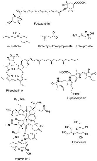
Fucoxanthin (carotenoids)

Sargassum siliquastrum

50 and 100 μM

H2O2-induced cell damage in kidney fibroblast cells

Attenuates oxidative stress

n.d.

↓ROS level

[69]

Fucoxanthin


5, 10, and 50 μM

H2O2 induced BV2 microglial cells

Antioxidation

Antioxidant pathway

↓ROS

↑SOD and GSH

[36]

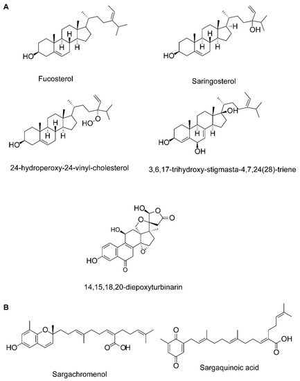
Fucosterol, 3,6,17-trihydroxy-stigmasta-4,7,24(28)-triene and 14,15,18,20-diepoxyturbinarin (sterols)

Pelvetia siliquosa

A seven day-dose regimen at 30 mg/kg/day before carbon tetrachloride (CCl4) administration

Rat model

Antioxidation

n.d.

↑SOD, CAT, and GPx

[70][71]

Fucosterol

Eisenia bicyclis,

brown alga

25, 50, 100, 200, and 400 μM

RAW 264.7 murine macrophages

(t-BHP stimulated)

Protects against oxidative stress

n.d.

↓ROS generation

[71][72]

Fucosterol

Ecklonia stolonifera and Eisenia bicyclis;

Brown algae

25, 50, and 100 μM

tert-Butyl hydroperoxide- and tacrine-induced HepG2cell injury model

Antioxidation

n.d.

↓ROS generation

↑GSH level

[72][73]

Fucosterol

Sargassum

Binderi;

brown alga

3.125, 6.25, 12.5, 25, 50, and 100 μg /mL

Particulate matter-induced injury and inflammation in A549 human lung epithelial cells

Attenuates oxidative stress


↓ROS level

↑SOD, CAT, and HO-1 in the cytosol, and NRF2 in the nucleus

[73][74].

Glycoprotein

U. pinnatifida

SOD activity and Xox activity at a concentration of 5 mg/mL and 1 mg/mL, respectively

In vitro enzyme assay


↑SOD and↓Xox

[74][75]

Sulfated oligosaccharides

Ulva lactuca and Enteromorpha prolifera;

green algae

150 mg/kg·day

Aging model (male senescence-accelerated prone (SAMP8) and male senescence resistant (SAMR1) mice)

Antioxidantion

n.d.

↑GSH, SOD, CAT, telomerase levels,

↑Total antioxidant capacity,

↓MDA and AGEPs

[75][96]

Anti-inflammatory activity

Fucoxanthin


5, 10, and 50 μM

42-induced BV2 microglia cells

Anti-inflammation

MAPK pathway

↓iNOS, COX-2

↓TNF-α, IL-6, IL-1β, PGE2

↓JNK, ERK, and p38 MAPK phosphorylation

[36]

Fucoxanthin

-


LPS-activated BV-2 microglia

Anti-inflammation and antioxidation

Akt/NF-κB and MAPKs/AP-1 pathways;

PKA/CREB pathway

↓iNOS, COX-2, ↓TNF-α, IL-6, PGE2, NO, ROS

↓IL-6, TNF-α, iNOS, and COX-2 mRNA expression

↓Akt, NF-κB, ERK, p38 MAPK and AP-1 phosphorylation

↑Nrf2, HO-1

↑PKA, CREB

↑BDNF

[76][70]

Fucosterol

E. bicyclis;

brown alga

5–20 μM for NO

RAW 264.7 murine macrophages

(t-BHP 200 μM, LPS-1μM stimulated)

↓Inflammatory response

↓NF-κB pathway

↓NO production

↓iNOS and COX-2

[71][72]

Fucosterol

U. pinnatifida

10, 25, or 50 μM

LPS-induced RAW 264.7 macrophages and THP-1 human monocyte cell line

↓Inflammatory response

↓NF-κB pathway

↓iNOS, TNF-α, and IL-6

↓DNA binding

↓phosphorylation of NF-κB, MKK3/6 and MK2

[77][83]

Fucosterol

Hizikia fusiformis

1–10 μM

CoCl2 induced hypoxia in keratinocytes

↓Inflammatory response

n.d.

↓IL-6, IL-1β and TNF-α

↓pPI3K and pAkt and HIF1-α accumulation

[78][82]

Fucosterol

Panida. australis

0.004,0.2, and 10 μM

LPS or Aβ-induced BV2 (microglial) cells

Protects against LPS or Aβ-mediated neuroinflammation

n.d.

↓IL-6, IL-1β, TNF-α, NO, and PGE2

[79][85]

Fucosterol

S. Binderi;

brown alga

3.125, 6.25, 12.5, 25, 50, 100 μg/mL

Particulate matter-induced injury and inflammation in A549

human lung epithelial cells

↓Inflammatory response

n.d.

↓COX-2, PGE2, TNF-α and IL-6

[73][74]

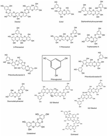
Dieckol (phlorotannin)

E. cava

50–300 µg/mL

LPS-stimulated murine BV2 microglia

Anti-inflammation and antioxidation

p-38 MAPK/ NF-κB pathway

↓NO and PGE2;

↓iNOS and COX-2;

↓IL-1β and TNF-α;

↓ROS

[80][86]

Phloroglucinol, eckol, dieckol, 7-phloroeckol, phlorofucofuroeckol A and dioxinodehydroeckol (phlorotannin)

E. bicyclis;

brown alga

5–20 μM for NO

LPS-stimulated RAW 264.7 murine macrophages

↓Inflammatory response

↓NF-κB pathway

↓NO production

[71][72]

Phlorofucofuroeckol A

E. stolonifera

20 μM

LPS-activated BV2 and primary microglial cells

Anti-inflammation

NF-κB, JNKs, p38 MAPK, and Akt pathways

↓NO and PGE2;

↓iNOS and COX-2;

↓IL-1β, IL-6 and TNF-α;

↓NF-κB activation and IκB-α degradation

↓JNK, p38, and Akt

[81][87]

Phlorofucofuroeckol B (phlorotannin)

E. stolonifera

10–40 µM

LPS-stimulated murine BV2 microglia

Anti-inflammation

IκB-α/NF-κB and Akt/ERK/JNK pathways

↓TNF-α, IL-1β and IL-6;

↓COX-2 and iNOS

↓NF-κB activation and IκB-α degradation

↓Akt, ERK, and JNK phosphorylation

[82][88]

8,8’-bieckol (phlorotannin)

E. cava


LPS-stimulated primary macrophages and RAW 264.7 macrophages

&

LPS-induced septic mice

Anti-inflammation; Protects mice from endotoxin shock

NF-κB pathway

↓NO and PGE2;

↓iNOS mRNA and protein expression;

↓IL-6;

↓Transactivation of NF-κB and nuclear translocation of the NF-κB p65 subunit

↓ROS

[83][90]

6,6′-bieckol (phlorotannin)

E.stolonifera


LPS-stimulated BV2 and murine primary microglial cells

Anti-inflammation

IκB-α/NF-κB and JNK/p38 MAPK/Akt pathways

↓COX-2 and iNOS;

↓NO and PGE2,

↓IL-6

↓Transactivation of NF-κB and nuclear translocation of the NF-κB p65 subunit

↓Akt, JNK and p38 MAPK phosphorylation

[84][89]

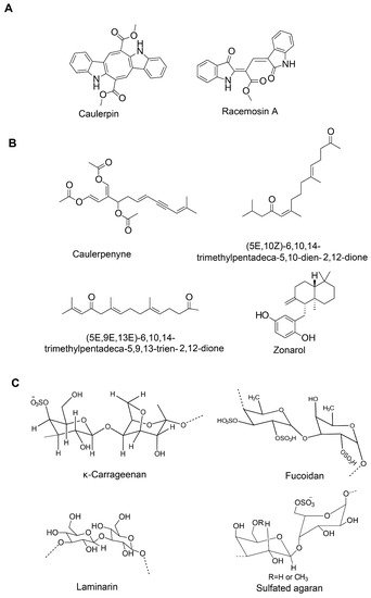
Fucoidan (sulfated polysaccharide)

Brown seaweed

25, 50, and 100 µg/mL

LPS-stimulated murine BV2 microglia

Anti-inflammation

NF-κB and JNK/p38 MAPK/Akt pathways

↓NO and PGE2;

↓COX-2, iNOS and MCP-1;

↓TNF-α and IL-1β;

↓NF-κB activation; ↓Akt, ERK, p38 MAPK and JNK phosphorylation

[85][92]

Fucoidan

-

125 µg/mL

LPS-activated primary microglia

Anti-inflammation

n.d.

↓TNF-α and ROS

[86][93]

κ-carrageenan oligosaccharides and desulfated derivatives

Red algae


LPS-activated microglia

Anti-inflammation

n.d.

↓TNF-α

[87][94]

Sulfated oligosaccharides

U. lactuca and E. prolifera;

green algae

150 mg/kg·day

Aging model (male senescence-accelerated prone (SAMP8) and male senescence resistant (SAMR1) mice)

↓Inflammatory response

n.d.

↓IFN-γ, TNF-α, and IL-6

[75][96]

Alginate-derived oligosaccharide

Brown algae

50–500 µg/mL

LPS/Aβ-stimulated BV2 microglia

Anti-inflammation

TLR4/NF-κB signaling pathway

↓NO and PGE2;

↓COX-2 and iNOS;

↓TNF-α, IL-6 and IL-12;

↓TLR4;

↑NF-κB/p65 subunit translocation

[88][97]

Seleno-polymannuronate

Brown algae

0.8 mg/mL

LPS-activated primary microglia and astrocytes;

mouse model of acute inflammation

Anti-inflammation

NF-κB and MAPK signaling

↓NO and PGE2;

↓COX-2 and iNOS;

↓TNF-α, IL-1β and IL-6;

↑IκB-α, p65, p38, ERK and JNK phosphorylation

[89][98]

Sargachromenol (plastoquinone)

Sargassum micracanthum

30.2 μM (IC50)

LPS-stimulated RAW 264.7 macrophages

Anti-inflammation

NF-κB signaling

↓NO and PGE2;

↓COX-2 and iNOS;

↑IκB-α

[90][99]

Sargaquinoic acid (plastoquinone)

Sargassum siliquastrum


LPS-stimulated RAW 264.7 macrophages

Anti-inflammation

NF-κB signaling

↓NO; ↓iNOS;

↑IκB-α; ↓nuclear translocation of NF-κB;

↓JNK1/2 MAPK

[91][100]

Floridoside (glycerol glycosides)

Laurencia undulate;

red alga

50 μM

LPS-stimulated murine BV2 microglia

Anti-inflammation

MAPK Signaling

↓NO, ROS;

↓iNOS and COX-2;

↓p38 MAPK and ERK phosphorylation

[92][101]

Glycoprotein

U. pinnatifida

COX-1 and COX-2 inhibition with IC50 values of 53.03 ± 1.03 μg/mL and 193.35 ± 3.08 μg/mL, respectively

LPS-stimulated RAW 264.7 macrophages

Anti-inflammation

n.d.

↓COX-1 and COX-2

↓NO

[74][75]

Caulerpin (bisindole alkaloid)

Caulerpa racemosa

100 µM/kg body wt

Capsaicin-induced ear edema and carrageenan-induced peritonitis

Inhibition of nociception

n.d.

n.d.

[93][130]

Caulerpenyne (sesquiterpene)

C. prolifera and

C. racemosa

5.1 μM

Lipoxygenase (LOX) enzyme activity assay

Inhibitory activity against LOX

-

Un-competitive type of inhibition

[94][131]

Aquamin (multi-mineral complex)

Lithothamnion corallioides; red alga


LPS-stimulated, glial-enriched primary cultures of rat cortex

Anti-inflammation

n.d.

↓TNF-α and IL-1β

[95][132]

Anticholinesterase activity

Fucosterol and 24-hydroperoxy 24-vinylcholesterol

E. stolonifera

IC50 values of 421.72 ± 1.43, 176.46 ± 2.51 µM, respectively

In vitro enzymatic assay

↓BChE activity

-

Selective inhibition of BChE

[96][114]

Fucosterol

Panida australis

inhibition against AChE (10.99–20.71%) and BChE (4.53–17.53%) with concentrations ≤ 56 μM,

In vitro enzymatic assay

↓AChE and BChE activities

-

Nonselective cholinesterase inhibition

[79][85]

Fucosterol

Sargassum horridum

-

In vitro enzymatic assay

↓AChE activity

-

Non-competitive inhibition

[97][115]

Fucoxanthin

-

IC50 value 1.97 mM

In vitro BChE activity assay

↓BChE activity


Mixed inhibition type

[98][116].

Fucoxanthin

Brown seaweed

IC50 value of 81.2 μM

In vitro AChE activity assay;

Molecular docking analysis

↓AChE activity

Fucoxanthin likely interacts with the peripheral anionic site within AChE

Non-competitive manner

[99][117]

α-Bisabolol

Padina gymnospora

IC50 value < 10 μg/mL

In vitro enzymatic assay

↓AChE and BChE activity

-

-

[100][118]

Glycoprotein

U. pinnatifida

AChE and BChE inhibitory activities with IC50 values of 63.56 ± 1.86 and 99.03 ± 4.64, respectively

In vitro enzymatic assay

↓AChE and BChE activity

-

-

[74][75]

Phloroglucinol,

dibenzo [1][4][1,4] dioxine-2,4,7,9-tetraol and eckol

Ecklonia maxima; Brown alga

IC50 value: 76.70 to 579.32 μM

In vitro AChE activity assay

↓AChE activity

-

-

[101][119]

Dieckol and phlorofucofuroeckol

E. cava


Ethanol-intoxicated memory impairment in mice

↓AChE activity

n.d.

↑Acetylcholine

[102][120]

Sargaquinoic acid and sargachromenol

(plastoquinones)

Sargassum sagamianum

IC50 value for anti-AChE: 23.2 and 32.7 μM, respectively;

IC50 value for anti-BChE of sargaquinoic acid 26 nm

In vitro ChE activity assay

Sargaquinoic acid shows potent inhibitory activity against BuChE and moderate inhibitory activity against AChE

-.

-

[103][121]

(5E,10Z)-6,10,14-trimethylpentadeca-5,10-dien-2,12-dione and (5E,9E,13E)-6,10,14-trimethylpentadeca-5,9,13-trien-2,12-dione

(Sesquiterpenes)

S. sagamianum

IC50 values of 65.0 and 48.0, and 34.0 and 23.0 μM, respectively

In vitro ChE activity assay

Moderate inhibitory activity against AChE and BuChE

-

-

[104][133]

Anti-amyloidogenic and aggregation inhibition activity

Fucoxanthin

E. stolonifera and U. pinnatifida


↓β-secretase activity;

Binding energy

(-7.0 kcal/mol)

-

mixed-type inhibition

[105][134]

Fucoxanthin

-

0.1–30 μM


Suppresses the formation of Aβ1-42 fibrils and Aβ1–42 oligomers, and inhibits Aβ aggregation

-

-

[106][135]

Fucoxanthin

-

2 μM

ThT assay

Inhibits Aβ1-42 fibril and aggregate formation

-

-

[107][136]

Fucosterol

E. stolonifera and U. pinnatifida

10–100 μM (IC50 value of 64.12 ± 1.0 μM)

In vitro enzyme assay;

In silico analysis

↓β-secretase activity;

Binding energy

(−10.1 kcal/mol)

-

Noncompetitive inhibition

[105][134]

α-Bisabolol

Padina gymnospora

5 μg/mL

Thioflavin T (ThT), Confocal laser scanning microscopy (CLSM) analysis, Transmission electron microscopy (TEM), Fourier transform infrared (FTIR) spectroscopic analysis and molecular dynamics simulation

Prevents oligomers formation as well as disaggregates the matured fibrils

-

-

[108][137]

Glycoprotein

U. pinnatifida

IC50 values of 73.35 ± 2.54 μg/mL

In vitro enzymatic assay

↓BACE1 activity

-

-

[74][75]

Cholesterol homeostasis and Aβ clearance activity

Fucosterol

-

100 and 200 μM (HEK293 cell cultures);

100 or 200 μM (macrophages and HepG2, H4IIE, and Caco2 cells)

HEK293 cell cultures (Reporter system);

THP-1-derived macrophages;

Caco-2 cells

HepG2 cells

Reverses cholesterol transport.

No accumulation of triglyceride in HepG2

n.d.

Dual-LXR agonist (LXR-α and LXR-β)

↑ABCA1, ABCG1, and ApoE;

↑Intestinal NPC1L1 and ABCA1;

↑Insig-2a, that delays nuclear translocation of SREBP-1c

[109][138]

Saringosterol

Sargassum fusiforme

30 μM

Luciferase reporter

assay system;

HEK293T, THP-1 monocytes, HepG2, RAW264.7, THP-1 macrophages and Caco-2 cells

n.d.

n.d.

Selective LXRβ agonist;

↑ABCA1, ABCG1, and SREBP-1c

[110][139]

Alginate-derived oligosaccharide

Marine brown algae


BV2 microglial cells

Microglial phagocytosis of Aβ

Toll-like receptor signaling

↑TLR4

[88][97].

Monoamine oxidase inhibition and affinity to dopaminergic receptors

Phlorofucofuroeckol-A and dieckol (phlorotannin)

-


In vitro enzyme assay and functional assay for GPCR screening; Docking analysis

hMAO activity;

D3R and D4R stimulation

-

-

[111][140].

Antiaging

Sulfated oligosaccharides

U. lactuca and E. prolifera;

green algae

150 mg/kg/day

Aging model (male senescence-accelerated prone (SAMP8) and male senescence resistant (SAMR1) mice)

Antioxidant and anti-inflammation

n.d.

↑GSH, SOD, CAT, telomerase levels,

↑Total antioxidant capacity,

↓MDA and AGEPs

↓IFN-γ, TNF-α, and IL-6

↑BDNF and ChAT;

↑Sirt1, ↑p53 and FOXO1

[75][96]

Fucosterol

Hizikia fusiformis

50 µg/mL

Culture model of C. elegans

Extends lifespan

↑Antioxidant mechanism

n.d.

[112][141]

n.d.: not defined; -: information not available.

4. Neuropharmacological Potentials of Marine Algae and Their Metabolites: Evidence from In Vivo Studies

The neuroprotective effects of some potential algal compounds that were reported in the in vitro conditions have successfully been translated into animal models, suggesting that these compounds could be potential candidates for further evaluation in the clinical trials.
Fucoidan is one of the algal compounds that has shown strong neuroprotection in several animal models. In the PD model of C57 / BL mice, fucoidan ameliorated MPTP-induced behavioral deficits, probably by elevating dopamine and its metabolite levels and increasing tyrosine hydroxylase expression [113][183]. In addition, fucoidan inhibited MPTP-induced lipid peroxidation and restored antioxidant capacity [113][183]. Similarly, fucoidan also improved behavioral capacity, by attenuating the loss of dopaminergic neurons and inhibited the deleterious activation of microglia in the substantia nigra pars compacta in LPS-induced neurotoxicity [86][93]. In an Aβ-induced rodent AD model, fucoidan ameliorated impaired memory, by reversing the decreased activity of ChAT, SOD, and GPx, increased activity of AChE, and rectifying the imbalance between apoptosis and pro-survival signals [114][193]. Fucoidan improved d-Gal-induced cognitive impairment in mice by mitigating OS and attenuating the caspase-dependent apoptosis pathway [115][185]. Wang and colleagues demonstrated that the supplementation of fucoidan alleviated Aβ-induced paralyzed phenotype in a transgenic C. elegans AD model [116][194]. Fucoidan reduced Aβ accumulation, probably by promoting proteasomal activity [116][194]. In another study, fucoidan-rich substances from Ecklonia cava improved trimethyltin-induced cognitive dysfunction by inhibiting Aβ production and Tau hyperphosphorylation [117][195]. Fucoidan also attenuated transient global cerebral ischemic injury in the gerbil hippocampal CA1 area through mitigating glial activation and oxidative stress [118][196].
Laminarin, another polysaccharide of brown algae, has shown to protect I/R injury in gerbil models. Intraperitoneal injection of laminarin (50 mg/kg) following 5 min I/R attenuated reactive gliosis (anti-inflammatory) in the hippocampal CA1 of young gerbils [119][197]. A similar study following the same experimental protocol, but with aged gerbils, showed that laminarin (50 mg/kg) attenuated ischemia-induced death of pyramidal neurons in the hippocampal CA1 of aged gerbils [120][198]. This neuroprotective effect of laminarin is attributed to its antioxidant and anti-inflammatory properties [120][198]. Oligo-porphyran, a synthetic product of porphyran (Pyropia yezoensis) ameliorated behavioral deficits in 6-OHDA-induced Parkinsonian mice model by protecting dopaminergic loss and activating the PI3K/Akt/Bcl-2 pathway that involved cellular signaling of anti-apoptosis and antioxidation [121][199]. Zhang and colleagues demonstrated that porphyran from Pyropia haitanensis improved the Aβ1-40-induced learning and memory deficits probably by elevating cerebral acetylcholine level [122][200].
Fucoxanthin is another significant algal metabolite that was found to be effective in a wide range of brain dysfunction (such as AD, ischemic stroke, and traumatic brain injury). Fucoxanthin ameliorated scopolamine-induced [106][135] and Aβ oligomer-induced [99][117] cognitive impairments in mice, possibly by inhibiting AChE activity and OS, modulating ChAT activity, and increasing BDNF expression. Fucoxanthin alleviated cerebral ischemic/reperfusion (I/R) injury, improved the neurologic deficit score, and downregulated the expression of apoptosis-linked proteins in brain samples [123][168]. Fucoxanthin also attenuated traumatic brain injury that involved the Nrf2-ARE and Nrf2-autophagy pathways-dependent neuroprotective mechanism [124][169].
Fucosterol co-infusion ameliorated sAβ1-42-induced cognitive deficits in aging rats by modulating BDNF signaling [125][172]. Dieckol and phlorofucofuroeckol raised the brain level of acetylcholine by inhibiting AChE and reduced the inhibition of latency in ethanol-intoxicated memory-impaired mice [102][120]. Yang and co-investigators demonstrated that stereotaxic injection of phloroglucinol promoted synaptic plasticity and improved memory impairment in 5XFAD (Tg6799) mice [126][177]. In a later study, the same group reported phloroglucinol (orally administered)-mediated amelioration of cognitive dysfunction that involved a reduction in the amyloid β peptide burden and pro-inflammatory mediators and restoration of reduction in the dendritic spine density in the hippocampus of 5XFAD mice [127][220]. Phlorofucofuroeckol improved ischemic brain damage in the rat MCAO model [128][180]. C-Phycocyanin improved the functional outcome and survival of gerbils on global cerebral I/R injury [129][201]. The in vitro neuroprotective effect of tramiprosate has been translated into in MCAO rat model in which it improved functional recovery following ischemic stroke [130][191]. Sulfated agaran, a sulfated polysaccharide from Gracilaria cornea, attenuated oxidative/nitrosative stress and ameliorates behavioral deficits in rat 6-hydroxydopamine Parkinson’s disease model [131][202]. It raised levels of dopamine, 3,4-Dihydroxyphenylacetic acid (DOPAC), GSH, and BDNF, decreased serotonin (5-HT) and thiobarbituric acid reactive substances (TBARS) levels, and decreased the expression of p65, IL-1β, and iNOS [131][202]. Glycoproteins isolated from Capsosiphon fulvescens ameliorated aging-induced spatial memory deficits by attenuating GSK-3β-mediated ER stress in rat dorsal hippocampus [132][221] and promoted probiotics-induced cognitive improvement in aged rat model [133][222]. Gracilariopsis chorda and its active compound arachidonic acid, given independently through oral route for 10 days, improved scopolamine-induced memory impairment in mice [134][150].
In addition, extracts from several marine algae have shown to either ameliorate memory impairment or enhance cognition in various in vivo models. For instance, Gelidiella acerosa attenuated Aβ25-35-induced cytotoxicity and memory deficits in mice [135][223], Sargassum swartzii improved memory functions in rats [136][224], Ishige foliacea [137][128], Undaria pinnatifida [138][225] ameliorated scopolamine-induced memory deficits in mice, Haematococcus pluvialis recovered Alzheimer’s disease in rats [139][226], and fermented Spirulina maxima prevented memory impairment in mice [140][227]. In addition, some marine algae have shown to attenuate ischemic injury in stroke models. For example, Ecklonia cava ameliorated transient focal ischemia in the rat MCAO model [141][228].

5. Conclusion and Future perspectives

The current reviework highlights several neuropharmacological attributes, such as antioxidant, anti-inflammatory, anti-cholinesterase, anti-amyloidogenic, antiaging, protein clearance, cholesterol homeostasis, and neuritogenic capacity of algae-derived metabolites that underlie their neuroprotective functions against a wide range of neurotoxic stimulii (Figure 5). The neuroprotective effects of marine algae and their metabolites do not necessarily depend on a single attribute, rather on the synergism of multiple of these pharmacological properties. As neurodegenerative disorders involve complex pathogenic mechanisms, they could be better managed with a single compound targeting two or more of the pathogenic mechanisms or multiple compounds with the complementary mechanism of action. In this context, algal compounds, such as fucoxanthin, fucosterol, and fucoidan that are known to target multiple pathogenic mechanisms could be potential candidates for future drug development. In addition, several metabolites, including laminarin, porphyran, saringasterol, α-bisabolol, and phlorotannins that exhibited encouraging neuroprotective roles, also deserve further attention.
candidates for future drug development. In addition, several metabolites, including laminarin, porphyran, saringasterol, α-bisabolol, and phlorotannins that exhibited encouraging neuroprotective roles, also deserve further attention.

Although neuroactive compounds were isolated from a range of algae, seaweed species under Phaeophyceae yield the highest number of compounds. However, species from other groups, for example, Gelidium amansii under Rhodophyceae that exhibited significant neuromodulatory effects, also could offer some promising metabolites. Moreover, a large number of species remain unexplored. While degenerating brains experience disruption of synaptic connectivity, compounds with neuritogenic capacity may potentially enhance the regeneration of damaged processes. Therefore, compounds, both neuroprotective and neurotrophic, are equally important. However, in contrast to neuroprotective compounds that potentially support neuronal survival, a few compounds showing neurite outgrowth potential have been discovered in marine algae. Compounds, including those that have already shown neuroprotective ability as well as those that have not yet been explored, therefore, need to be screened for their ability to promote neurite extension.

Despite a sizable collection of algae-based natural products with distinct neuroprotective functions, only sodium oligomannate has emerged as a successful drug for AD. This work, therefore, calls for intensive research on other potential compounds to translate the preclinical findings into clinical models. In addition, the factors that are responsible for the failure of a clinical trial need to be carefully reviewed. For example, the bioavailability of a candidate drug in the brain, including its ability to cross BBB, remains one of the barriers to therapeutic success. If the ADME (absorption, distribution, metabolism, and excretion) properties of a preclinically effective compound sufficiently guarantee its drug-likeliness, it is highly likely that the compound may succeed in clinical trials. This is why the ongoing strategy requires a rational reformation incorporating modern approaches, such as virtual screening and system biology, to strengthen the algae-based drug development process. The computational study will provide some crucial information on the ADME properties of potential leads and its interaction and binding affinity to molecular targets while system biology knowledge will identify the potential interaction of target molecules and cellular signaling pathways at the systemic level. With the constant discovery of new compounds, all these strategies will accelerate the designing and development of algae-based future drugs.

Despite a sizable collection of algae-based natural products with distinct neuroprotective functions, only sodium oligomannate has emerged as a successful drug for AD. This review, therefore, calls for intensive research on other potential compounds to translate the preclinical findings into clinical models. In addition, the factors that are responsible for the failure of a clinical trial need to be carefully reviewed. For example, the bioavailability of a candidate drug in the brain, including its ability to cross BBB, remains one of the barriers to therapeutic success. If the ADME (absorption, distribution, metabolism, and excretion) properties of a preclinically effective compound sufficiently guarantee its drug-likeliness, it is highly likely that the compound may succeed in clinical trials. This is why the ongoing strategy requires a rational reformation incorporating modern approaches, such as virtual screening and system biology, to strengthen the algae-based drug development process. The computational study will provide some crucial information on the ADME properties of potential leads and its interaction and binding affinity to molecular targets while system biology knowledge will identify the potential interaction of target molecules and cellular signaling pathways at the systemic level. With the constant discovery of new compounds, all these strategies will accelerate the designing and development of algae-based future drugs.

Video Production Service