Rotavirus (RV) and norovirus (NoV) are the leading causes of acute gastroenteritis (AGE) worldwide. Histo-blood group antigens (HBGAs) have a role in NoV and RV infections since their presence on the gut epithelial surfaces is essential for the susceptibility to many NoV and RV genotypes. A second factor that influences enteric viral infections is the gut microbiota of the host. In vitro and animal studies have determined that the gut microbiota limits, but in some cases enhances enteric viral infection. The ways that microbiota can enhance NoV or RV infection include virion stabilization and promotion of virus attachment to host cells, whereas experiments with microbiota-depleted and germ-free animals point to immunoregulation as the mechanism by which the microbiota restrict infection.
- rotavirus
- norovirus
- gut microbiota
1. Enteric Viruses and Their Impact on Human Health
2. Glycobiology Mediates Enteric Virus/Host Interactions
Carbohydrate binding is a common method many viruses and other microorganisms use to attach to their host cells. As for RV and NoV, several studies demonstrate that histo-blood group antigens (HBGAs) act as their receptors [15,16][3][4]. These complex carbohydrates are linked to proteins or lipids on the surface of red blood cells and mucosal epithelia of the respiratory, genitourinary, and digestive tracts, or as free oligosaccharides in biological fluids such as saliva [17][5]. HBGAs are synthesised from precursors by stepwise addition of monosaccharides, catalyzed by a set of glycosyltransferases coded by three major HBGA gene families: secretor, Lewis, and ABO [17][5]. The secretor gene codes for an α-1,2 fucosyltransferase (FUT2), the Lewis gene codes for an α-1,3 or α-1,4 fucosyltransferase (FUT3), while the ABO family codes for two glycosyltransferases (A and B enzymes) [18][6]. The type-1 (galactose-β-1→3-N-acetyl-glucosamine, lacto-N-biose) and the type-2 (galactose-β-1→4-N-acetyl-glucosamine, N-acetyl-lactosamine) precursors act as a substrate of the FUT2 enzyme, which modifies them by the addition of an L-fucose on the galactose moiety through an α-1→2 linkage, generating type-1 and type-2 H antigens, respectively. However, if it is the FUT3 enzyme that modifies the precursors, Lea (type-1 precursor) and Lex (type-2 precursor) antigens are generated. This modification consists of the addition of an L-fucose to N-acetyl-glucosamine with an α-1→4 linkage in the case of the type-1 precursor or an α-1→3 linkage in the case of the type-2 precursor. The FUT3 enzyme can also act on the H type-1 and -2 antigens generating Leb and Ley antigens, respectively. H type-1 and -2 antigens are also substrates of A and B enzymes, giving A and/or B blood groups as a result [17,18][5][6] (Figure 1).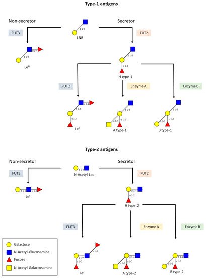
2.1. HBGAs and RV
2.2. HBGAs and NoV
The P2 subdomain found in the P-domain of the VP1 protein from NoVs interacts with HBGAs. Several studies have been made in order to elucidate the recognition pattern of NoVs. Some of them are based on the expression of the P-domain in vitro, which results in dimerization (P dimer) and the formation of P particles that retain HBGA-binding function, while others used virus-like particles (VLPs). These studies have utilized ELISA or haemagglutination-based assays using saliva, human milk, red blood cells, or synthetic oligosaccharides as HBGAs sources. The prototype Norwalk virus (GI.1) recognizes the type A and H secretors, but does not interact with type B secretors and non-secretors; Va387 (GII.4) binds to A, B, and O secretors; MOH (GII.5) and Hiro (GII.12) bind to A and B secretors; and Va207 (GII.9) recognizes Lewis positive secretors and non-secretors (Lex and Ley) [18,51][6][24]. As for the GII.4 strains Den Haag_2006b and Sydney_2012, Carmona et al. demonstrated that they did not recognize any HBGAs [52][25]. By contrast, these strains may recognize heparan sulphate or citrate since they are all capable of binding human NoV and may potentially play a role in NoV pathogenesis as cellular receptors/co-factors [53][26]. GI.3 NoV VLPs show strong binding to blood type A salivary HBGAs, slightly lower binding to blood type O salivary HBGAs, and weakly binding or none to blood type B and AB salivary HBGAs [54][27].3. The Role of Bacteria in RVs and NoVs Infection
3.1. Bacteria against Enteric Viral Infections
Several studies demonstrate the beneficial effect of probiotic bacteria against enteric virus infections and many other diseases [66,67,68,69][28][29][30][31]. Probiotics protect the host from viral infection by modulating gut microbiota composition, enhancing intestinal barrier function, and promoting mucosal immunity [70][32]. Additionally, they interfere with the binding of the virus to their target cells by competitive exclusion by blocking viral receptors and binding viruses on the surface to promote their elimination in faeces [71][33].3.2. Microbiota and Promotion of Enteric Viral Infections
Despite the significant evidence available about the role of intestinal-derived bacteria in the inhibition of viral infections, several investigations argued for a role of microbiota in promoting virus infection [81][34]. This was first demonstrated by Kuss et al. [82][35] and Kane et al. [83][36] when using poliovirus, reovirus, and mouse mammary tumour virus (MMTV) for infecting germ-free or antibiotic-treated mice. In these cases, it was observed that a substantial attenuation of infection occurred when compared to infection of microbially-colonized mice. Reconstitution of intestinal microorganisms into antibiotic-treated mice was enough to restore poliovirus pathogenesis [82][35]. Moreover, intestinal titres of reovirus were substantially reduced in antibiotic-treated, compared with control mice [83][36]. Similar findings were reported with RV and NoV when antibiotic-treated or germ-free mice were used [84[37][38],85], suggesting that microbiota enhances the pathogenesis of multiple families of enteric viruses.3.3. Microbiota and Restriction of Enteric Viral Infections
Recently, it has been shown that microbiota ablation with antibiotics in mice allows for infection with the human RV strain Wa (G1P[8]), which replicates very inefficiently in animals with normal microbiota [96][39]. These results are in conflict with earlier experiments which demonstrated that microbiota eradication by antibiotics results in reduced infection of murine RV (EC strain), as shown by lower viral shedding in adult mice and diminished diarrhea incidence in mice pups [84][37]. Nevertheless, viral clearance lasted longer in this model. Furthermore, recent experiments with the murine EDIM strain confirmed that antibiotic treatment and the consequent decrease in intestinal bacterial loads or the use of germ-free mice results in increased RV infection [97][40], which argues against a positive effect of the microbiota in RV infection. In the experiments with the Wa strain, animals with ablated microbiota and subsequent subjection to self-transplantation of intestinal microbiota partially recovered the resistance to infection, which allowed the identification of bacterial taxa that likely participate indirectly or directly in the restriction of Wa infection in mice [96][39]. Thus, bacteria belonging to lactobacilli, Mucispirillum, Oscillospira, and Bilophila genera were negatively linked to RV infection in mice. Faecal material transplantation with infants as donors did not restrict infectivity in this model, suggesting that the microbiota from the donors was not able to control RV infection in this model and that mice autochthonous bacteria were needed for the process [96][39].4. Microbiota and Enteric Viruses, Studies in Humans
Microbiota composition varies depending on the population [105][41] since it is affected by many factors including nutrition [106][42], sex, age, genetics, and health status [107][43], and these vary greatly between low-income and high-income countries. Such differences could be some of the reasons why RVVs have significantly lower efficacy in low-income countries [108,109][44][45].
A recent study evaluated whether microbiota modification by the use of broad- and narrow-spectrum antibiotics had an effect on immunization with Rotarix in adults [116][46]. Although the experimental groups did not differ in terms of total IgA produced, the narrow spectrum group showed a boost in IgA at day seven post-vaccination (basal levels of anti-RV IgA were high in the vaccination group) and the viral shedding was increased in both groups treated with antibiotics. Differences in the microbiota composition in faeces were evident between the groups and correlations between enrichment in Bacteroides populations at the boost at day seven were observed and several taxa (Prevotellaceae, Cloacibacillus everynsis, and Proteobacteria members such as Escherichia and Shigella) were associated with increased viral shedding. Antibiotic treatment had no effect on the immunogenicity of other systemic vaccines applied (pneumococcal and tetanus vaccine) [116][46]. These results highlight the fact that targeting the microbiota could be an alternative strategy to enhance RVVs efficacy, although the effectiveness in children still needs further investigation.
An ex vivo study analysed the bacterial groups that were interacting with RV in stool samples from children suffering RV (G1P[8]) diarrhea by flow cytometry followed by 16S rDNA sequencing [80][47]. This study also allowed the identification of Ruminococcus as RV-interacting bacteria. As already mentioned, a species of this group (R. gauvreauii) was shown to inhibit RV infection in vitro [80][47]. This, together with the correlation Ruminococcus-anti-RV IgA in humans and the fact that higher Ruminococcus numbers are found in healthy children compared to children with RV diarrhea [121][48], postulates these bacteria as likely players in the cross-talk bacteria-virus-host. Similar studies conducted with individuals suffering AGE caused by NoV will certainly aid in identifying bacterial taxons that interact with these viruses in stools. However, whether this interaction has some relevance in the infection process needs further investigation. All these findings may help to improve RVVs performance in such a way that they have higher efficacy in low-income countries, preventing tens of thousands of RV-related deaths per year. However, differences in the conclusions drawn from the microbiota analyses are evident, and standardized and controlled methods (e.g., sampling and DNA extraction techniques, bacterial 16S rDNA sequencing platforms, microbial composition analysis methods, etc.) are needed to get a clearer picture.References
- World Health Organization (WHO). The Top 10 Causes of Death. Available online: http://www.who.int/en/news-room/fact-sheets/detail/the-top-10-causes-of-death (accessed on 10 September 2021).
- Donaldson, E.F.; Lindesmith, L.C.; Lobue, A.D.; Baric, R.S. Norovirus pathogenesis: Mechanisms of persistence and immune evasion in human populations. Immunol. Rev. 2008, 225, 190–211.
- Jiang, X.; Liu, Y.; Tan, M. Histo-blood group antigens as receptors for rotavirus, new understanding on rotavirus epidemiology and vaccine strategy. Emerg. Microbes Infect. 2017, 6, e22.
- Shanker, S.; Czakó, R.; Sapparapu, G.; Alvarado, G.; Viskovska, M.; Sankaran, B.; Atmar, R.L.; Crowe, J.E.; Estes, M.K.; Prasad, B.V.V. Structural basis for norovirus neutralization by an HBGA blocking human IgA antibody. Proc. Natl. Acad. Sci. USA 2016, 113, E5830–E5837.
- Marionneau, S.; Cailleau-Thomas, A.; Rocher, J.; Le Moullac-Vaidye, B.; Ruvoën, N.; Clément, M.; Le Pendu, J. ABH and Lewis histo-blood group antigens, a model for the meaning of oligosaccharide diversity in the face of a changing world. Biochimie 2001, 83, 565–573.
- Tan, M.; Jiang, X. Histo-blood group antigens: A common niche for norovirus and rotavirus. Expert. Rev. Mol. Med. 2014, 16, e5.
- Barbé, L.; Le Pendu, J.; Echasserieau, K.; Ruvoën-Clouet, N.; Bernardeau, K.; Le Moullac-Vaidye, B.; Bovin, N.; Nordgren, J.; Carton, T.; Svensson, L. Histo-blood group antigen-binding specificities of human rotaviruses are associated with gastroenteritis but not with in vitro infection. Sci. Rep. 2018, 8, 12961.
- Marionneau, S.; Ruvoën, N.; Le MoullacVaidye, B.; Clement, M.; CailleauThomas, A.; RuizPalacois, G.; Huang, P.; Jiang, X.; Le Pendu, J. Norwalk Virus binds to histo-blood group antigens present on gastroduodenal epithelial cells of secretor individuals. Gastroenterology 2002, 122, 1967–1977.
- Ayouni, S.; Sdiri-Loulizi, K.; de Rougemont, A.; Estienney, M.; Ambert-Balay, K.; Aho, S.; Hamami, S.; Aouni, M.; Neji-Guediche, M.; Pothier, P.; et al. Rotavirus P infections in persons with secretor and nonsecretor phenotypes, Tunisia. Emerg. Infect. Dis. 2015, 21, 2055–2058.
- Arias, C.F.; López, S. Rotavirus cell entry: Not so simple after all. Curr. Opin. Virol. 2021, 48, 42–48.
- Böhm, R.; Fleming, F.E.; Maggioni, A.; Dang, V.T.; Holloway, G.; Coulson, B.S.; Von Itzstein, M.; Haselhorst, T. Revisiting the role of histo-blood group antigens in rotavirus host-cell invasion. Nat. Commun. 2015, 6, 5907.
- Liu, Y.Y.; Huang, P.; Tan, M.; Biesiada, J.; Meller, J.; Castello, A.A.; Jiang, B.; Jiang, X. Rotavirus VP8*: Phylogeny, host range, and interaction with histo-blood group antigens. J. Virol. 2012, 86, 9899–9910.
- Xu, S.; McGinnis, K.R.; Liu, Y.; Huang, P.; Tan, M.; Stuckert, M.R.; Burnside, R.E.; Jacob, E.G.; Ni, S.; Jiang, X.; et al. Structural basis of P rotavirus evolution and host ranges under selection of histo-blood group antigens. Proc. Natl. Acad. Sci. USA 2021, 118, e2107963118.
- Huang, P.; Xia, M.; Tan, M.; Zhong, W.; Wei, C.; Wang, L.; Morrow, A.; Jiang, X. Spike protein VP8* of human rotavirus recognizes histo-blood group antigens in a type-specific manner. J. Virol. 2012, 86, 4833–4843.
- Xu, S.; Ahmed, L.U.; Stuckert, M.R.; McGinnis, K.R.; Liu, Y.; Tan, M.; Huang, P.; Zhong, W.; Zhao, D.; Jiang, X.; et al. Molecular basis of P major human rotavirus VP8* domain recognition of histo-blood group antigens. PLoS Pathog. 2020, 16, e1008386.
- Fix, J.; Chandrashekhar, K.; Perez, J.; Bucardo, F.; Hudgens, M.G.; Yuan, L.; Twitchell, E.; Azcarate-Peril, M.A.; Vilchez, S.; Becker-Dreps, S. Association between Gut Microbiome Composition and Rotavirus Vaccine Response among Nicaraguan Infants. Am. J. Trop. Med. Hyg. 2020, 102, 213–219.
- Gozalbo-Rovira, R.; Ciges-Tomas, J.R.; Vila-Vicent, S.; Buesa, J.; Santiso-Bellón, C.; Monedero, V.; Yebra, M.J.; Marina, A.; Rodríguez-Díaz, J. Unraveling the role of the secretor antigen in human rotavirus attachment to histo-blood group antigens. PLoS Pathog. 2019, 15, e1007865.
- Liu, Y.; Xu, S.; Woodruff, A.L.; Xia, M.; Tan, M.; Kennedy, M.A.; Jiang, X. Structural basis of glycan specificity of P VP8*: Implications for rotavirus zoonosis and evolution. PLoS Pathog. 2017, 13, e1006707.
- Lee, S.-K.; Oh, S.J.; Choi, S.; Choi, S.H.; Shin, S.-H.; Lee, E.J.; Cho, E.-J.; Hyun, J.; Kim, H.S. Relationship Between Rotavirus P Infection in Korean Neonates and Histo-Blood Group Antigen: A Single-Center Study. Ann. Lab. Med. 2021, 41, 181–189.
- Liu, Y.; Ramelot, T.A.; Huang, P.; Liu, Y.; Li, Z.; Feizi, T.; Zhong, W.; Wu, F.-T.; Tan, M.; Kennedy, M.A.; et al. Glycan Specificity of P Rotavirus and Comparison with Those of Related P Genotypes. J. Virol. 2016, 90, 9983–9996.
- Hu, L.; Crawford, S.E.; Czako, R.; Cortes-Penfield, N.W.; Smith, D.F.; Le Pendu, J.; Estes, M.K.; Prasad, B.V.V. Cell attachment protein VP8* of a human rotavirus specifically interacts with A-type histo-blood group antigen. Nature 2012, 485, 256–259.
- Liu, Y.; Huang, P.; Jiang, B.; Tan, M.; Morrow, A.L.; Jiang, X. Poly-LacNAc as an Age-Specific Ligand for Rotavirus P in Neonates and Infants. PLoS ONE 2013, 8, e78113.
- Hu, L.; Sankaran, B.; Laucirica, D.R.; Patil, K.; Salmen, W.; Ferreon, A.C.M.; Tsoi, P.S.; Lasanajak, Y.; Smith, D.F.; Ramani, S.; et al. Glycan recognition in globally dominant human rotaviruses. Nat. Commun. 2018, 9, 2631.
- Huang, P.; Farkas, T.; Zhong, W.; Tan, M.; Thornton, S.; Morrow, A.L.; Jiang, X. Norovirus and Histo-Blood Group Antigens: Demonstration of a Wide Spectrum of Strain Specificities and Classification of Two Major Binding Groups among Multiple Binding Patterns. J. Virol. 2005, 79, 6714–6722.
- Carmona-Vicente, N.; Vila-Vicent, S.; Allen, D.; Gozalbo-Rovira, R.; Iturriza-Gómara, M.; Buesa, J.; Rodríguez-Díaz, J. Characterization of a Novel Conformational GII.4 Norovirus Epitope: Implications for Norovirus-Host Interactions. J. Virol. 2016, 90, 7703–7714.
- Almand, E.A.; Moore, M.D.; Jaykus, L.-A. Norovirus Binding to Ligands Beyond Histo-Blood Group Antigens. Front. Microbiol. 2017, 8, 2549.
- Zheng, L.; Zhang, H.; Ma, J.; Liu, J.; Ma, S.; Wang, M.; Huo, Y. Phylogenetic and biological characterizations of a GI.3 norovirus. Infect. Genet. Evol. 2020, 85, 104554.
- Hao, Q.; Lu, Z.; Dong, B.R.; Huang, C.Q.; Wu, T. Probiotics for preventing acute upper respiratory tract infections. Cochrane Database Syst. Rev. 2015, 2, CD006895.
- Ahmadi, E.; Alizadeh-Navaei, R.; Rezai, M.S. Efficacy of probiotic use in acute rotavirus diarrhea in children: A systematic review and meta-analysis. Casp. J. Intern. Med. 2015, 6, 187–195.
- Wu, Y.; Zhang, Q.; Ren, Y.; Ruan, Z. Effect of probiotic Lactobacillus on lipid profile: A systematic review and meta-analysis of randomized, controlled trials. PLoS ONE 2017, 12, e0178868.
- Rees, C.M.; Hall, N.J.; Fleming, P.; Eaton, S. Probiotics for the prevention of surgical necrotising enterocolitis: Systematic review and meta-analysis. BMJ Paediatr. Open 2017, 1, e000066.
- Lei, S.; Twitchell, E.; Yuan, L. Pathogenesis, Immunity and the Role of Microbiome/Probiotics in Enteric Virus Infections in Humans and Animal Models. In Mechanisms Underlying Host-Microbiome Interactions in Pathophysiology of Human Diseases; Springer: Boston, MA, USA, 2018; pp. 55–78.
- Monedero, V.; Rodríguez-Díaz, J. Intestinal microbiota and susceptibility to viral infections: Role of probiotics. Probiotics Prebiotics Synbiotics 2016, 813–826.
- Domínguez-Díaz, C.; García-Orozco, A.; Riera-Leal, A.; Padilla-Arellano, J.R.; Fafutis-Morris, M. Microbiota and Its Role on Viral Evasion: Is It with Us or Against Us? Front. Cell. Infect. Microbiol. 2019, 9, 256.
- Kuss, S.K.; Best, G.T.; Etheredge, C.A.; Pruijssers, A.J.; Frierson, J.M.; Hooper, L.V.; Dermody, T.S.; Pfeiffer, J.K. Intestinal microbiota promote enteric virus replication and systemic pathogenesis. Science 2011, 334, 249–252.
- Kane, M.; Case, L.K.; Kopaskie, K.; Kozlova, A.; MacDearmid, C.; Chervonsky, A.V.; Golovkina, T.V. Successful Transmission of a Retrovirus Depends on the Commensal Microbiota. Science 2011, 334, 245–249.
- Uchiyama, R.; Chassaing, B.; Zhang, B.; Gewirtz, A.T. Antibiotic treatment suppresses rotavirus infection and enhances specific humoral immunity. J. Infect. Dis. 2014, 210, 171–182.
- Jones, M.K.; Watanabe, M.; Zhu, S.; Graves, C.L.; Keyes, L.R.; Grau, K.R.; Gonzalez-Hernandez, M.B.; Iovine, N.M.; Wobus, C.E.; Vinje, J.; et al. Enteric bacteria promote human and mouse norovirus infection of B cells. Science 2014, 346, 755–759.
- Gozalbo-Rovira, R.; Santiso-Bellón, C.; Buesa, J.; del Campo, A.R.; Vila-Vicent, S.; Muñoz, C.; Yebra, M.J.; Monedero, V.; Rodríguez-Díaz, J. Microbiota Depletion Promotes Human Rotavirus Replication in an Adult Mouse Model. Biomedicines 2021, 9, 846.
- Schnepf, D.; Hernandez, P.; Mahlakõiv, T.; Crotta, S.; Sullender, M.E.; Peterson, S.T.; Ohnemus, A.; Michiels, C.; Gentle, I.; Dumoutier, L.; et al. Rotavirus susceptibility of antibiotic-treated mice ascribed to diminished expression of interleukin-22. PLoS ONE 2021, 16, e0247738.
- Gupta, V.K.; Paul, S.; Dutta, C. Geography, Ethnicity or Subsistence-Specific Variations in Human Microbiome Composition and Diversity. Front. Microbiol. 2017, 8, 1162.
- Srivastava, V.; Deblais, L.; Huang, H.-C.; Miyazaki, A.; Kandasamy, S.; Langel, S.N.; Paim, F.C.; Chepngeno, J.; Kathayat, D.; Vlasova, A.N.; et al. Reduced rotavirus vaccine efficacy in protein malnourished human-faecal-microbiota-transplanted gnotobiotic pig model is in part attributed to the gut microbiota. Benef. Microbes 2020, 11, 733–751.
- Desselberger, U. Differences of Rotavirus Vaccine Effectiveness by Country: Likely Causes and Contributing Factors. Pathogens 2017, 6, 65.
- Armah, G.E.; Sow, S.O.; Breiman, R.F.; Dallas, M.J.; Tapia, M.D.; Feikin, D.R.; Binka, F.N.; Steele, A.D.; Laserson, K.F.; Ansah, N.A.; et al. Efficacy of pentavalent rotavirus vaccine against severe rotavirus gastroenteritis in infants in developing countries in sub-Saharan Africa: A randomised, double-blind, placebo-controlled trial. Lancet 2010, 376, 606–614.
- Madhi, S.A.; Cunliffe, N.A.; Steele, D.; Witte, D.; Kirsten, M.; Louw, C.; Ngwira, B.; Victor, J.C.; Gillard, P.H.; Cheuvart, B.B.; et al. Effect of Human Rotavirus Vaccine on Severe Diarrhea in African Infants. N. Engl. J. Med. 2010, 362, 289–298.
- Harris, V.C.; Haak, B.W.; Handley, S.A.; Jiang, B.; Velasquez, D.E.; Hykes, B.L.; Droit, L.; Berbers, G.A.M.; Kemper, E.M.; van Leeuwen, E.M.M.; et al. Effect of Antibiotic-Mediated Microbiome Modulation on Rotavirus Vaccine Immunogenicity: A Human, Randomized-Control Proof-of-Concept Trial. Cell Host Microbe 2018, 24, 197–207.e4.
- Gozalbo-Rovira, R.; Rubio-Del-campo, A.; Santiso-Bellón, C.; Vila-Vicent, S.; Buesa, J.; Delgado, S.; Molinero, N.; Margolles, A.; Yebra, M.J.; Collado, M.C.; et al. Interaction of intestinal bacteria with human rotavirus during infection in children. Int. J. Mol. Sci. 2021, 22, 1010.
- Dinleyici, E.C.; Martínez-Martínez, D.; Kara, A.; Karbuz, A.; Dalgic, N.; Metin, O.; Yazar, A.S.; Guven, S.; Kurugol, Z.; Turel, O.; et al. Time Series Analysis of the Microbiota of Children Suffering from Acute Infectious Diarrhea and Their Recovery After Treatment. Front. Microbiol. 2018, 9, 1230.