Your browser does not fully support modern features. Please upgrade for a smoother experience.
Please note this is a comparison between Version 1 by Shan Li and Version 2 by Rita Xu.
The vascular bundle is an important structural unit that determines the growth and properties of bamboo. A high-resolution X-ray microtomography (μCT) was used to observe and reconstruct a three-dimensional (3D) morphometry model of the vascular bundle of the Qiongzhuea tumidinoda node due to its advantages of quick, nondestructive, and accurate testing of plant internal structure.
- Qiongzhuea tumidinoda
- structure of bamboo node
- three-dimensional reconstruction
1. Introduction
The node is the basic characteristic of bamboo plants and plays a key role in the overall performance of the hollow structure of bamboo culms [1][2][1,2]. The vascular bundle serves as a mechanical support to improve the mechanical properties of bamboo [2][3][2,3] and perform the function of transporting water and nutrients [4]. Numerous reports are available on the microstructure of the bamboo culm, mainly on the internodes [5][6][7][5,6,7]. However, the research concerning the structure of bamboo nodes due to its disordered and relatively complicated arrangement of vascular bundles is limited. The research on the structure of bamboo nodes began in the 1960s [8] and mainly relied on optical microscopes. In the late 20th century, the researches related to the anatomy of bamboo nodes gradually increased [4][9][10][11][4,9,10,11]. Grossor and Liese [11] studied the characteristics of vascular bundles of bamboo nodes through enlarged and spliced images of microsections. Xiong et al. studied the shape of bamboo nodes and conducted a preliminary discussion on the 3D characteristics of vascular bundles [10][12][10,12]. Ding and Liese [4] reconstructed a complex 3D structure of vascular bundles of bamboo nodes by observing continuous longitudinal and transverse slices. Currently, the research of the bamboo microstructure mainly relies on two-dimensional (2D) image characterization techniques such as optical microscope and field emission scanning electron microscope. However, the aforementioned methods damaged the samples, leading to the inaccurate results. The complexity and fragility of sample preparation with traditional techniques make it difficult to meet the requirements of nondestructive, high-efficiency visualization. Therefore, a higher-resolution nondestructive testing method is needed.
With the continuous development of research methods, X-ray microscopy has strong penetrating power, high resolution, and nondestructive property, resulting in fast detection and intuitive results, and indicating advantages of bamboo node analysis. Peng et al. [13] and Xiang et al. [14] used X-ray microtomography (μCT) as a tool for 2D and 3D complex network structures of the vascular bundle in the bamboo node. Huang et al. quantified the difference in the density distribution and the porosity of bamboo nodes based on 2D images [15][16][15,16]. Palombinifl et al. [17][18][19][17,18,19] analyzed the relative density and cell shape of different tissues of a cutting region of bamboo nodes, evaluated the mechanical behavior of parenchyma by finite element analysis, and first obtained the 3D reconstruction model of the complex vascular bundle system of monocot nodes. The volume, porosity, relative density, and distribution of nodes were also characterized. The aforementioned research by X-ray technique had two limitations: (1) The conducting tissue and the fibers of the completed node’s vascular bundle were not separated to separately observe the morphology of them. (2) The complex distribution of transverse vascular bundles in the diaphragm was neglected.
Qiongzhuea tumidinoda Hsueh et T.P.Yi. belongs to the genus Qiongzhuea in the Gramineae family, with an enlarged node and a peculiar-shaped culm. The species reaches approximately 2.5–6 m in height, and its culm reaches 1–3 cm in diameter. Q. tumidinoda, only found in a restricted area in S.W. Sichuan and N.E. Yunnan, is a rare and endangered bamboo species, which is a Class 3 protected plant on the national key protection list in China. Among them, especially in Daguan County, Yunnan province, this bamboo accounts for more than 50% of the total global resources, with 0.014 million hectares. The bamboo culm is mainly used for crafts and production of round bamboo furniture with extremely high economic and ornamental value. However, this bamboo at more than 5-years of age will gradually begin to wither and die. The most obvious feature is that the node automatically disconnects. Furthermore, the culm is more prone to regular fracture at the nodal ridge of the node, even less than 5-year-old bamboo. The presence of the bamboo node makes it possible for the lateral transport of water and nutrients [4], and there is little research on what happened to its structure before the node fell off.
2. Materials
Five 2-year-old Q. tumidinoda bamboo plants with good growth and straight stems were collected from Yinji Village, Daguan County, Yunnan Province (103°99′ N, 28°13′ E). The culm was cut every 2 m from the base to the top of it, marked, and transported to the laboratory. After natural air drying, complete bamboo nodes with 5.74% moisture content from the middle of the culm were selected (Figure 1b) and were ready to be used for the subsequent experiments. The selected node was approximately 1.83 m of the culm, which was 3.7 m in height and 1.2 cm in diameter.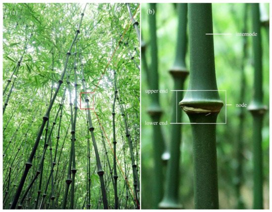
Figure 1.
Preparation of μCT sample. (
a
) Bamboo. (
b
) Sampling location (white wireframe area).
3. Image Processing and 3D Reconstruction
Images were processed and analyzed with the open-source Fiji software (National Institutes of Health, Bethesda, MD, USA) and the AVIZO software (FEI Hillsboro, OR, USA), the processing steps are shown in Figure 2. The slices were combined and reconstructed from the projections with a noniterative filtered backprojection algorithm in NRecon software and then exported as a stack of TIFF images. A total of 1290 slices with serial numbers 329–1618 were obtained. The original TIFF stack (Figure 2a) was first denoised with the adaptive median filtering technique combined with the non-local mean filtering algorithm (Figure 2b), and the contrast was adjusted (Figure 2c). Figure 2a1,b1,c1 demonstrates details of the same slice before and after noise reduction and contrast adjustment, respectively. The denoised intensity images had clear boundaries for easy segmentation.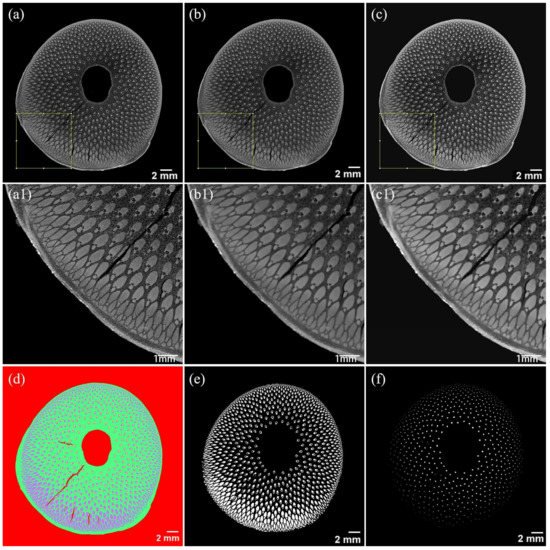
Figure 2. Image processing of μCT. (a) Original μCT slice and its details (a1). (b) Filter processing and its details (b1). (c) Contrast adjusting and its details (c1). (d) Segmentation of the conducting tissue (yellow), fibers (purple), parenchyma (green), and air (red). (e) Binary images of fibers. (f) Binary images of conducting tissue.
4. Morphology of the Bamboo Node
The morphological diagram of the node of Q. tumidinoda is shown in Figure 3a, which consists of a sheath scar, nodal ridge, diaphragm, and the intra-node between the nodal ridge and sheath scar. The rare swollen node was seen in Q. tumidinoda bamboo, the nodal ridge was more swollen than the sheath scar. This might be related to the different speeds of cell division and growth around the intercalary meristem [12].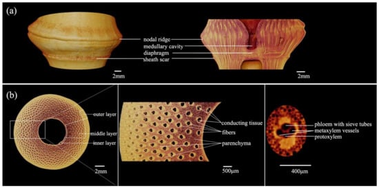
Figure 3. Morphology of the node in Q. tumidinoda. (a) Longitudinal section view. (b) Cross-section and details.
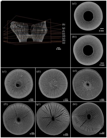
Figure 4. X-ray microtomography slices of the Q. tumidinoda node. The sequence number of the transversal slices is 329–1618 from the lower end to the upper end (details of a–h present in a1–h1). (a1) Slice 339, end of the node. (b1) Slice 387, lower end of the node. (c1) Slice 579, lower end of the sheath scar. (d1) Slice 669, middle of the diaphragm. (e1) Slice 849, upper end of the diaphragm. (f1) Slice 944, lower end of the nodal ridge. (g1) Slice 1129, middle of the nodal ridge, and (h1) Slice 1429, upper end of the nodal ridge.