Your browser does not fully support modern features. Please upgrade for a smoother experience.
Please note this is a comparison between Version 1 by Domenico Baldini and Version 2 by Catherine Yang.
Intracytoplasmic sperm injection (ICSI) is the method that has definitely revolutionized the field of ART since normal fertilization and ongoing pregnancies can be achieved even with low quality sperm samples and affected spermatozoa. By injecting a single sperm cell in the oocyte, the technique bypasses several biological barriers that naturally select the gametes to achieve optimal embryonic and fetal development.
- assisted reproductive technology (ART)
- in vitro fertilization (IVF)
- ICSI
1. Natural Sperm Selection
In nature, the process that leads to the selection of the best sperm cells, capable of fertilizing the oocyte in the female genital tract, is very selective and it involves high morphologic qualities and dynamic features. Leaving the seminal plasma in the vagina, sperm cells perform the first step that leads to this process.
Only a small portion of the sperm cells that swim from the uterus to the oviduct can be collected in niches, where cells interact with unknown receptors to form a sperm reserve, as in some animal species the sperm cells can stay in the fallopian tubes even for months before ovulation. However, in the human species, the fertilization window is reduced, and the sperm cells can survive no longer than 5/6 days [1][14]. This process allows the sperm to reach complete capacitation, which is based on plasma membrane cholesterol ultrastructure changes; it leads to an increase in concentration of intracellular ions, such as Ca2+, that switch the motility patterns of tails to hyper-activation. Moreover, phosphorylation on tyrosine residues determines the preparation for the acrosomal reaction event [2][15]; subsequent interactions with molecules, such as secreted protein or hormones, from the female reproductive tract, modulate the swim, by a chemotactic and thermotactic effect, towards the oocyte.
All current methodologies improve sperm cells quality, but none have been associated with a significant increase in clinical results [3][17]. Methods for sperm cells’ selection are categorized into classic techniques based on sperm motility or density, and advanced methods that rely on membrane surface charge, high-resolution morphology, and nuclear or membrane integrity. Nowadays, the most used techniques in the ART lab are SU and DGC [4][18]. Here below the Table 1 represent a summary of all the sperm cell preparation techniques with advantages and disadvantages of each of them.
Table 1.
Advantages and disadvantages of sperm cells’ selection techniques.
| Procedures | Advantages | Disadvantages |
|---|
| Swim-up |
|
| ||||||
| Centrifugation on density gradient |
|
| ||||||
| HOST |
|
| ||||||
| Polarization microscopy |
|
| ||||||
| LAISS |
|
| ||||||
| MACS |
|
| ||||||
| PICSI and selection with hyaluronic acid |
|
| ||||||
| Zeta potential |
|
| ||||||
| IMSI |
|
| ||||||
| Microfluidic separation |
|
| ||||||
| Horizontal | Sperm migration |
|
|
2. Advanced Methods
2.1. Selection Methods for Sperm Cells with Reduced Motility
When the sperm sample is retrieved by testicular aspiration (TESE) [50][73] or epididymal aspiration (MESA) [51][74], the spermatozoa might appear immotile due to the lack of complete maturation that takes place in the final tract of the epididymis [52][75].
It is important to underline that in these cases, the SU and DGC techniques, that exploit dynamic characteristics for separation, are inadequate.
An alternative selection technique is the hypo-osmotic swelling test (HOST) [14][28]. This method (Figure 12) assumes that the tails of viable spermatozoa swell and bend if they are introduced into a hypoosmotic environment, due to the activity of the osmo-sensitive calcium membrane channels [53][54][78,79]. HOST can be used to estimate the percentage of integrity in chromatin.
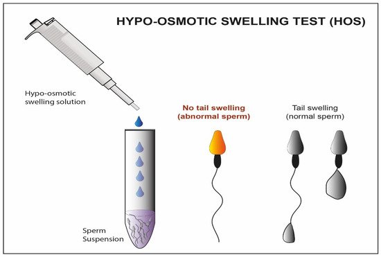
Figure 12. Schematic representation Hypo-osmotic swelling Test. In this figure, we observe the difference between the abnormal sperm with not swelled tail and the normal sperm with swelled tail after treatment with a hypo-osmotic solution.
Using polarized light implemented in the microscope (Figure 23) is another possible strategy for the selection of immotile sperm cells [19][33]. Some research teams have proposed its use to verify the birefringence of sperm cell head as an index of suitability. As a matter of fact, the sub-acrosomal protein filaments extend themselves longitudinally, giving a typical pattern of birefringence to the sperm cell head [20][34]. In some published studies, the selection of non-motile spermatozoa with birefringent heads resulted in an increase in clinical pregnancy and implantation rate (58% vs. 9% and 42% vs. 12%), compared to a control group with immotile spermatozoa (where polarized light was not used) [55][81], and an increased implantation rate when the HOST technique was used instead as selection method (45% vs. 11%) [56][82].
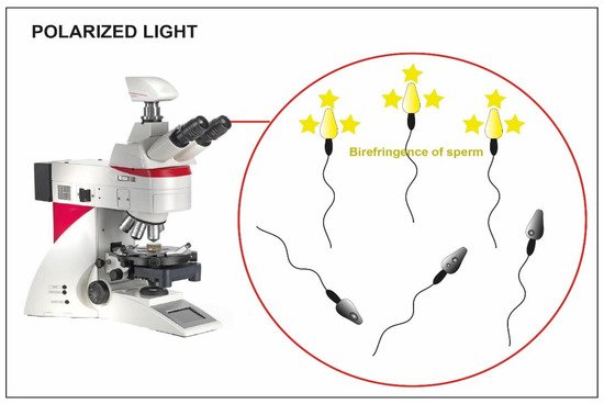
Figure 23. Microscope implemented with polarized light. The birefringence of the heads is clear in the viable sperm (yellow heads) compared to the not viable one where the birefringence is absent (dark heads).
Unfortunately, even this technique presents some disadvantages: the polarized light implemented in the microscope is expensive and there is a lack of data regarding the integrity of the sperm DNA. Furthermore, the success of the latter is based largely on the experience of the operator.
A group of researchers has shown another potential method to recover immotile but suitable sperms for ICSI. It exploits the chemical inducers of motility (Figure 34), that belong to the class of phosphodiesterase inhibitors, such as pentoxifylline (PTF), dimethylxanthines and papaverines [57][90], allowing the motility reactivation of spermatozoa recovered from the testicular and epididymal level [58][91].
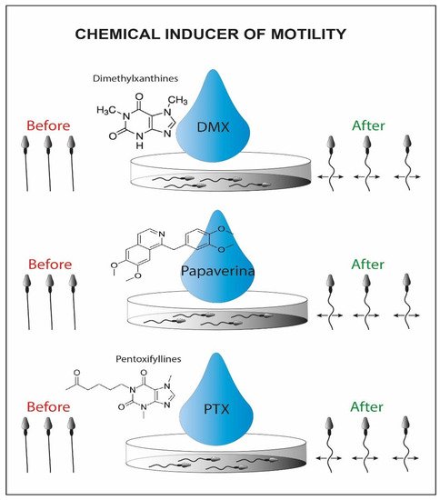
Figure 34. Schematic representation of chemical inducers of motility. On the left we can recognize the initial immotile sperms and on the right side the viable motile sperms activated by the chemical inducer.
Alternatively, motility can be further reactivated by using a laser incorporated in the magnification system that is shot at the immotile sperm cell tail (LAISS laser-assisted immotile sperm selection) (Figure 45) [21][35]. Laser irradiation causes the release in cytosol of second messengers, such as Ca2+ or ROS, and an increase in the synthesis of ATP, which could generate a slight movement of the tail [59][98]. The sperm cell is then considered viable when its tail is coiled after the laser shot [22][36]. On the other hand, some authors claim that high laser doses cause an excess of potentially toxic ROS [25][39]. Furthermore, more Ca2+ influx induces the hyperactivity of Ca2+-ATPase calcium channels and exhausts the ATP reserves of the cell. This process could lead to depletion of cell channels activity, correlated with an increase in internal osmotic pressure causing the swelling of the sperm cell and subsequently the rupture of its plasma membrane [26][40]. Contrary, other authors suggest that this technique does not damage the spermatic membrane and does not affect the percentage of fragmentation of the genetic material [60][99].
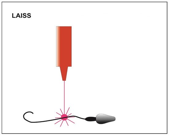
Figure 45. Schematic representation of LAISS (laser-assisted immotile sperm selection). The laser irradiation generates a slight movement of the tail in those viable sperms initially immotile.
It has also been demonstrated that the use of ATP/MgSO4 generates an increase in motility in testicular seminal samples, generating a weak contraction of the flagellum (Figure 56). The results of the study have shown that using ATP as a solute, the induced motility increases significantly, and viable sperm cells can be captured and used for injection through ICSI [61][102].
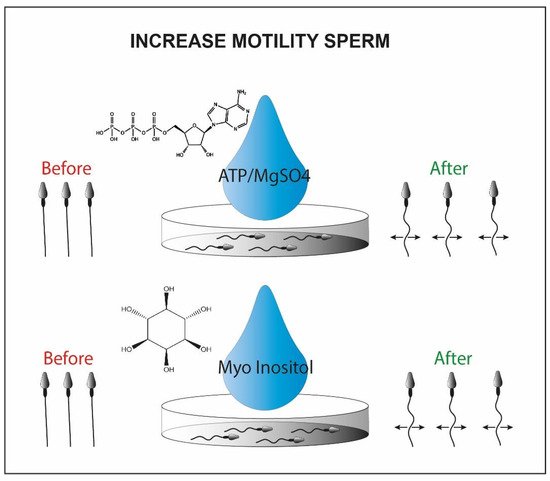
Figure 56. Sperm motility enhanced by ATP/MgSo4 or myo-inositol. Briefly in the picture we observe the immotile sperm before the treatment with the chemical inducer (left side) and the motile sperm cells after the chemical exposure (right side).
Alternatively, the myoinositol can be used to isolate viable sperm cells for ICSI. It represents the most abundant stereoisomer of the inositol class modulating the intracellular concentration of Ca2+ [62][103]. This is synthesized in two steps by two enzymes, myo-1-phosphate synthase and myo-monophosphatase-1, located in high concentration in the testicular mesenchymal tissue [63][64][104,105]. By incubating sperm cells frozen and then thawed from oligoasthenospermic patients with myoinositol, some research groups were able to recover a portion of sperm cells with significantly increased motility [65][101].
2.2. Sperm Cells Selection by Membrane Characteristics
The outer sperm membrane is critical for their functionality since it is firstly involved in many aspects of the fertilization such as capacitation, oocyte binding and acrosome reaction [66][67][109,110]. In order to select high-quality sperm cells, methods that gain benefits from the characteristics of their membrane have been studied.
A further method that takes advantage of the membrane characteristics is the magnetic activated cell sorting (MACS). This methodology allows the selection of the non-apoptotic portion from a sample of interest [68][114]. It involves the use of magnetic microspheres conjugated to Annexin V (AV-MACS) (Figure 67) [69][115], that have a high affinity for phosphatidyl-serine. The latter is normally exposed on the outer side of the membrane when the sperm cells are in an apoptotic state [70][116]. The seminal sample of interest passes through a column containing microspheres, to which the annexin has adhered. The non-viable spermatozoa remain trapped inside the column, while the viable fraction is eluted, improving the vitality characteristics of the starting sample [27][28][41,42].
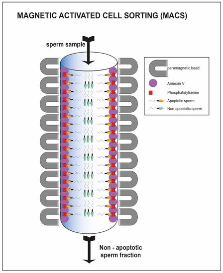
Figure 67. Sperm cell passing through column with annexin V. The sperm cells able to pass through the column represent the viable one (green heads) while the apoptotic fraction remain trapped inside the column (yellow heads).
Hyaluronic acid is the main component of the extracellular matrix surrounding the cumulus-oocyte complex [71][119]. Only the sperm cells that have successfully completed spermatogenesis and maturation are able to show the polysaccharide-binding receptors on their outer membrane [32][33][46,47]. Moreover, they are usually characterized by a normal morphology and a low percentage of fragmentation of the nuclear material [34][48]. Exploiting these characteristics, a methodology for sperm cell selection has been devised where spermatozoa are incubated in plates or in media containing hyaluronic acid; only the sperm cells able to bind the molecule are then used to perform a modified version of ICSI: physiological intracytoplasmic sperm injection (PICSI) [72][73][120,121]. The selection should result in an increased fertilization rate, but in this case, the clinical data are conflicting. Some studies suggest that both the fertilization rate and the percentage of top-quality embryos benefit from the technique (92 vs. 86; 36 vs. 24%) [74][122], but other research groups do not confirm these improvements [35][49]. Studies show that a negative charge is present on the sperm cell membrane [75][123]. The Zeta method (Figure 78) uses this feature to separate sperm cells containing the Y chromosome from those containing the X chromosome [36][50]. Two distinct research groups have developed two different methodologies, one using a positively charged centrifuge tube [40][54], the other using migration in an electrophoretic field [41][55], which allow the collection of live spermatozoa with normal morphology and a high percentage of the integrity of the genetic material [37][38][76][77][51,52,124,125].
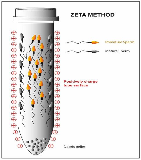
Figure 78. Schematic representation of the Zeta method. The high negative charge of the mature sperm cell membrane reacts with the positive charge of the tube while the immature fraction is detached form the tube.
2.3. Selection Based on Morphology–IMSI
The analysis of semen quality has for a long time been associated with the morphological evaluation of spermatozoa. The introduction of digital microscopy has made its possible to analyze the ultrastructural characteristics of motile spermatozoa (MSOME; motile sperm organelle morphology examination) [78][126]. The integration of the MSOME into the ICSI method allows a high-magnificence micro-injection (IMSI) (Figure 89) [79][127]. The selection of motile spermatozoa with few vacuoles and normal nuclear morphology is possible through a 6000× magnification system integrated with the micromanipulation system [80][128].
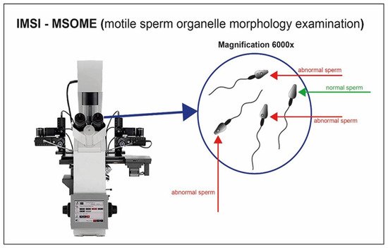
Figure 89. Sperm cell visualization with magnification system 6000×. Sperm cells with appropriate morphology are well selected through the high magnification.
2.4. Dynamic Selection of Spermatozoa
The three main mechanisms that control the movements of spermatozoa within the oviducts, rheotaxis, chemotaxis and thermotaxis, have been exploited as methods for sperm cell selection, to improve the ART outcomes:
-
Human spermatozoa orient their motion, by rheotaxis, against the fluid stream directed towards them [81][129]. Rheotaxis is defined as the tendency of certain living beings to move in response to the mechanical stimulus of a current of water. Some studies have confirmed the presence of a flow-directed towards the uterus in the oviductal region, capable of increasing its intensity after intercourse, which is able to attract the sperm cells [82][130]. In the technique developed by Nagata et al. [83][131], using bull sperm as a mammalian model, a flow is created through a series of microchannels towards a well where the sperm sample is delivered. In response to the flow, the spermatozoa swim towards it passing through the microchannels and finally reaching a receptive well where they can be collected for ART applications. Another study was conducted on normospermic patients [84][132] to select spermatozoa using rheotaxis: compared to an untreated sample and a sample subjected to density gradient centrifugation, the recovered spermatozoa showed higher chromatin compactness (99% vs. 71% vs. 83%).
-
Progesterone plays the main role as a chemo-attractor for the navigation of spermatozoa through the environment surrounding the cumulus-oocyte complex [85][134]. Only capacitated spermatozoa possess the receptors to recognize and bind this type of molecule [85][134]. Some sperm cell selection techniques exploit this feature to distinguish capacitated spermatozoa from non-capacitated ones. Several studies have been conducted on different types of sperm populations, using a device where a progesterone concentration gradient is constituted (sperm selection assay) [86][135]. In this method, two wells are connected by a 2 mm length per 2.5 mm diameter tube. One of the wells is filled with a media containing the chemoattractant molecule in solution (progesterone for instance) and the other well is filled with the human sperm sample. The chemoattractant diffuses through the tube generating a gradient and the spermatozoa respond by moving towards the higher concentration and accumulating in the initial well free of cells where they can be used for ART applications. The recovered spermatozoa from this technique exhibit better morphology, less DNA fragmentation and a reduced rate of apoptosis as compared to those selected with density gradient centrifugation [87][136]. Although the results are also promising in this case, further studies need to be conducted to confirm the improvements in the clinical field.
-
The spermatozoa can direct their motion according to the variation in temperature, moving from cold areas to warmer areas [88][137]. Several studies show that this mechanism underlies the movement of sperm cells from the fallopian tubes to the ampulla [89][90][138,139]. Even in this case, however, only the capacitated spermatozoa can respond to the temperature gradient, making this motion a mechanism for the selection of sperm cells with better fertilizing characteristics. The method of sperm selection by thermotaxis was developed by Pérez-Cerezales et al. [91][140].
2.5. Microfluidics Applied to Sperm Selection
Technologies related to microfluidics are rapidly growing within ART laboratories. Among the first experiments with this methodology, Smith and Takayama [46][60] have published a series of articles demonstrating the efficiency of the method for the selection of high-quality spermatozoa.
By controlling fluid dynamics (Figure 910), within millimeter diameter capillaries, it is possible to mimic the physiological conditions of pH and temperature of the female genital tract [92][141]. Hence, we could potentially select spermatozoa with increased motility through flows [93][142], chemical gradients [94][143] or electrophoretic fields [95][144]. The method of Smith and Takayama [46][60] uses two parallel laminar flow channels. While the motile spermatozoa can move through the flows and be eluted separately, the debris and immotile cells are passively transported from the entrance to the exit of the capillary canal.
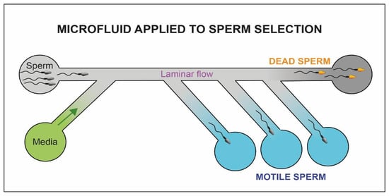
Figure 910. Schematic representation of the microfluidic system. The motile fraction of sperm cells is able to swim through the flow and be collected in separate chambers (blue chambers) while the immotile sperm cells (yellow heads) and debris reach the exit of the microfluidic system (dark chamber).
The application of optical systems to microfluidics could further improve the selection system [96][148]. Raman spectroscopy allows the discernment of sperm cells with high nuclear integrity from sperm cells with fragmented DNA [97][149]. By coupling the three-dimensional imaging to the selected channels, it is possible to accurately identify the different types of flagellar movement using a digital sensor, consisting of a semi-conductive material, which can virtually reconstruct the cell volume by recording the differences in the types of interference between a reference light wave and a scatter from the sample [98][150]. Using this method, De Wagenaar B. et al., were able to trace the profile of the flagellar beat of hyper-activated spermatozoa [99][151].
2.6. Horizontal Sperm Migration
During ICSI, an adequate number of spermatozoa reaches the distal edge of the furthest drop; some of these can be recovered from the injection needle, to be moved to the PVP and carefully selected for fertilization (Figure 110).
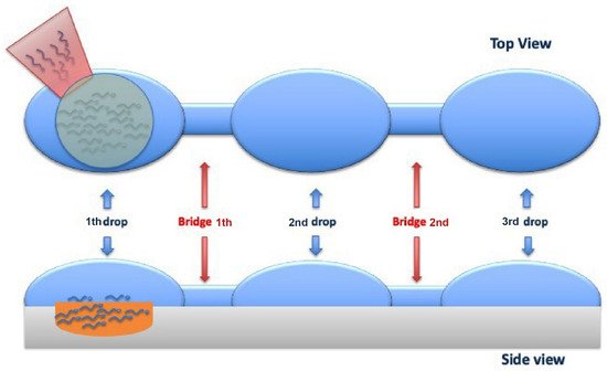
Figure 101. Top and Side viewing of the sperm cells horizontal migration from the first drop (where the cells are added) to the third drop (where the sperm cells are aspirated) through 2 bridges that link them.
The technique allows the recovery of spermatozoa with high motility, normal morphology and minimal damage to the DNA, using a fast, safe, and economical procedure. Comparing the clinical results obtained from this new selection method with the classic swim-up technique, no significant differences were found in terms of fertilization and implantation rate. On the other hand, segmentation and blastocyst rates are higher in the horizontal swim-up, suggesting that this process that generates a lower quantity of harmful oxygen reactants is correlated with better seminal quality and a lower DNA fragmentation.