SALL Proteins in Cancer: Comparison
Please note this is a comparison between Version 1 by Roxana Pincheira and Version 2 by Peter Tang.

SALL proteins are a family of four conserved C2H2 zinc finger transcription factors that play critical roles in organogenesis during embryonic development. They regulate cell proliferation, survival, migration, and stemness; consequently, they are involved in various human genetic disorders and cancer. SALL4 is a well-recognized oncogene; however, SALL1–3 play dual roles depending on the cancer context and stage of the disease.

  • SALL1
  • SALL2
  • SALL3
  • SALL4
  • cancer
  • epigenetic regulation
  • Wnt
  • PTEN
  • biomarker

1. Introduction

SALL proteins are transcription factors that belong to the Spalt-like (Sall) family, broadly conserved through evolution. They are present in nematodes, flies, planarians, bilaterians, and vertebrates. They were first identified in Drosophila melanogaster, which harbors two paralogs: spalt major (salm) and spalt-related (salr). Both proteins play a role in the homeotic specification of the embryonic termini, wing patterning, sensory organ development, and photoreceptors specification [1][2][1,2].
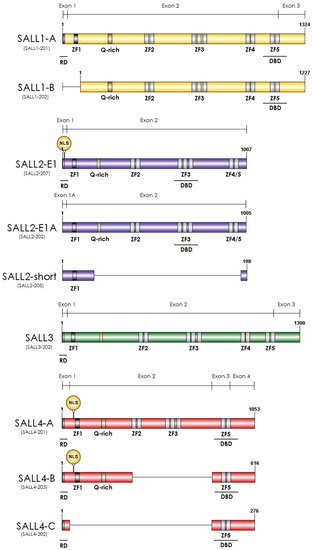
Vertebrate genomes harbor four paralogs, SALL1–SALL4, apparently originated by several duplication events of the spalt locus and evolved from one ancestor more closely related to Drosophila (salm ortholog) [2]. A phylogenetic analysis of SALL proteins indicates that SALL1 and SALL3 derived from one common ancestor and SALL4 derived from a more distant one. SALL2 shares the least homology, being the most dissimilar member of the SALL family, especially in the C-terminal region [3][4]. Multiple zinc finger domains characterize them throughout the protein, a glutamine-rich (poly-Q) region important for protein-protein interactions, and a conserved twelve-amino-acid domain at the N-terminal region responsible for the repression activity of SALL proteins, mediated by an interaction with the Nucleosome Remodeling and Deacetylase (NuRD) complex [4][5] (Figure 1).
Figure 1. Schematic representation of the main SALL protein isoforms. The colors represent the different SALL proteins; yellow, blue, green, and red for SALL1, SALL2, SALL3, and SALL4. Dark grey rectangles at the N-terminal region represent the C2HC-type Zinc Finger Motif (ZF1). Light grey rectangles represent C2H2-type Zinc Finger Motifs 2–5 (ZF2–ZF5); all of them are in SALL1-A, SALL1-B, and SALL3. SALL4 lacks ZF4, and SALL2 has a motif that differs from the others, located between ZF4 and ZF5 (depicted as ZF4/5). The pink rectangle at the N-terminal region represents the conserved 12-amino-acid region that binds to the NuRD complex, named repression domain (RD). The RD is in SALL1A, SALL2 E1, SALL3, SALL4 A, B, and C. The yellow rectangle between ZF1 and ZF2 corresponds to the conserved Glutamine-rich (Q-rich) region. The circle at the N-terminal region shows the nuclear localization sequences (NLS) described only for SALL2 E1A and SALL4 A and B. The Ensembl transcript ID is under each isoform name in brackets. Exon representation is above each SALL protein, and the protein length is at the end of each isoform. ZF: Zinc Finger; Q-rich: Glutamine-rich; RD: Repression Domain; DBD: DNA Binding Domain; NLS: Nuclear Localization Sequence.

2. Essential Roles of SALL Genes during Development

Vertebrate SALL proteins participate in the development of extremities and organs, including the brain, kidney, eye, and heart. Accordingly, SALL genes are implicated in human genetic disorders [1][2][5][3][4][6][1,2,3,4,5,16]. Mutations of SALL1 cause the Townes–Brocks syndrome (TBS), a rare autosomal malformation syndrome characterized by anal, renal, limb, and ear anomalies (Reviewed in [1][2][1,2]). Similarly, SALL4 mutations cause the Okihiro/Duane-radial ray syndrome (DRRS), an autosomal dominant condition characterized by upper-limb, ocular, and renal anomalies in some cases [7][17]. Meanwhile, SALL2 deficiency causes recessive ocular coloboma [8][18]. SALL3 deficiency is associated with ocular anomalies and facial dysmorphism of the human 18q deletion syndrome [9][19].
Functional studies using knockout mice confirmed the essential roles of Sall1, Sall3, and Sall4 during development. Loss of function of these genes results in perinatal or neonatal lethality due to organ alterations during embryonic development. The organ alterations include kidney agenesis or dysgenesis, abnormal cranial nerve morphology, and exencephaly [10][11][12][20,21,22]. Two Sall2 knockouts (KO) models exist; the first Sall2KO model did not show an essential role for Sall2 in embryonic or kidney development [13][23]. However, the second Sall2KO model showed severe neural tube defects and defects in the optic fissure closure, similar to the phenotype of coloboma patients [8][14][18,24]. Differences between the phenotype of Sall2KO models might be related to the different genetic backgrounds of the mice strains used [13][14][23,24]. Sall2KO did not show spontaneous tumor formation [13][23], but when crossed with tumor-susceptible mice p53−/−, it exhibited significantly accelerated tumorigenesis, tumor progression, and mortality rate among Sall2+/+/p53−/− mice. The Sall2−/− or Sal2−/+/p53−/− mice showed thymus T-cell lymphoma that metastasized to the liver, lung, kidney, marrow, peripheral blood, and central nervous system, while in most Sall2+/+/p53−/− mice, the lymphoma was limited to the thymus and adjacent organs such as the lung [15][25]. Moreover, supporting a tumor suppressor function, the immortalized Sall2−/− MEFs showed an enhanced growth rate, foci formation, and anchorage-independent growth compared to the immortalized Sall2+/+ MEFs [16][26].
In development, common findings on SALL proteins include direct interaction with chromatin remodeling complexes, such as the SWI/SNF or NuRD complexes, and an association with the canonical Wnt/β-catenin pathway [4][17][18][19][20][21][5,10,27,28,29,30]. Particularly relevant for the SALLs function is their interaction with the NuRD complex. NuRD is involved in global transcriptional repression and specific developmental processes [20][22][29,31]. SALLs interact with the NuRD complex through the conserved 12-amino-acid motif. This motif is not present in some SALL isoforms (Figure 1), suggesting that NuRD is essential in differentiating their function. However, there is a lack of studies addressing this issue. Most of the studies have focused on the functional relationship of NuRD with SALL1 or SALL4 in development and cancer. In kidney development and leukemogenesis, the function of SALL4 through the NuRD complex relies on the repression of PTEN and SALL1 [23][32]. In other contexts, the interaction between SALL4 and NuRD impacts different genes. SALL4 was involved in spermatogonial differentiation. SALL4/NuRD repressed the expression of the tumor suppressor genes Foxl1 and Dusp4, associating SALL4 function with the maintenance of undifferentiated spermatogonial activity and stem cell-driven regeneration [24][33]. However, in the context of pluripotent cell transcriptional programs, free SALL4 regulates transcription independently of NuRD [25][34].
The SALL1/NuRD complex is also involved in kidney development, inhibiting the premature differentiation of nephron progenitor cells. The disruption of SALL1/NuRD interaction in vivo resulted in the accelerated differentiation of nephron progenitors and bilateral renal hypoplasia [19][28]. In Xenopus embryos, SALL1 interaction with NuRD directly repressed Gbx2, a transcription factor for cell pluripotency and differentiation in the embryo [26][35]. Interestingly, SALL1 association with NuRD is disrupted by the protein kinase C phosphorylation at serine 2 of the repression motif, suggesting that this kinase regulates the NuRD-dependent repression function of SALL1 [26][35]. SALL1 phosphorylation by PKC may also be involved in breast cancer [27][36]. SALL1/NuRD inhibited breast cancer cell growth, proliferation, and metastasis, and a phosphomimetic mutation of SALL1 impaired its tumor suppressor function. Whether Gbx2 is associated with the tumor suppressor function of SALL1 is currently unknown.
As with many genes that play essential roles in organogenesis during embryonic development, SALL genes are cancer-related. Developmental pathways are crucial for the cellular processes required during embryonic stages, such as epithelial-mesenchymal transition (EMT), coordinated migration, and cell proliferation, which are also essential at different stages of tumor progression [28][37].

3. Common Cellular Functions and Targets of the SALL Proteins in Cancer

In recent years, the number of identified SALL proteins’ target genes has increased. They are associated with diverse cellular events such as proliferation, cell death, migration, invasion, and stemness.

3.1. Cell Proliferation

Several studies have established the role of SALL proteins in cell proliferation, acting as oncogenes or tumor suppressors under different pathological contexts. For instance, ectopic SALL2 expression inhibited SKOV3 ovarian cancer cell proliferation by a mechanism that involves the positive transcriptional regulation of cell cycle inhibitors such as p21 and p16 [29][30][38,39]. Accordingly, SALL2 depletion increased A2780 ovarian carcinoma cell proliferation [31][40]. The loss of Sall2 in mouse embryonic fibroblasts (MEF) enhanced cell proliferation and showed faster postmitotic progression through the G1 and S phases. The mechanism is related to the transcriptional derepression of two SALL2 targets, cyclins D1 and E1 [16][26]. On the contrary, SALL4 accelerated cell cycle progression in cervical, lung, and breast cancer cells and esophagus squamous cell carcinoma and glioma [32][33][34][35][36][41,42,43,44,45]. The opposite roles of SALL2 and SALL4 in cell proliferation agree with the regulation of c-MYC, a transcription factor involved in cell growth and cell cycle control. SALL2 directly binds to the nuclease hypersensitive element in the promoter of c-MYC, repressing its expression [37][46]. However, SALL4 indirectly increases the levels of c-MYC by activating the Wnt pathway. SALL4 enhanced the proliferative capacity of HeLa and SiHa cervical cancer cells through the positive transcriptional regulation of CTNNB1 [32][41]. CTNNB1 encodes β-catenin, a transcriptional cofactor of TCF/LEF in the Wnt signaling pathway. Additionally, SALL4 is directly bound to β-catenin, which activates the Wnt pathway in AML (acute myeloid leukemia) [38][8]. In both studies, Wnt pathway activation by SALL4 increased c-MYC and cyclin D1, related to increased proliferation, survival, EMT, and metastasis [38][32][8,41].
SALL1 is also associated with increased β-catenin expression in human primary AML samples. The inhibition of SALL1 resulted in decreased cell proliferation and AML engraftment in NSG (NOD SCID gamma) mice [39][9]. Interestingly, similar to SALL4, SALL1 interacts with β-catenin in human kidney BOSC23 cells derived from HEK293T cells [40][47], suggesting a common mechanism for Wnt pathway activation via the interaction of β-catenin with SALL1 and/or SALL4. However, in contrast to the AML context, SALL1 over-expression in MDA breast cancer cells inhibited tumor cell growth and proliferation. It promoted cell cycle arrest by increasing Cyclin A2, Cyclin B1, Cyclin E1, CDK2, and CDK4, which are essential for checkpoint regulation in the G1-S transition and S phases [27][36]. These findings suggest a dual role for SALL1 in cancer, depending on the cell context.

3.2. Apoptosis and Cell Survival

SALL proteins’ target genes are also associated with the regulation of apoptosis, indicating that SALL2 and SALL4 play opposite roles in this process. Using chromatin immunoprecipitation followed by microarray hybridization in the human acute promyelocytic leukemia cell line NB4, Yang and collaborators validated the SALL4 upregulation of anti-apoptotic genes, such as Bmi-1, BCL2, DAD1, TEGT, BIRC7, and BIRC4, and the negative regulation of pro-apoptotic genes, such as TNF, TP53, PTEN, CARD9, CARD11, ATF3, and LTA. Moreover, the inhibition of SALL4 induced apoptosis in NB4 cells, increasing DNA fragmentation and caspase-3 and annexin V levels [41][48]. On the other hand, the apoptotic cell response to genotoxic stress and Trichostatin A (TSA) treatment required SALL2 [42][43][44][49,50,51]. In response to doxorubicin- and etoposide-induced genotoxic stress, SALL2 induced pro-apoptotic genes such as BAX and PMAIP1 (also known as NOXA) in human ovarian surface epithelial (HOSE) cells and MEFs. Particularly noteworthy is the pro-apoptotic role of SALL2, which was independent of p53 expression, suggesting a critical role of SALL2 in the response of cancer cells to therapy in p53 inactive cancer contexts [42][43][49,50].

3.3. Cell Migration and Invasion

Migratory and invasive cell capacities increase during tumor development, which are strongly associated with metastasis in advanced stages of cancer. There are several mechanisms by which tumor cells acquire these characteristics of malignancy. One of the central mechanisms is the inhibition of PTEN, a phosphatase that blocks the PI3K signaling pathway, inhibiting cell migration, proliferation, and survival [45][52]. SALL4 repressed PTEN expression through its interaction with the NuRD complex and favored the development of AML in mice [23][32]. In ICC-9810 cholangiocarcinoma cells, SALL4 inhibited migratory and invasive capacities through the repression of PTEN and the upregulation of Bmi-1 [46][53]. Similarly, SALL1 inhibition increased PTEN expression in AML cell lines and primary samples and downregulated mTOR, β-catenin, and NF-қB expression [39][9]. SALL1 is bound to the NuRD complex in breast cancer; thus, it is likely that SALL1 and SALL4 share a similar repressive mechanism for PTEN regulation. However, no changes in PTEN expression were detected in breast cancer cells with SALL1 over-expression, suggesting that the regulation of PTEN by SALL1 is tissue-specific [27][36].
Meanwhile, SALL2 induces PTEN expression. In breast cancer cells, SALL2 silencing activated the AKT/mTOR pathway via the downregulation of PTEN. The mechanism involves the positive regulation of PTEN through the direct binding of SALL2 to canonical GC-rich consensus elements in the PTEN promoter [47][54]. Although this study did not associate SALL2-dependent PTEN regulation with cell migration, previous studies demonstrated that SALL2 expression correlated with impaired cell migration in human ovarian and esophageal carcinoma cell lines [31][48][40,55]. Additionally, the CDH1 and VIM genes, involved in migration, invasion, and EMT, are common targets for SALL1 and SALL4 in breast cancer (discussed below).

3.4. Stemness

Maintenance of stemness is another essential feature of the heterogeneous cell population within the tumor, increasing its complexity by conferring the ability to differentiate into many unrelated cell types.
The role of SALL proteins in stemness maintenance is relevant during embryo development and cell fate. For instance, SALL2 and SALL4 are necessary factors for the self-renewal of hematopoietic stem cells (HSC) [15][49][50][25,56,57]. Interestingly, several studies suggest that SALL1 and SALL4 act as stemness or differentiation factors, depending on the development stage and the cell type involved. SALL1 is required for the stem cell maintenance of kidney, heart, and spermatogonial progenitors [18][51][52][53][27,58,59,60]. However, SALL1 also participates in the heart and odontoblast lineage differentiation [52][54][59,61].
Similarly, SALL4 plays opposite roles in postnatal spermatogenesis and embryonic germ cells [55][62]. During spermatogonia differentiation, SALL4 sequesters Plzf, a factor required to maintain adult stemness. This interaction leads to the expression of the differentiation marker KIT and the repression of SALL1 [55][62]. SALL1 expression in the germline is specific for spermatogonia progenitor cells. It was proposed as one of the factors involved in spermatogonial stem cell self-renewal [51][58].
SALL4 was proposed as a crucial factor for maintaining pluripotency in embryonic stem cells (ESCs) by directly interacting with the core master regulators SOX2 and OCT4 [56][63]. Recent research has revealed that SALL4 maintains the pluripotent state in ESCs by regulating a set of AT-rich genes that promote neuronal differentiation. Worthy of note here is that the AT-rich gene pull-down by SALL4 depends on the C2H2 zinc-finger cluster 4 (ZFC4) domain, also found in SALL1 and SALL3, but not in SALL2 [57][64].
Interestingly, the putative tumor suppressor SALL2 was identified as one of the critical transcription factors necessary for maintaining the tumor propagating cells in glioblastoma. SALL2 interacted with SOX2, OCT4, and Nanog in this specific context, promoting stemness and aggressive behavior [58][65]. Similarly, SALL1 can interact with SOX2 and Nanog, but not with OCT4, inducing an undifferentiated state. SALL1 also suppresses ectodermal and mesodermal differentiation. Meanwhile, SALL1 overexpression was found to inhibit the induction of gastrulation markers (T brachyury, Goosecoid, and Dkk1) and neuroectodermal markers (Otx2 and Hand1) [59][66]. Recently, SALL3 was identified as part of a small set of transcription factors, including SOX2 and SALL2, that interact with the Mediator complex in neural stem cells [60][67]. Altogether, these studies identified SOX2 as a common SALL protein partner, relevant for the maintenance of stemness.

4. Common Regulatory Mechanisms for SALL Proteins in Cancer
The regulation of SALL proteins is an open field of study and involves several different mechanisms for each family member. These include genetic alterations and specific transcriptional, posttranscriptional, and posttranslational regulation. However, a common regulatory mechanism for all family members relates to epigenetic modifications, including chromatin modifications and microRNAs (miRNAs).
The loss of heterozygosity (LOH) was reported in overlapping regions of SALL genes in several independent cancer studies and was associated with poor prognosis and metastatic recurrence. These regions include SALL1 (16q12.1) [61][62][68,69], SALL2 (14q11.1–12) [63][64][65][66][67][68][69][70,71,72,73,74,75,76], and SALL3 (18q23) [69][70][71][76,77,78]. On the other hand, chromosomal amplifications were found in the SALL4 region (20q13.2) [72][73][74][75][76][77][78][79,80,81,82,83,84,85].
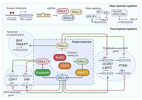
Specific transcription factors regulate the expression of SALL genes. SALL2 is transcriptionally activated by AP4 and Sp1 [44][79][51,86] and repressed by WT1, p53, and FosL1 [80][81][82][87,88,89]. On the other hand, TCF/LEF, STAT3, and CDX1 are transcriptional activators of SALL4 [17][83][84][10,90,91] (Figure 2). Remarkably, SALL4 controls their expression and represses SALL1 and SALL3, thus regulating the stemness of ES cells [56][63]. In murine transgenic models, SALL4 represses SALL1 and PTEN through the NuRD repressor complex, leading to pathologies such as cystic kidney and myeloid leukemia, respectively [23][32].
Figure 2. Common upstream regulation, partners, genes, and cellular functions of the SALL family. Epigenetic changes, including gene hypermethylation, hypomethylation, and miRs, are common mechanisms of SALL regulation. SALL1–3 promoters are hypermethylated in several cancers [47][48][85][86][87][88][89][90][54,55,92,93,94,95,96,97]. In contrast, the SALL4 promoter is hypomethylated in AML and MDS [91][92][98,99] and regulated by miRNAs in multiple cancer types [93][100]. Additionally, SALL1 and SALL2 are regulated by miRs (Table 1). Regulation by specific transcription factors depicted for SALL2 and SALL4. SALL proteins interact with specific partners to perform their functions; shared protein partners among the family include β-catenin and DNMT3. In addition, the four SALLs interact with the NuRD complex and with SOX2. Common transcriptional targets of SALLs are associated with cell proliferation and migration/invasion. SALL2 and SALL4 oppositely regulate CCDN1, c-MYC, and PTEN. Similarly, SALL1 and SALL4 oppositely regulate CDH1 (E-cadherin) and VIM (vimentin). However, SALL1 and SALL4 are both negative regulators of PTEN. Moreover, apoptosis-associated genes, such as BAX and PMAIP1 (NOXA), are regulated by SALL2. Green lines: positive regulation. Red lines: negative regulation. Dotted lines: proposed association.
Table 1. Summary of all microRNAs known to regulate SALLs.

Cancer Type/Cellular Model

microRNA

Target

SALL Status/Key Findings

Experimental Approach

Ref.

Glioma/Glioblastoma

miR-302/367 cluster

SALL2

miR-302/367 cluster can reprogram tumor cells, generating more benign phenotypes by suppressing OCT3/4, SOX2, KLF4, c-MYC, POU3F2, OLIG2, and SALL2

Undescribed

qRT-PCR, cytokine array analysis

Expression correlated with lower overall survival of NSCLC patients

[94]

[101]

Oncogene

[

115

]

[122]

Glioma/Glioblastoma

miR-16

SALL4

miR-16 inhibits proliferation, migration, and invasion in glioma cells by directly targeting SALL4

qRT-PCR and Luciferase reporter assay

Lung

[

95

SALL2

]

[102]

Low

LOH

Glioma/Glioblastoma

miR-103/miR-195/miR-15-B

SALL4

miR-103, miR-195, and miR-15-B inhibit proliferation, migration, and invasion and promote apoptosis in glioma by directly targeting SALL4

qRT-PCR, Western blot, and Luciferase reporter assay

[96]

[103]

Undescribed

Undescribed

[64]

[71]

Lung

SALL4

High

Undescribed

Expressed in 88% of the lung cancer samples

May be used as a diagnostic marker

Oncogene

[116]

[123]

Glioma/Glioblastoma

miR-107

SALL4

miR-107 inhibits proliferation and promotes apoptosis in glioma cells by directly targeting SALL4

qRT-PCR, Western blot, and Luciferase reporter assay

Lung

SALL4

[

High

97

Undescribed

]

SALL4 knockdown inhibits cell proliferation by cell cycle arrest at the GO/G1 phase

Loss of SALL4 function inhibits migration, invasion and reduces the size of the transplanted tumor in an in vivo model.

[104]

Oncogene

[

34

]

[43]

Glioma/Glioblastoma

miR-181b

Lung

SALL4

SALL4

High

miR-181b inhibits proliferation, migration, and invasion and promotes apoptosis in glioma by directly targeting SALL4

Undescribed

qRT-PCR, Western blot, and Luciferase reporter assay

SALL4 silencing sensitizes cells to cisplatin, carboplatin, and paclitaxel treatment

[98]

[105]

Oncogene

[

117

]

[124]

Gastric cancer

miR188-5p

SALL4

miR-188-5p promotes proliferation and migration by upregulating SALL4 expression, nuclear translocation, and transcription

qRT-PCR, Western blot, and Luciferase reporter assay

Esophageal

[

99]

[106]

SALL1

Low

Hypermethylation

SALL1, ADHFE1, EOMES, and TFPI2 are proposed as part of a tumor suppressors panel with diagnostic relevance

Tumor suppressor

[86][118]

[93,125]

Gastric cancer

miR-16

Esophageal

SALL2

SALL4

Low in radioresistant ESCC cell lines

miR-16 inhibits proliferation and migration in GC by directly targeting SALL4

Hypermethylation

qRT-PCR and Luciferase reporter assay

SALL2 overexpression enhances apoptosis after radiation and decreases migration, viability, and cisplatin resistance in TE-1/R and Eca-109/R cell lines

[100]

[107]

Tumor suppressor

[

48

]

[55]

Colorectal cancer

miR-181a-2 *

Esophageal

SALL4

SALL1

High

miR-181a-2 * correlates with a trend of repression of SALL1 and high methylation status of the SALL1 promoter

qRT-PCR and bisulfite modification followed by quantitative methylation- specific PCR (qMSP)

Undescribed

SALL4 silencing in ESCC cells is associated with suppressing cell migration, invasion, viability, and drug resistance in vivo

SALL4 knockdown reduces epithelial-mesenchymal transition by targeting the Wnt/β-catenin signaling pathway

[101]

[108]

Oncogene

[

33

][119]

[42,126]

Colorectal cancer

miR-219-5p

Bladder

SALL4

SALL2

miR-219-5p inhibits proliferation, migration, and invasion, reduces drug resistance, and promotes apoptosis in CRC by directly targeting SALL4

Low

LOH

qRT-PCR, Western blot, and Luciferase reporter assay

Undescribed

[102]

[109]

Tumor suppressor

[

63

]

[70]

Colorectal cancer

miR-3622a-3p

Bladder

SALL4

SALL3

miR-3622a-3p inhibits proliferation, cell cycle, migration, invasion, and stemness features and promotes apoptosis by targeting SALL4

Low

Hypermethylation

qRT-PCR, Luciferase assay, RNA immunoprecipitation (RIP) assay, and pull-down assay

SALL3, CFTR, and TWIST1 are proposed as disease recurrence predictors

[103]

[110]

Tumor suppressor

[

120

][121]

[127,128]

Embryonic stem cell

miR15-B

SALL4

Anti-miR-15b enhances expansion of HSC in vitro by targeting SALL4

qRT-PCR

Testicular tumors

SALL4

High

Undescribed

SALL4 is a novel sensitive and specific marker for testicular germ cell tumors

[104]

[111]

Oncogene

[

122

]

[129]

Embryonic stem cell

Kidney

miR-294 and let-7 miRNAs

SALL4

Let-7 miR family inhibits self-renewal genes, and miR-294 indirectly induces self-renewal genes, including SALL4

SALL1

qRT-PCR, Western blot, and Luciferase reporter assay

Low

miR-942

SALL1 inhibition plays a potential role in sunitinib resistance in RCC patients

[105]

[112]

Tumor suppressor

[

108

]

[115]

Oral squamous cell carcinoma

Wilms’ tumor

miR-103

SALL1

SALL4

High

miR-103 inhibits cell proliferation and invasion by downregulating SALL4 mRNA in Tca8113 cells

Undescribed

Luciferase reporter assay

Undescribed

[106]

[113]

Oncogene

[

123

][124]

[130,131]

Breast cancer

SNHG12 and miR-15a-5p

Wilms’ tumor

SALL4

Long non-coding RNA (lncRNA) small nucleolar RNA host gene 12 (SNHG12) promotes proliferation, migration, and invasion and inhibits apoptosis in breast cancer by upregulating SALL4 expression via sponging miR-15a-5p; SALL4 is a direct target of miR-15a-5p

SALL2

High

qRT-PCR, Western blot, and Luciferase reporter assay

Undescribed

SALL2 was identified as one of the 27 signature genes highly expressed by comparing tumor samples with normal fetal kidneys

[107]

[114]

Oncogene

[

125

]

[132]

Renal cell carcinoma

miR-942

SALL1

Kidney

SALL3

miR-942 affects the survival of patients with renal cell carcinoma by negatively regulating the expression of SALL1

RNA-seq and qRT-PCR

[108]

[115]

Low

Methylation

SALL3 downregulation may contribute to genome hypermethylation similar to VHL

Tumor suppressor

[126]

[133]

Prostate cancer

miR-4286

Wilms’ tumor

SALL4

SALL1

High

miR-4286 regulates proliferation and apoptosis in PCa cells by directly targeting the 3′UTR of SALL1 mRNA

qRT-PCR and Luciferase reporter assay

Undescribed

[109]

[116]

Undescribed

Oncogene

[127]

[134]

Lung cancer

HOXA11-AS and miR-3619-5p

SALL4

lncRNA homeobox A11 antisense (HOXA11-AS) promotes proliferation, migration, invasion, and glycolysis in non-small cell lung cancer (NSCLC) cells by upregulating SALL4 expression via sponging miR-3619-5p; SALL4 is a direct target of miR-3619-5p

qRT-PCR, Western blot, and Luciferase reporter assay

[110]

[117]

Osteosarcoma

ZEB2-AS1 and miR-107

SALL4

lncRNA ZEB2-AS1 (ZEB2-AS1) promotes proliferation, invasion, and metastasis and inhibits apoptosis in osteosarcoma cells by upregulating SALL4 expression via sponging miR-107; SALL4 is a direct target of miR-107

qRT-PCR, Luciferase assay, and RNA pull-down assay

[111]

[118]

Hepatocellular carcinoma

miR-296-5p

SALL4

miR-296-5p inhibits stemness potency of hepatocellular carcinoma (HCC) cells via the Brg1/Sall4 axis; Brg1 binds to the SALL4 promoter

qRT-PCR, Western blot, Luciferase reporter assay, and Chromatin immunoprecipitation (ChIP) assay

[112]

[119]

Hepatocellular carcinoma

miR-15a

SALL4

Exosomal miR-15a reduces proliferation, migration, invasion, and survival by directly targeting SALL4

qRT-PCR, Western blot, and Luciferase reporter assay

[113]

[120]

Promoter hypermethylation is frequent for SALL1, SALL2, and SALL3, and the regulation of 3′UTR by miRNAs appears as a typical regulatory mechanism for SALL4. Epigenetic modifications on SALL genes are consistent with their prominent roles as tumor suppressors or oncogenes. For instance, associated with their tumor suppressor role, the SALL1, and SALL2 promoters' hypermethylation was described in breast cancer and esophageal squamous cell carcinoma (ESCC) [47][48][85][86][54,55,92,93]. The hypermethylation of the SALL2 promoter was associated with aggressive and tamoxifen-resistant breast cancer phenotypes [47][54]. In oral squamous cell carcinoma (OSCC), SALL2 promoter hypermethylation positively correlates with SALL1 and SALL3 promoter methylation status and aggressive tumor behavior [87][94].
The SALL promoters are also aberrantly methylated in HPV-related cancers. Several studies indicate that the hypermethylation of SALL1 and SALL3 promoters correlate with poor outcomes and recurrence in head and neck squamous cell carcinoma (HNSCC) [88][89][90][95,96,97]. However, SALL4 is upregulated in this type of cancer, and its expression correlates with disease recurrence and decreased disease-free survival. High SALL4 expression positively correlated with DNA methyltransferase 3 alpha (DNMT3A) expression and the increased methylation rate of 11 tumor suppressor genes. Still, there was no significant correlation between SALL4 expression and SALL1, SALL2, and SALL3 methylation status [114][121]. Aberrant hypomethylation of the SALL4 promoter is described as a common event in AML and myelodysplastic syndrome (MDS) [91][92][98,99].

5. SALL Proteins in Cancer

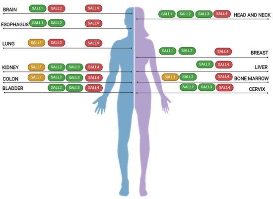
SALL proteins are altered in various cancer types (Table 2 and Figure 3). Alterations include deregulation in gene expression, isoform expression, and genetic aberrations. We focused our analysis on those cancer types with the most considerable data on SALL family members.
Figure 3. SALL proteins in cancer. SALL proteins are deregulated in major cancer types, including lung, colon, and breast cancers. As shown above, independent studies identified alterations in more than one family member in specific cancer types. According to genetic alterations, isoform expression, and changes in their expression, they are classified as oncogenes (red), tumor suppressors (green), or genes with a dual role in cancer (yellow).
Table 2. Deregulation of SALLs in other cancers.

Cancer Type

SALL Member

Expression Levels

Genetic Alteration/Regulation

Association With Cancer/Biological Process

Proposed Cancer Role

Ref.

Lung

SALL1

High

6. Targeting SALLs for Cancer Therapy

Considering the evidence on the role of the SALL family members in cancer initiation and progression, targeting SALLs provides a unique therapeutic opportunity for cancer treatments.
Even though transcription factors have remained challenging drug targets, several in vitro and in vivo approaches targeting SALL4 protein activity have already been investigated.
The HDAC-1 and -3 inhibitor Entinostat was identified as a potential treatment for SALL4-expressing cancers. The study used a panel of 17 lung cancer cell lines with varied SALL4 expression levels, showing that cells expressing high levels of SALL4 were more sensitive to Entinostat treatment [128][182]. However, HDAC inhibitors are not selectively targeting SALL4 expressing cells. Pharmacological peptides that exclusively target cancer cells expressing SALL4 were tested as potential cancer therapeutic agents. A 12-amino-acid peptide that disrupts the interaction between SALL4 and the NuRD complex comprising HDAC1 and HDAC2 was tested in AML and HCC. The peptide disrupted the interaction between SALL4 and HDAC, which blocked the NuRD-mediated SALL4 repression function [129][130][155,174]. Similarly, another peptide, PEN-FFW, was recently designed to target SALL4 in HCC cell lines. The peptide disrupted the SALL4–NuRD interaction via the blocking of the SALL4 interaction with RBBp4, specifically inhibiting the transcription-repressor function of SALL4. Treatment of HCC cells with the PEN-FFW peptide-induced apoptosis enhanced cell adhesion and dramatically inhibited xenograft tumor growth [131][183]. The use of miRNAs targeting SALL4-associated HCC has also been proposed. Let-7/miR-98-induced SALL4 depletion decreased the expression of MMP2/9, Fibronectin, n-cadherin, and increased E-cadherin, which correlated with reduced migration/invasion and EMT in an HCC in vivo cancer model [93][100].
Alternative strategies are the use of drugs that can induce the degradation of SALL4. Small immunomodulatory drugs (IMiDs), such as thalidomide and derivatives, induce ubiquitination and the proteasomal degradation of zinc finger transcription factors. The mechanism involves recruiting C2H2 zinc finger (ZnF) domains to Cereblon (CRBN), the substrate receptor of the CRL4CRBN E3 ubiquitin ligase [132][184]. Thalidomide induced the robust degradation of SALL4 in the neuroblastoma (Kelly and SK-N-DZ) cell lines and the MM1 multiple myeloma cell line [133][185]. However, because of the critical role of SALL4 in limb development, treatment with thalidomide or analogous drugs during pregnancy could contribute to severe birth developmental abnormalities [7][133][134][135][17,185,186,187].
Additional therapeutic strategies to restore tumor suppressor function could be related to the modulation of SALL1 or SALL2 protein levels by altering their ubiquitylation and subsequent proteasome degradation or by inhibiting still unknown negative regulators of the transcription factor expression. Thus, efforts should also focus on identifying novel SALL regulators and partners.
ScholarVision Creations