The genus Aeromonas has been recognised as an important pathogenic species in aquaculture that causes motile Aeromonas septicaemia (MAS) or less severe, chronic infections. This study compares the pathogenicity of the different Aeromonas spp. that were previously isolated from freshwater fish with signs of MAS. A total of 124 isolates of Aeromonas spp. were initially screened for the ability to grow on M9 agar with myo-inositol as a sole carbon source, which is a discriminatory phenotype for the hypervirulent A. hydrophila (vAh) pathotype. Subsequently, LD50 of six selected Aeromonas spp. were determined by intraperitoneal injection of bacterial suspension containing 10^3, 10^5, and 10^7 CFU/mL of the respective Aeromonas sp. to red hybrid tilapias. The kidneys, livers and spleens of infected moribund fish were examined for histopathological changes. The screening revealed that only A. dhakensis 1P11S3 was able to grow using myo-inositol as a sole carbon source, and no vAh strains were identified. The LD50–240h of A. dhakensis 1P11S3 was 10^7 CFU/mL, while the non-myo-inositol utilizing A. dhakensis 4PS2 and A. hydrophila 8TK3 was lower at 10^5 CFU/mL. Similarly, tilapia challenged with the myo-inositol A. dhakensis 1P11S3 showed significantly (p < 0.05) less severe signs, gross and histopathological lesions, and a lower mortality rate than the non-myo-inositol A. dhakensis 4PS2 and A. hydrophila 8TK3. These findings suggested that myo-inositol utilizing A. dhakensis 1P11S3 was not a hypervirulent Aeromonas sp. under current experimental disease challenge conditions, and that diverse Aeromonas spp. are of concern in aquaculture farmed freshwater fish. Therefore, future study is warranted on genomic level to further elucidate the influence of myo-inositol utilizing ability on the pathogenesis of Aeromonas spp., since this ability correlates with hypervirulence in A. hydrophila strains.
1. Introduction
Aquaculture is one of the most important sectors that provides valuable sources of protein, aside from generating income for certain countries. FAO
[1] reported a new height in global aquaculture production in 2018, with 114.5 million tonnes in live weight, generating USD 263.6 billion of farmgate sale value. Malaysia has been relying on the aquaculture sector as an important source of revenue, and FAO
[2] also states that fish is an essential food in Southeast Asian nations, such as Malaysia, Indonesia, Myanmar, Thailand, Vietnam, Philippines, and Cambodia. In 2018, it was reported that the red hybrid tilapia (
Oreochrommis niloticus × O. mossambicus) and black tilapia (
Oreochromis spp.) accounted for 30.7% of total freshwater production in Malaysia, wherein red hybrid tilapia alone constituted about 97% of total tilapia production
[3]. Tilapia farming is preferred because of their resilience in many environmental conditions, disease resistance, marketability, and easier-to-produce market-sized fish using a varied range of feed, ranging from natural organisms to humanmade pellets
[4][5][4,5].
However, disease outbreak is one of the main problems which resulted in economic, environmental and social impacts on aquaculture. Fish farms are vulnerable to losses due to outbreaks of bacterial infections such as motile
Aeromonas septicemia (MAS)
[6]. This disease is caused by members of the genus
Aeromonas, such as
Aeromonas hydrophila,
A. veronii,
A. dhakensis,
A. jandaei,
A. sobria and
A. caviae [7][8][9][10][11][12][7,8,9,10,11,12]. The disease was described as having two forms, the acute hemorrhagic septicemia and the chronic ulcerative syndrome
[13][14][15][13,14,15]. The genus
Aeromonas plays an important role in diseases of aquaculture, with
Aeromonas spp. showing a ubiquitous distribution in aquatic habitats including freshwater, seawater, estuaries and even in chlorinated water
[16][17][16,17]. The ability of
Aeromonas spp. to thrive in a wide range of temperatures, from 4 °C to 42 °C, and tolerate up to 5.5 g/L NaCl and in a range of pH from 5 to 10 contributes to their widespread distribution
[18][19][18,19].
Aeromonas spp. are generally opportunistic pathogens that are normal residents of the fish gut microbiota
[20].
Recently, hypervirulent vAh strains were identified in diseased carp species in China and from diseased channel catfish from the USA, which were found to cause acute persistent MAS
[21]. It was reported as a primary pathogen
[22][23][22,23] among grass carp (
Ctenopharyngodon idella) and channel catfish (
Ictalurus punctatus) suffering from mass mortalities without concurrent infection
[21][24][25][21,24,25]. Most of the affected fish were of market size
[23][26][23,26], while another study reported the strain in juvenile fish
[27].
The vAh strains have an apparently unique ability among
A. hydrophila strains to utilize
myo-inositol as a sole carbon source and Hossain et al.
[26] further reported that
myo-inositol utilizing gene cluster was part of epidemic associated region in vAh strains. Therefore, this characteristic has been used to screen vAh isolates among
A. hydrophila [24][26][28][24,26,28]. Nevertheless, the only known vAh strains are among
A. hydrophila, while
A. dhakensis,
A. hydrophila,
A. jandaei,
A. veronii,
A. sobria and
A. caviae were believed to be unable to utilize inositol
[10][29][30][31][10,29,30,31]. However, with more recent data, there is a possibility that horizontal gene transfer may have happened between vAh and other notorious virulent
Aeromonas spp., such as
A. dhakensis.
2. Characterization of Aeromonas spp.
Out of the 124
Aeromonas isolates tested in this study, one isolate, the
A. dhakensis 1P11S3 was found to utilize
myo-inositol. The growth on M9 agar was observed as early as 12 h post-incubation and could clearly be seen after 24 h, while the M9 broth became turbid after 24 h of incubation with
A. dhakensis 1P11S3 (
Figure A1). All isolates (
n = 124) produced complete haemolysis (β-haemolysis) on 7% blood agar (
n = 124).
3. Clinicopathological Changes of Infected Fish
In general, all
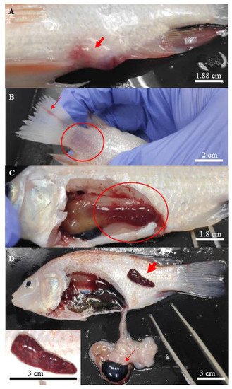
Aeromonas-infected fish showed various degrees of typical gross lesions and clinical signs of aeromoniasis. These symptoms included irregular breathing and lethargy, reduced feed consumption, displayed erratic movement with loss of balance, isolating and swimming to the surface of the water. The gross lesions included swollen and haemorrhagic site of injection (
Figure 1A), scale loss that exposed the underlying skin at the caudal fin (
Figure 1B) and necrosis of the fins (
Figure 1B).
Figure 1. Clinical signs found on the hybrid red tilapias infected with Aeromonas. (A) focal haemorrhage on the site of injection, (B) scale loss (circle) at the caudal peduncle with rotting caudal fin (thin arrow), (C) liver with deep haemorrhage (circle), and (D) haemorrhage with multiple white spots observed on the enlarged spleen (thick arrow), enlarged gallbladder (thin arrow), with a close-up picture of the affected spleen at the bottom left.
Post-mortem examinations revealed hepatomegaly (
Figure 1C) in a majority of the infected fish, while multifocal splenic infarction (
Figure 1D) was observed in fish infected with
A. hydrophila 8TK3. Some infected fish had a haemorrhagic spleen and swollen gall bladder (
Figure 1D). Overall, fish infected by
A. hydrophila 8TK3 and
A. dhakensis 4PS2 showed more severe gross lesions than other isolates. All control fish exhibited neither clinical sign nor gross lesion.
4. Lethal Dose of Aeromonas spp. in Red Hybrid Tilapia
The rates of mortality following infection by different isolates of
Aeromonas were summarized in
Table 1. Mortalities were observed as early as 24 h post-challenge in all infected groups, except for
A. jandaei 7KL3 and lasted for 216 h. By 240 h, the cumulative mortality of fish challenged with 10
3 CFU/mL of bacteria ranged between 0% and 20%, between 20% and 60% with 10
5 CFU/mL and between 35% and 95% with 10
7 CFU/mL. The two highest cumulative mortalities (95% and 90%) involved non-
myo-inositol utilizing
A. dhakensis 4PS2 (10
7 CFU/mL) and
A. hydrophila 8TK3 (10
7 CFU/mL), which was higher than the
myo-inositol utilizing
A. dhakensis 1P11S3 (10
7 CFU/mL) with 55% cumulative mortality.
Table 1. The number of mortalities of red hybrid tilapias infected with six strains of Aeromonas spp.
| Species |
Bacterial Concentration |
Number of Deaths at Specific Time (Hour Post Infection) |
Sample Number |
Cumulative Mortality (%) |
LD50–240h
(CFU/mL) |
| (CFU/mL) |
24 |
48 |
72 |
96 |
120 |
144 |
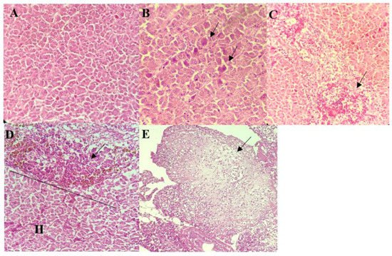
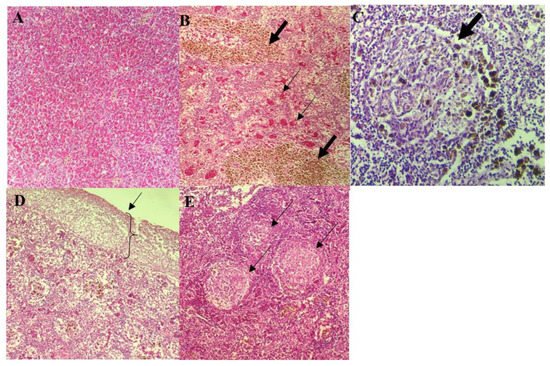
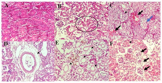
A. dhakensis 1P11S3 caused mild to moderate histopathological changes in liver, spleen and kidney of the red hybrid tilapia.
Table 2. Summary of histopathological changes scoring in liver, spleen and kidney of red hybrid tilapias following experimental infection by Aeromonas spp.
| Species |
Organ |
| 168 |
|---|
| Liver | 192 |
Spleen |
Kidney | 216 |
240 |
| A. dhakensis 1P11S3 * |
103 |
2 |
0 |
0 |
0 |
0 |
0 |
0 |
0 |
0 |
0 |
20 |
2 (10) |
1 × 10⁷ |
| |
105 |
2 |
1 |
1 |
0 |
0 |
0 |
0 |
0 |
0 |
0 |
20 |
4 (20) |
|
| |
107 |
2 |
4 |
1 |
4 |
0 |
0 |
0 |
0 |
0 |
0 |
| A. dhakensis 1P11S3 (107 CFU/mL) * |
1.0 ± 0.0 a |
2.0 ± 0.1 a |
1.0 ± 0.0 a |
| 20 |
11 (55) |
| A. dhakensis 4PS2 (107 CFU/mL) |
2.0 ± 0.1 c |
2.0 ± 0.0 a |
3.0 ± 0.0 c |
|
| A. hydrophila 8TK3 (107 CFU/mL) |
3.0 ± 0.0 b |
3.0 ± 0.0 b |
3.0 ± 0.0 b |
A. dhakensis 4PS2 |
103 |
0 |
0 |
0 |
1 |
0 |
0 |
0 |
0 |
0 |
0 |
20 |
| A. veronii | 1 (5) |
6TS5 (107 CFU/mL) | 1 × 10 | 5 |
| 1.0 ± 0.0 | a |
2.0 ± 0.0 a |
1.0 ± 0.1 a |
|
105 |
1 |
5 |
6 |
8 |
0 |
0 |
0 |
0 |
0 |
0 |
20 |
8 (40) |
|
| A. caviae 7X11 (107 CFU/mL) |
1.0 ± 0.0 a |
2.0 ± 0.0 a |
1.0 ± 0.0 a |
|
107 |
11 |
0 |
3 |
4 |
1 |
| A. jandaei 7KL3 (107 CFU/mL) | 0 |
0 |
0 |
0 |
0 |
20 |
19 (95) |
1.0 ± 0.0 a |
1.1 ± 0.1 c |
2.1 ± 0.1 | |
| d |
A. hydrophila 8TK3 |
103 |
0 |
3 |
0 |
0 |
0 |
0 |
0 |
0 |
0 |
0 |
20 |
3 (15) |
1 × 10⁵ |
| Control (sterile TSB) |
0.0 ± 0.0 d |
0.0 ± 0.0 d |
0.0 ± 0.0 e |
|
105 |
0 |
2 |
6 |
2 |
1 |
0 |
1 |
0 |
0 |
0 |
20 |
12 (60) |
|
| |
107 |
7 |
2 |
6 |
1 |
1 |
0 |
1 |
0 |
0 |
0 |
20 |
18 (90) |
|
| A. veronii 6TS5 |
103 |
0 |
1 |
0 |
0 |
1 |
0 |
0 |
0 |
0 |
0 |
20 |
2 (10) |
1 × 10⁷ |
| |
105 |
2 |
3 |
1 |
0 |
0 |
0 |
0 |
0 |
0 |
0 |
20 |
6 (30) |
|
| |
107 |
3 |
0 |
1 |
3 |
0 |
0 |
1 |
0 |
1 |
0 |
20 |
9 (45) |
|
| A. caviae 7X11 |
103 |
0 |
0 |
0 |
0 |
0 |
0 |
0 |
0 |
0 |
0 |
20 |
0 (0) |
1 × 10⁷ |
| |
105 |
2 |
0 |
0 |
1 |
0 |
2 |
0 |
1 |
0 |
0 |
20 |
6 (30) |
|
| |
107 |
1 |
3 |
3 |
1 |
1 |
1 |
0 |
0 |
0 |
0 |
20 |
10 (50) |
|
| A. jandaei 7KL3 |
103 |
0 |
2 |
0 |
1 |
1 |
0 |
0 |
0 |
0 |
0 |
20 |
4 (20) |
1 × 10¹¹ |
| |
105 |
0 |
3 |
0 |
0 |
0 |
2 |
0 |
0 |
0 |
0 |
20 |
5 (25) |
|
| |
107 |
0 |
3 |
2 |
0 |
0 |
0 |
1 |
1 |
0 |
0 |
20 |
7 (35) |
|
| Control (sterile TSB) |
0 |
0 |
0 |
0 |
0 |
0 |
0 |
0 |
0 |
0 |
0 |
20 |
0 (0) |
|