MicroRNAs in Migraine: Comparison
Please note this is a comparison between Version 1 by Lara Ahmad and Version 2 by Amina Yu.

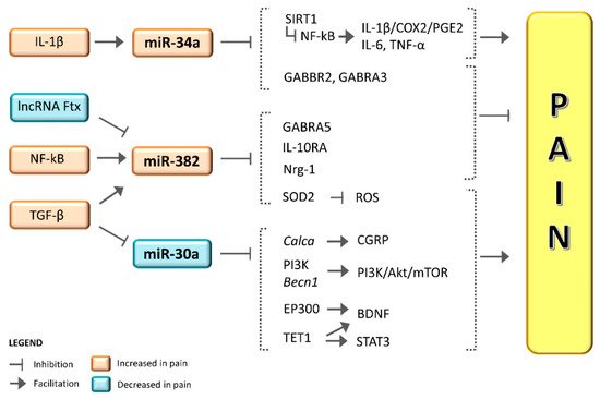
Preliminary but convergent findings suggest a role for microRNAs (miRNAs) in the generation and maintenance of chronic pain and migraine. Initial observations showed that serum levels of miR-382-5p and miR-34a-5p expression were increased in serum during the migraine attack, with miR-382-5p increasing in the interictal phase as well. By contrast, miR-30a-5p levels were lower in migraine patients compared to healthy controls. Of note, antimigraine treatments proved to be capable of influencing the expression of these miRNAs. Altogether, these observations suggest that miRNAs may represent migraine biomarkers, but several points are yet to be elucidated. A major concern is that these miRNAs are altered in a broad spectrum of painful and non-painful conditions, and thus it is not possible to consider them as truly “migraine-specific” biomarkers.  We feel that Tthese miRNAs may represent useful tools to uncover and define different phenotypes across the migraine spectrum with different treatment susceptibilities and clinical features, although further studies are needed to confirm theour hypothesis. In this narrative entry , review we provide an update and a critical analysis of available data on miRNAs and migraines was provided,  iin order to propose possible interpretations. TheOur main objective is to stimulate research in an area that holds promise when it comes to providing reliable biomarkers for theoretical and practical scientific advances.

  • miRNA
  • biomarker
  • pain
  • headache
  • CGRP

1. Current evidence on miRNA expression in migraine.

Table 1. Current evidence on miRNA expression in migraine.
miRNA Specimen Population Summary of Study Results Reference
Treatment Posology and Duration Population miRNAs Modifications and Timing Reference
miR-382-5p Serum 8 migraine patients without any medication

12 migraine patients with normal medication habits

8 healthy controls
4.1-fold increase expression during migraine attack.

Higher expression during the interictal period in migraine patients vs. healthy subjects.
Andersen et al. (2016) [1]Andersen et al. (2016) [46]
Detoxification 7-day standardized detoxification protocol in hospitalized patients: abrupt withdrawal of overused drugs associated to intravenous therapy twice daily with isotonic 0.9% NaCl saline 500 mL + cyanocobalamin 2500 mcg + folic acid 0.70 mg + nicotinamide 12 mg + ascorbic acid 150 mg + sodic glutathione 600 mg + delorazepam 0.5 mg 28 patients with CM–MOH Significant reduction of miR-382-5p and miR-34a-5p two months after detoxification in CM–MOH. Greco et al. (2020) [2]Greco et al. (2020) [51]
Peripheral blood mononuclear cells 27 patients with EM

28 patients with CM–MOH
Higher levels in CM–MOH vs. EM.

Positive correlation with CGRP levels.
Erenumab 70 mg One administration s.c. every 28 days for a total of three administrations 7 patients with CM

33 patients with CM–MOH
Greco et al. (2020) [2]Greco et al. (2020) [51]
Reduction of miR-382-5p and miR-34a-5p in the overall study population after three months of treatment with erenumab.

No differences between 30% Responders and NON-responders after three months of erenumab treatment. De Icco et al. (2020) [6]De Icco et al. (2020) [52] miR-34a-5p Serum
NSAIDs and long-term magnesium8 migraine patients without any medication

Chronic treatment with magnesium (400 mg/day for three months) + Abortive treatment with acetaminophen (15 mg/kg) or ibuprofen (10 mg/kg)12 migraine patients with normal medication habits

8 healthy controls
24 children and adolescents affected by migraine without aura divided into two groups (treated, and untreated) and 12 healthy controls9-fold increase expression during migraine attack. Decreased expression of miR-34a-5p in migraine patients treated with NSAIDs and long-term magnesium vs. untreated migraine patients.

Decreased expression in healthy controls vs. untreated migraine patients.

Comparable expression between migraine patients treated with NSAIDs and long-term magnesium and healthy controls.
Andersen et al. (2016) [1]Andersen et al. (2016) [46]
Gallelli et al. (2019) [7]Gallelli et al. (2019) [50] Peripheral blood mononuclear cells 27 patients with EM and 28 with CM–MOH Higher levels in CM–MOH vs. EM.

Positive correlation with CGRP levels.
Greco et al. (2020) [
Biphasic ketogenic diet2]Greco et al. (2020) [51]
Phase 1: 3 weeks with <30 g of carbohydrates miR-30a Serum Patients with migraine with or without aura (sample size not defined) Significant lower expression in patients with migraine vs. healthy controls. Zhai et al. (2018) [3]Zhai et al. (2018) [93]
Other miRNAs Peripheral blood mononuclear cells 15 female patients with migraine without aura

13 healthy controls
Reduced miR-181a, let-7b and miR-22 levels in EM vs. HC.

Increased miR-27b levels in EM vs. HC.
Tafuri et al. (2015) [4]Tafuri et al. (2015) [94]
Serum 8 migraine patients without any medication

12 migraine patients with normal medication habits

8 healthy controls
Increased miR-29c-5p and miR-26b-3p expression in EM patients. Andersen et al. (2016) [1]Andersen et al. (2016) [46]
Serum 30 patients with EM

30 healthy controls
Increased expression of miR-155, miR-126, and let-7 in EM vs. HC. Chen et al. (2018) [5]Chen et al. (2018) [95]
Legend: EM: episodic migraine. CM: chronic migraine. MOH: medication overuse headache. HC: healthy controls. CGRP: calcitonin gene-related peptide.
Table 2. Treatment driven modifications of miRNAs expression in migraine.


Phase 2: 3 weeks with <120 g of carbohydrates
6 female obese patients with migraine Reduction of has-miR-590-5p and has-miR-660-3p expression after a 6-week ketogenic diet Cannataro et al. (2020) [8]Cannataro et al. (2020) [96]
Legend: CM: chronic migraine. MOH: medication overuse headache. CGRP: calcitonin gene-related peptide. NSAIDs: nonsteroidal anti-inflammatory drugs.

2. miR-382-5p and miR-34a-5p in Migraine

There is mounting evidence that miR-382-5p and miR-34a-5p are dysregulated in migraine (Table 1 and Table 2).
Andersen et al. first detected the aberrant serum expression of 32 miRNAs in migraine patients in 2016 and identified a nine-fold increase in miR-34a-5p expression with a four-fold increase in miR-382-5p expression during the migraine attack when compared to the interictal phase [1][46]. Intriguingly, they also detected a significant higher miR-382-5p expression in migraine patients during the interictal period when compared to healthy subjects, and thus proposed this miRNA as a possible actor in migraine pathophysiology and a specific biomarker of the disease [1][46]. A main limitation of the study was that the migraine population was not well characterized, thus it was not possible to correlate the results obtained with the migraine phenotype (episodic or chronic) and with headache features.
A previously suggested hypothesis was that miR-382-5p serum expression was explained by increased blood–brain barrier permeability, which was suggested to occur during the migraine attack in a pre-clinical model [9][47], but this hypothesis has never been confirmed in humans and needs further confirmation.
Previous studies revealed that both miR-382-5p and miR-34a-5p negatively modulate target genes linked to GABAergic facilitating signaling, a neurotransmitter involved in trigeminal pain analgesia [1][46]. This may represent a protective compensatory mechanism in migraine self-limitation; indeed, higher GABA levels have been detected in the cerebrospinal fluid and in the saliva during migraine attacks [10][11][48,49].
miR-382-5p is also involved in Interleukin (IL)-10 anti-inflammatory signaling, where it acts via a negative modulation of the interleukin 10 receptor alpha subunit (IL-10RA), an endogenous inhibitor of pro-inflammatory IL-1β signaling [1][46].
The seminal work of Gallelli et al. paved the way for the identification of miRNAs as potential peripheral biological markers of drug response. They demonstrated lower miR-34a-5p and miR-375-5p expression in serum and saliva of children and adolescents treated with magnesium and acute nonsteroidal anti-inflammatory drugs (NSAIDs) compared to untreated subjects with migraine without aura [7][50]. It is noteworthy that in the first cohort of patients, the expression of these specific miRNAs was comparable to a group of healthy controls [7][50].
A recent study specifically analyzed miR-382-5p and miR-34a-5p expression in subjects with EM or CM and MOH (CM–MOH) [2][51]. The authors described a significantly higher interictal expression of miR-34a-5p and miR-382-5p in the plasma of CM–MOH subjects [2][51]. This analysis detected for the first time the association between specific miRNAs and the migraine phenotype as well as disease severity [2][51]. Notably, in this study CGRP plasma levels positively correlated with miR-382-5p and miR-34a-5p expression [2][51].
Although analyzed as a secondary endpoint, miR-382-5p and miR-34a-5p expression was reduced two months after detoxification from acute medications (in-hospital detoxification program), regardless of the clinical outcome [2][51]. This observation suggests the possibility of a direct role for acute medications in the expression of both miRNAs.
miR-382-5p and miR-34a-5p were also negatively modulated by erenumab, a monoclonal antibody directed against the CGRP receptor, in a population of difficult-to-treat CM patients with and without MOH [6][52]. The expression of each miRNA was reduced after three monthly s.c. administrations of erenumab, irrespective of the clinical outcome, which suggests a direct role for CGRP in miRNA regulation [6][52]. By contrast, central sensitization improved only in migraine patients who responded to erenumab treatment with an at least 30% reduction in monthly migraine days [6][52].

3. miR-382-5p and miR-34a-5p in Pain and Putative Mechanisms

miR-34a expression was reported to be altered in several human diseases besides migraine, including osteoarthritis [12][13][53,54], Alzheimer’s disease [14][55], cancer [15][16][56,57] and leukemia [17][58]. The connection between miR-34a and these pathologies resides in its strong contribution to the regulation of cell cycles and apoptosis, with miR-34a being a major transcriptional target of p53 [18][19][59,60]. The involvement of miR-34a in several painful and non-painful conditions clearly suggests that, when considered alone, it cannot represent a migraine-specific biomarker. Nonetheless, it may still play a role as part of a multi-biomarker panel signature of migraine [20][7].
Regarding pain-related disorders, miR-34a plays a role in the modulation of the inflammatory response [21][22][61,62]. Upregulation of mir-34a has been reported in osteoarthritic tissues specimens and in patients with low back pain related to intervertebral disc degeneration [23][24][25][63,64,65]. miR-34a expression was enhanced by IL-1β release [12][53], although miR-34a itself can up-regulate the IL-1β/cyclooxygenase 2 (COX-2)/Prostaglandin E2 (PEG2) inflammation pathway [26][66] by targeting the deacetylase silencing information regulator 1 (SIRT1) [26][27][28][66,67,68]. miR-34a also inhibits SIRT1 by increasing the acetylation of the nuclear factor kappa B (NF-κB) P65 subunit, ultimately enhancing the transcription of inflammatory mediators, including IL-1β, IL-6 and tumor necrosis factor-α [27][29][67,69]. In an animal model of inflammatory pain, Chen et al. reported an increase in miR-34a expression in the spinal cord and showed that its inhibition by a miR-34a antagonist exerted analgesic effects by increasing spinal SIRT1 expression [28][68]. The inhibition of SIRT1 was reported to enhance the release of CGRP in rat trigeminal ganglia in vitro, probably as a consequence of IL-1β/COX-2/PGE2 pathway activation [26][66]. Other genes coding for proteins that are involved in chronic pain pathways have been validated as targets of miR-34a, such as sodium voltage-gated channel beta subunit 2 (Scn2b) and vesicle-associated membrane protein 2 (VAMP2) [30][70]. This latter belongs to the SNARE (soluble N-ethyl-maleimide-sensitive factor attachment protein receptor) protein complex that regulates presynaptic neurotransmitter release as well as neurotransmitter receptor trafficking and synaptic plasticity [31][71], whose variants have been reported to be linked with migraine susceptibility [32][72]. It must be noted that miR-34a was found to be down-regulated in rats’ dorsal root ganglions 12 days after the experimental induction of neuropathic pain, with the concomitant up-regulation of VAMP2 [30][70]. Since the increase of synaptic proteins is related to higher glutamate release [33][73], this may ultimately lead to an enhancement of nociception. Nonetheless, miR-34a was predicted to target the genes coding for specific GABA receptors subunits (GABBR2, Gamma-aminobutyric acid type B receptor subunit 2; GABRA3, Gamma-aminobutyric acid receptor subunit alpha-3) [1][46]. In this context, an up-regulation of this microRNA, as reported in serum of migraine patients, could contribute to the reduction of the GABA inhibitory signal [1][46]. Although some controversies exist, it is reasonable to assert that the role of miR-34a may vary across different pain disorders according to the time of evaluation and, more importantly, depending on specimen collection. However, together these observations strongly point towards the contribution of miR-34a to the regulation of pain-related neurotransmission.
Like miR-34a, miR-382-5p was predicted to target the GABRA5 (Gamma-aminobutyric acid receptor subunit alpha-5) GABA receptor subunit and also IL-10RA [1][46]. Thus, the observed up-regulation in the serum levels of miR-382-5p in migraine patients may be explained by reduced GABAergic and anti-inflammatory signaling [1][34][35][46,74,75]. The increased miR-382-5p serum levels in migraine patients may be also related to high protein levels of transforming growth factor β (TGF-β). In agreement with this hypothesis, increased TGF-β levels were found in the plasma and cerebrospinal fluid of patients with EM [36][37][76,77]. TGF-β is able to increase the expression of miR-382-5p [38][39][40][78,79,80] by inhibiting transcription factors’ repressors [41][81]. TGF-β modulated miR-382-5p in vitro (human CD34+ stem cells), resulting in the increased accumulation of reactive oxygen species (ROS). ROS accumulation was explained by miR-382-5p-induced inhibition of superoxide dismutase 2 (SOD2), a direct target of this mRNA [39][40][79,80]. This latter issue appears to be relevant in the context of migraine pain, since oxidative stress and mitochondrial dysfunction was suggested as actors in migraine pathophysiology [42][43][44][82,83,84]. miR-382-5p regulation is also influenced by the long non-coding RNA Ftx (lncRNA Ftx), which exerts its action as a competing endogenous RNA (ceRNA), competing with miR-382-5p at targets level [45][85]. An increase in miR-382-5p expression together with a down-regulation of lncRNA Ftx and neuregulin-1 (Nrg-1) was reported in an animal model of neuropathic pain [45][85]. The lack of inhibition exerted by lncRNA Ftx on miR-382, is supposed to increase the inhibition of Nrg-1, thus facilitating pain and inflammation [45][85]. The involvement of Nrg-1 in pain was also reported in an animal model of inflammatory pain. Its reduction has been found in the dorsal root ganglia and spinal cord of mice, contributing to hypersensitivity [46][86]. Notably, increasing Nrg-1 expression facilitates analgesia and tissue repair by modulating glial cells proliferation, central nervous system injury repair and the reduction of the release of pro-inflammatory cytokine [45][47][48][49][85,87,88,89]. Increased miR-382-5p expression in pain conditions may also be related to nuclear factor kappa B (NF-κB) signaling, implicated in migraine pain [50][51][52][90,91,92]. NF-κB acts as a transcription factor for many miRNAs [3][93], including miR-382 [4][94]. Indeed, the inhibition of NF-κB significantly suppressed miR-382 in vitro [4][94].
These observations suggest that the activation of the inflammatory pathways may lead to an up-regulation of miR-34a and miR-382 which in turn modulate the release of inflammatory and pain mediators (Figure 1).
Figure 1. Schematic representation of putative mechanisms through which miR-34a, miR-382 and miR-30a interfere with pain. Legend: in pain conditions, miR-34a and miR-382 were upregulated, while miR30a expression was downregulated. Other molecules are also implicated in the inhibition or enhancement of miRNA expression in pain disorders. The physiological activity of miRNAs is the downregulation of target mRNAs, thus the final effects depend on the role of the mRNAs involved. High levels of miR-34a and miR-382 promote the inhibition of pathways that reduce pain (e.g., GABA signaling). These miRNAs may also downregulate up-stream gene regulators, which in physiological conditions would have blocked pain pathways (e.g., SIRT1). The downregulation of miR30a in pain conditions leads to a reduction of its inhibitory effects. This would lead to an increase in pain-related pathways, since miR-30a targets genes that facilitate pain (like Calca). miRNA = microRNA; mRNA = messenger RNA; BDNF = brain-derived neurotrophic factor; Becn1 = Beclin 1 gene; Calca = Calcitonin related polypeptide alpha gene; CGRP = calcitonin gene-related peptide; COX-2 = cyclooxygenase 2; EP300 = E1A Binding Protein P300; GABBR2 = Gamma-aminobutyric acid type B receptor subunit 2; GABRA3 = Gamma-aminobutyric acid receptor subunit alpha-3; GABRA5 = Gamma-aminobutyric acid receptor subunit alpha-5; IL-10RA = interleukin 10 receptor alpha subunit; IL-1β = Interleukin 1β; IL-6 = Interleukin 6; lncRNA Ftx = long non-coding RNA Ftx; NF-κB = nuclear factor kappa B; Nrg-1 = neuregulin-1; PGE2 = Prostaglandin E2; PI3K = phosphatidyl Inositol 3-kinase; ROS = reactive oxygen species; Scn2b = voltage-gated channel beta subunit 2; SIRT1 = silencing information regulator 1; SNARE = soluble N-ethyl-maleimide-sensitive factor attachment protein receptor; SOD2 = superoxide dismutase 2; STAT3 = Signal transducer and activator of transcription 3; TET1 = ten-eleven translocation methylcytosine dioxygenase 1; TGF-β =Transforming growth factor β; TNF-α = tumor necrosis factor-α; VAMP2 = vesicle-associated membrane protein 2.

4. Role of miR-30a in Migraine and Pain

Like many other miRNAs, miR-30a can target several different genes [53][54][55][56][97,98,99,100], of which many are connected with pathways related with pain transmission/modulation. Zhai and Zhu reported lower serum miR-30a levels in migraine patients compared to healthy subjects [3][93] (Table 1 and Table 2). Interestingly, the authors also showed that miR-30a overexpression can degrade calcitonin/alpha-CGRP (Calca) gene in vitro, thus reducing CGRP levels [3][93].
TGF-β is known to downregulate miR-30a [57][58][101,102]. Given that high protein levels of TGF-β were found in migraine patients [34][35][74,75], it is worth speculating that, in contrast to what happens with miR-382, the increased TGF-β levels might negatively regulate miR-30a expression. This would lead to increased Calca expression and, consequently, to augmented CGRP levels. This pathway appears to be relevant for migraine, as it was hypothesized that Calca degradation may represent a protective mechanism in migraine progression [3][93].
In line with the above-mentioned observation, low levels of miR-30a were also found in animal models of neuropathic pain [59][60][103,104]. The involvement of miR-30a in pain modulation resides in its ability to regulate different pathways contributing to pain. For instance, miR-30a directly binds phosphatidyl Inositol 3-kinase (PI3K) [61][105] and Becn1 [62][106], the gene coding for Beclin 1, a core subunit of the PI3K complex. Up-regulation of miR-30a inhibits the PI3K/Akt/mTOR signaling pathway, thus resulting in a reduced central sensitization and pain relief [63][64][65][107,108,109]. In addition, miR-30a targets ten-eleven translocation methylcytosine dioxygenase 1 (TET1) [66][110], which is involved in the DNA demethylation of some genes linked to pain hypersensitivity, like brain-derived neurotrophic factor (BDNF) [67][68][111,112]. Accordingly, miR-30a levels were also found to be reduced in rats with spinal cord injuries, thus confirming the hypothesis that miR-30a may have a protective role when it comes to neuropathic pain [60][104].
Taken together, these findings support the role of miR-30a in pain modulation and suggest that a down-regulation of miR-30a in pain conditions may lead to an up-regulation of different pro-nociceptive effectors possibly involved in migraine pain. The overexpression of miR-30a may be a useful pathway to counteract pain in general and possibly also in migraine pain (Figure 1).

Conclusion

 The initial evidence prompts a possible role for miRNA profiling in migraine research. Clinical data on miR-382-5p, miR-34a-5p and miR-30a seem promising, but their scientific strength is limited by the lack of consistency across studies due to: (i) inhomogeneity or poor definition of migraine cohorts (EM, CM with and without MOH) and related comorbidities, (ii) small sample sizes and (iii) different types of specimens (e.g., blood, saliva, CSF) collected for miRNAs analysis. Further studies are therefore needed to confirm and expand on the role of miRNAs in migraine pathophysiology and management. In the future, it will be important to tackle the different components of migraine in their dynamic manifestations. Indeed, migraine is a complex disorder characterized by recurring attacks, each defined by a sequence of phases: a prodromal phase, a pain phase either preceded or not by aura symptoms and a postdrome phase. The prodromal and postdrome phases have attracted the attention of the scientific community as they may represent the true timeframe for the study of the mechanisms involved in the origin and cessation of the migraine attack. The availability of reliable and validated human migraine models will facilitate the study of miRNA expression in the different phases of the migraine cycle, prompting the possibility to set experimentally controlled conditions.
Video Production Service