Anticancer Properties of Eugenol: Comparison
Please note this is a comparison between Version 1 by Khalid Rehman Hakeem and Version 2 by Conner Chen.

Clove (Syzygium aromaticum L.) (Family Myrtaceae) is a highly prized spice that has been historically utilized as a food preservative and for diverse medical uses. It is reckoned amongst the valued sources of phenolics. Among diverse active components, eugenol, the principal active component of S. aromaticum, has optimistic properties comprising antioxidant, anti-inflammatory, and anticancer actions. Eugenol (4-allyl-2-methoxyphenol) is a musky oil that is mainly obtained from clove. It has long been utilized all over the world as a result of its broad properties like antioxidant, anticancer, anti-inflammatory, and antimicrobial activities. Anticancer effects of eugenol are accomplished by various mechanisms like inducing cell death, cell cycle arrest, inhibition of migration, metastasis, and angiogenesis on several cancer cell lines. Besides, eugenol might be utilized as an adjunct remedy for patients who are treated with conventional chemotherapy. This combination leads to a boosted effectiveness with decreased toxicity.

  • eugenol
  • clove oil
  • anticancer properties

1. Lung Cancer

Additionally, indication implies that eugenol may act as both an antioxidant, stopping mutation, and a pro-oxidant in cancer cells, affecting signal pathways and destroying tumor cells. The molecular process is thought to comprise varied phases: decreasing cyclooxygenase-2 activity, hindering NF-κB stimulation, downregulation of prostaglandin production, activating arrest of cell cycle S-phase, and triggering apoptosis by reducing inflammatory cytokine levels [1][2][20,55]. Chemotherapeutic activities were observed in eugenol against human lung cancer based on the Fangjun and Zahijia study [1][20]. Through inhibition of the PI3K/Akt pathway and prevention of MMP (matrix metalloproteinase) action, an in-laboratory study using lung cancer adenocarcinoma cells A549 and human embryonic lung fibroblast MRC-5 demonstrated that a low concentration of this compound inhibited carcinogenic cell migration and incursion, hampered lung cancer cell sustainability, and stopped metastasis. At greater concentrations (1000 μM), eugenol exhibited lethal influences on normal and lung cancer cells. Lung cancer has become one of the principal reasons for death worldwide in males. Furthermore, it is increasing in females at a disturbing degree. The goal of the research of Choudhury et al. was to utilize eugenol properties to restrict cancer progress in NDEA-triggered lung carcinogenesis. Eugenol was used to find the chemo-preventive mechanism behind the NDEA-triggered mouse lung carcinogenesis model in a vital efficient method and was confirmed in the A549 human lung cancer cell line. Choudhury et al. [3][30] explored the drug-resistant and the strongest cancer cells called CSCs by investigating their controller molecule β-catenin. The nontoxic concentration of eugenol was demonstrated to induce apoptosis, concurrently repressing cell invasion in the lung tissue of carcinogen-treated mice while it did not influence the normal animals. Coalescing cellular apoptosis and proliferation, eugenol exhibited an outstanding chemoprotective capability in this lung cancer. It powerfully constrained the lung carcinoma in the trifling dysplastic stage as a chemoprotective agent. The molecular investigation greatly showed β-catenin nuclear transportation restriction. The diminished β-catenin pool and activated N-terminal Ser37 phosphorylation form after eugenol administration caused its cytoplasmic degradation. Subsequently, CSC markers like Oct4, CD44, EpCAM, and Notcht1, whose expression is reliant on β-catenin, were considerably reduced, as confirmed through ICC, IHC, and WB analysis both in vivo and in vitro. The in vitro secondary sphere-forming assay also verified the significantly suppressed CSC population, and thus the virulency. Furthermore, eugenol was verified to greatly boost β-catenin degradation after exposure to the CK1α inhibitor D4476 in vitro via Western blot. CK1α in the Wnt/β-catenin pathway has a vital function for labelling with the N-terminal Ser45 phosphorylation of β-catenin, which eventually unlocks a spot for critical phosphorylation via the means of GSK3β at the Ser37 residue to happen. Therefore, the decisive killing of CSCs was linked with reappearance owing to the handling failure. This may accomplish an extended and healthier life quality using a natural cheap method [3][30].

2. Colon Cancer

Essential oils are a complicated blend of volatile and hydrophobic components produced from aromatic plants, usually found in our food. Various studies have presently implied probable anticancer effects of several essential oil compounds. Essential oils show their anticancer potential against many human neoplastic cell lines, either alone or in combination with anticancer medicines [4][58]. Petrocelli et al. used cinnamaldehyde (essential oil obtained from cinnamon) and eugenol (essential oil obtained from bud clove) in his study on NCM-460 cells (epithelial colon) and anticancer action against colorectal cancer (CRC) cell lines [5][25]. Petrocelli et al. [5][25] distinguished cinnamaldehyde and eugenol as ingredients with a definite antitumor activity selectively targeting the transformed colonic cells. After 72 h of treatment, cell death, necrosis, and cell cycle slowing were activated by both cinnamaldehyde (75 µM) and eugenol (800 µM) in Caco-2 and SW-620 cells but not by NCM-460 cells. These two ingredients can possibly prevent or cure colorectal cancer if they are linked with targeted delivery to the colon.
This compound powerfully decreased the vitality of CRC cells and mere cytotoxic influences on healthy cells. The influence of eugenol (800 µM) was inspected in cells after 72 h. Eugenol inhibited G2 in Caco-2 cells, while it favored G1 accumulation in SW-620 cells. Eugenol showed no effect on the cell cycle of NCM-460. Such findings displayed that a specific dosage of eugenol can decline the proliferation of CRC cells by either shrinking the G2 phase or promoting the G1 phase. Besides, the Annexin V-FITC assessment via flow cytometry exhibited a substantial rise in necrosis and late apoptosis in CRC cells exposed to eugenol and did not affect NCM-460. The absence of influences in normal cells implies that eugenol can be used for CRC remedies or to prevent cancer. Furthermore, eugenol was effectual as an anticancerous agent under lab situations, given alone, or combined with conventional medicines such as doxorubicin [6][7][76,77]. The superior role of cinnamaldehyde against SW-620, regarding Caco-2 cells, shows its tremendous potential for metastases. Regrettably, this potential fades upon increasing the duration of treatment [5][25]. One more remarkable characteristic of cinnamaldehyde and eugenol is their display of antioxidant and pro-oxidant action in varied circumstances. Antioxidant potential has been designated as a defensive counter to carcinogenesis. After the establishment of cancer, the pro-oxidant influences can activate death of cancer cells through cell-signaling [8][9][39,89]. Moreover, the comparatively longer time duration required for the transition of a normal colon epithelial cell to become cancerous motivates us to test eugenol and cinnamaldehyde in an appropriate preparation to interrupt and decrease CRC instigation. However, the formulation of chemotherapeutical drugs and cinnamaldehyde and eugenol, previously exhibited, may also provide a novel pharmacologic chance to expand the anticancer influences in stubborn CRC types, redeeming the healthy colon epithelial cells.

3. Gastric Cancer

The combination of apoptosis stimulation and inhibition of cancer cell proliferation and angiogenesis conduct may be useful for cancer chemoprevention. Manikandan et al. [10][78] assessed the chemopreventive influences of eugenol on N-methyl-N′-nitro-N-nitrosoguanidine (MNNG)-triggered gastric cancer in Wistar rats. MNNG-treated rats established gastric carcinogenesis, showing avoidance of apoptosis along with promotion of proinvasive and angiogenic elements. Eugenol administration triggered apoptosis by the mitochondrial conduit through modulation of Bcl-2 family proteins, Apaf-1, cytochrome C, and caspase, and hindering cell proliferation and angiogenesis, as proved via alternations in MMPs actions and MMP-2 and -9, VEGF, VEGFR1, TIMP-2, and RECK expression. Therefore, plant secondary metabolites like eugenol, which can manipulate the balance of pro and antiapoptotic proteins, besides the subtle equilibrium of initiators and inhibitors of angiogenesis and invasion, are desirable contenders for stopping cancer development [10][78].
Modulation of the intracellular signaling pathway of NF-κB included in the downregulation of cell invasion and cell cycle monitoring molecules is a practical method of chemoprevention. Eugenol is recognized to have interesting healing properties. Manikandan et al. examined the modulating influences of eugenol on NF-κB signaling in gastric carcinoma due to N-methyl-N′-nitro-N-nitrosoguanidine (MNNG) in a rat model by analyzing the expression of NF-κB family members ((NF-κB, p50, and p65), kappaB alpha inhibitor (IκBα), phosphorylated IκBα (p-IκBα), IκB kinase β (IKKβ)), and the NF-κB target genes that enhance (e.g., cyclin D1, cyclin B, and PCNA) or hinder (e.g., p21, p53, and Gadd45) cell proliferation and cell survival. MNNG-triggered gastric tumors were distinguished by activation of NF-κB, which was associated with the upregulation of IKKβ and the phosphorylation and disintegration of IκBα. Moreover, the upregulation of cyclins and PCNA with the downregulation of p21, p53, and Gadd45 implied that the proliferative benefit in gastric carcinomas depends on the activity of NF-κB. Eugenol treatment greatly decreased the occurrence of MNNG-triggered gastric tumors by overpowering NF-κB stimulation and modulation of the translation of NF-κB target genes that control cell proliferation and cell survival. The modification of the NF-κB signaling pathway via eugenol can have a substantial effect on chemopreventive and remedial methods of cancer [11][79].

4. Cervical Cancer

Cervical cancer is a general gynecological tumor that mostly leads to death in women [12][90]. It results from sustained HPV type 16 and 18 infections. It is a metastatic tumor owing to its ability to attack and grow at distant places and result in metastases. Therefore, the chief cause of the high prevalence and mortality of cervical cancer is metastasis to other body parts. The epithelial-mesenchymal transition (EMT), particularly EMT type 3, is involved in cervical cell carcinoma metastasis [13][14][91,92]. Snail-1 (zinc-finger transcription factor that is controlled via the PI3K/Akt signaling pathway) increases EMT through downregulation of E-cadherin and upregulation of vimentin [15][16][93,94]. EMT causes epithelial cells to transform into mesenchymal cells, triggering damage of cell adhesion and polarity [17][95]. Migration ability, invasion, apoptosis resistance, and extracellular matrix formation all increase as a result of these changes in features [18][96].
Recently, chemotherapy is the most used treatment for metastatic cervical cancer; however, it can have the most serious side effects since it proves toxic to other normal cells surrounding the tumor, leading to necrosis. Furthermore, the fee of chemotherapy treatment is excessive [19][97]. Thus, treatments that are both more effective and less expensive are required. Permatasari et al. showed that eugenol’s antitumor mechanisms trigger cell death in cancer cells via activation of p53 protein levels [20][98]. To evaluate the antimetastatic properties of eugenol against cervical cancer, Permatasari et al. examined the effect of eugenol on cell migration. Specifically, eugenol exposure to HeLa cells was given at varying doses in scratched wells [21][29]. In HeLa cells, eugenol boosts apoptosis. It had a cytotoxic influence on HeLa cells at concentrations of 50–200 μM. In particular, caspase-3 and p53 protein expression are involved in this apoptotic incidence [20][98]. One of eugenol’s anti-carcinogenic influences in cervical cancer was to hinder cell migration. As eugenol concentrations increased, cell invasion was repressed; HeLa cells exposed to eugenol (200 μM) displayed the most efficient cell migration suppression [21][29].
E-cadherin is a cell adhesion molecule that also serves as a marker for EMT. EMT is linked to E-cadherin expression loss, which commonly happens during cancer spread [22][99]. Studies were also achieved to examine the activity of methyl eugenol and cisplatin versus cervical cancer cells. The medicines were utilized discretely and in permutation. Methyl- eugenol united with cisplatin boosted the anticancer influence by triggering apoptosis and damaging HeLa cells in contrast to the medicine influences. Cell numbers in the G0/G1 phase, caspase-3 action, and mitochondrial membrane potential damage were considerably boosted in joint action contrary to the separate administration [23][27].

5. Melanoma

Melanoma (malignant melanoma) progresses from melanocytes and is a kind of skin cancer [24][100]. Amongst all types of skin cancers, melanoma is responsible for just 4%. Nevertheless, it is accountable for an elevated death rate, with over 80% of the deaths due to skin cancer [25][101]. Pisano et al. examined the anti-invasive influence of eugenol against melanocytes [26][80]. Eugenol seizes the cell cycle and induces apoptosis. In addition, Miyazawa and Hisama investigated the influence of eugenol on a B16 xenograft model [27][81]. Eugenol works through the synthesis of ROS [28][82], which leads to DNA synthesis inhibition, hence postponing cancer progress. A 40% decrease was documented in tumor size via eugenol activity [29][30][83,84]. In another study regarding the anticancer action of eugenol on human melanoma cells (WM1205Lu), eugenol showed an induction of apoptosis and arrested the cell cycle at the S-phase [29][83]. Ghosh et al. [31][85] explored eugenol and isoeugenol (an isomer of eugenol) for anti-melanoma action. They concluded that eugenol, but not isoeugenol, exhibited anticancer action against melanocytes [28][82].
Eugenol and six of its derivatives were examined by Pisano et al. [26][80] for their anti-proliferative action on primary melanoma cell lines. They deduced that the biphenyl eugenol derivative enantiomer (S)-6,6′-dibromo-dehydrodieugenol (S7-S) possesses the capability to trigger apoptosis in neuroblastoma and melanoma as compared with other eugenol derivatives. Eugenol treatment caused apoptosis of G361 cells with the probable caspases 3 and 6 association. Furthermore, substrates of caspases, such as PARP, DFF45, and lamin A, were slashed throughout eugenol-triggered apoptosis in G361 cells. Gosh et al. [31][85] implied that eugenol might be established as an E2F-targeting factor to treat melanoma. Eugenol hindered the proliferation of osteosarcoma (HOS) cells in both dose- and time-dependent ways. Elevated caspase-3, p53, and PARP cleavage levels are associated with eugenol-triggered apoptosis in HOS cells [32][86].
Nanotechnology approach has an important capacity in melanoma treatment, since it allows researchers to precisely target the tumour cells with anticancer medicines [33][34][35][26,102,103]. Mishra and colleagues used a solvent injection approach to create hyaluronic acid (HA)-coated liposomes that were laden with an efficient blend of dacarbazine and eugenol (antimelanoma drugs) [36][104]. With just a 0.5 µg/mL dacarbazine dose, coated-dacarbazine eugenol liposomes displayed 95.08% cytotoxicity as compared to 10.20% cytotoxicity by dacarbazine solution at the identical dose. Besides, coated-dacarbazine eugenol liposomes demonstrated a remarkable increased decline in cell invasion and proliferation. The suppression of the antiapoptotic protein survivin, which is highly expressed in melanoma cells and causes them resistance to apoptosis, is thought to be responsible for this performance. Eugenol addition led to survivin protein downregulation, thus allowing dacarbazine to achieve its role with dramatically increased cytotoxicity, elevated apoptosis, and significantly reduced cancer cell migration and proliferation. Therefore, surface-functionalized dacarbazine- and eugenol-laden liposomes possess substantial potential against resilient and violent metastatic melanoma [33][26].
Dwivedi et al. compared the anticancer actions of three distinct clove (S. aromaticum) extracts against varied cancer cell lines in vitro. Water, ethanol, and oil extracts were tested against various cancer cell lines, such as MCF-7 (ER+ve), MDA-MB-231 (ER−ve) breast cancer, HeLa (cervical cancer), DU-145 prostate cancer, TE-13 esophageal cancer cell lines, as well as normal human peripheral blood lymphocytes in order to evaluate their anti-proliferative activities. The MTT assay was used to assess cell proliferation inhibition. The extracts inhibited cell growth in a variety of ways in the five cancer cell lines studied, with the oil extract having the most cytotoxic effect. Cell disruption with membrane rupture caused cytotoxicity, according to morphological examination and DAPI staining. Clove oil at 300 μL/mL caused maximum cell death in TE-13 cells after 24 h, with 80 percent cell death, but DU-145 cells showed low cell death. However, no appreciable cytotoxicity was detected in human peripheral blood mononuclear cells (PBMCs) at an identical dose [37][105].

6. Breast Cancer

Kumar et al. studied if cloves have any cytotoxic action on MCF-7 human breast cancer cell lines. A brine shrimp lethality test (BSLT) and an MTT assay were used to examine the anticancer capability of varied doses of water extract, ethanol extract, and clove essential oil in vitro. The essential oil of cloves demonstrated the most cytotoxic influence, followed by ethanol and water extract. The LD50 concentration of clove essential oil in the 24 h BSLT was 37 μg/mL. Besides, the clove essential oil’s IC50 values in the 24- and 48-h MTT assays were 36.43 and 17.6 μg/mL, respectively. Therefore, they concluded that cloves are a prospective source for the creation of anticancer drugs [38][106].
Breast cancer is a serious life-threatening health problem for millions of women around the world every year. It is classified as the second most common cancer in women, and is ranked fourth for cancer-related deaths globally [39][37]. In women, mammary epithelial cells are controlled by keeping a balance between the cell cycle and apoptosis. Disruption of this causes an increase in mammary epithelial cells, lastly leading to breast cancer [40][107]. The majority of chemotherapy agents that are presently utilized as a means of treatment of this malady are extremely lethal and have long-term side effects. Thus, new anticancer medicines with greater efficacy and specificity are immediately required. The influence of eugenol on apoptotic and procarcinogenic proteins, both in vitro and in tumor xenografts, was evaluated via immunoblotting and RT-PCR was utilized to determine the influence of eugenol on E2F1 and survivin mRNA levels. Al-Sharif et al. examined the influence of eugenol on cell proliferation by means of a real-time electronic cell detection system. Low-dose eugenol (2 µM) has explicit toxicity against several breast cancer cells. This lethal influence was chiefly facilitated by induction of the internal apoptotic pathway and potent downregulation of E2F1 and its downstream antiapoptosis target, surviving, regardless of p53 and ERa status. Eugenol also inhibited many other breast cancer-related oncogenes, like NF-κB and cyclin D1. Furthermore, eugenol upregulated the multipurpose cyclin-dependent kinase inhibitor p21WAF1 protein and inhibited the proliferation of breast cancer cells in a p53-independent manner. Significantly, these antiproliferative and pro-apoptotic influences have also been noticed in vivo in xenografted human breast cancers. Therefore, eugenol displays anticancer characteristics both in vitro and in vivo, demonstrating that it might be utilized to strengthen adjuvant breast cancer treatment by targeting the E2F1/surviving pathway, particularly for the less-sensitive triple-negative subtype of the disorder [41][87]. Abdullah et al. investigated the probability of using eugenol as an antimetastatic and antiproliferative compound against MDA-MB-231 and SK-BR-3 breast cancer cells. Treatment with 4 and 8 μM eugenol for 48 h substantially hindered cell proliferation of MDA-MB-231, with an inhibition rate of 76.4%, while 5 and 10 μM of eugenol for 48 h substantially hindered the proliferation of SK-BR-3 cells with an inhibition rate of 68.1%. Eugenol-treated cells demonstrated substantially reduced expression of MMP2 and MMP9 and an insignificant rise in the expression of TIMP1 in HER2-positive and triple-negative breast cancer cells. Eugenol greatly raised the proportion of MDA-MB-231 and SK-BR-3 cells in late apoptosis and elevated Caspase3, Caspase7, and Caspase9 expression [42][88]. Vidhya and Devaraj [43][108] verified that breast cancer cells (MCF-7) experience the potent antimutagenic action of eugenol. Eugenol is both time and dose dependent when defeating the proliferation of MCF-7 cells [43][44][108,109]. Furthermore, Pisano et al. [26][80] described the antiproliferative activity of eugenol-related biphenyl (S)-6,60-dibromo-dehydrodieugenol, via induction of apoptosis.
The modification of autophagy may enhance either the survival or apoptosis of cancer cells. Abdullah et al. treated triple-negative (MDA-MB-231) and HER2-positive (SK-BR-3) breast cancer cell lines with varied eugenol doses. They examined apoptosis via a flow-cytometry technique, whereas autophagy via acridine orange. They investigated the influence of eugenol on the gene and protein expression levels of autophagy and apoptotic genes. Treating cells with varying doses of eugenol substantially hindered cell proliferation. The protein levels of AKT serine/threonine kinase 1 (AKT), forkhead box O3 (FOXO3a), cyclin-dependent kinase inhibitor 1A (p21), cyclin-dependent kinase inhibitor (p27), and Caspase-3 and -9 increased considerably in eugenol-treated cells. Furthermore, eugenol triggered autophagy through upregulation of microtubule-associated protein 1 light chain 3 (LC3) expression levels and downregulation of nucleoporin 62 (NU p62) expression. Eugenol is a potential natural anticancer agent against triple-negative and HER2-positive breast cancer. It seems to act via targeting of the caspase pathway and via triggering of autophagic cell death [45][28]
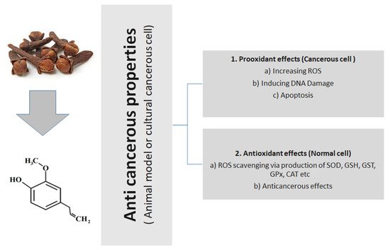
Figure 1 presents the general action of eugenol, tried and tested in animal or cultural cancerous cell models.
Figure 1.
 General anticancerous action of eugenol (tried and tested in animal or cultural cancerous cell models).
Video Production Service