Salivary Volatile Metabolites in Oral Diseases: Comparison
Please note this is a comparison between Version 1 by Jorge Pereira and Version 2 by Peter Tang.

Oral diseases (ODs) are highly prevalent affecting over 3.5 billion people, particularly in low- and middle-income countries (LMICs). Saliva is often described as the "mirror of the body" and so different omics approaches such us proteomics, metabolomics and more recently, volatomics are being employed to explore the potential of this biofluid towards the non-invasive diagnosis of ODs.

  • saliva
  • volatile organic compounds (VOCs)
  • oral diseases (ODs)
  • biomarkers

1. Introduction

Worldwide, oral diseases (ODs) are highly prevalent affecting over 3.5 billion people, particularly in low- and middle-income countries (LMICs) [1]. ODs compromise very seriously the overall health and wellbeing, causing a myriad of conditions to the patient, as pain, sepsis and reduced life quality. Additional side effects, as loss of school days, family disruption, decreased work productivity or dental treatment costs, have a significant impact in the healthcare systems and society. According to Peres et al. [1], dental caries (tooth decay), periodontal disease, tooth loss, and cancers of the lips and oral cavity are among the most prevalent ODs. Although different procedures have been described for early detection in order to cope with these diseases, the diagnostic efficiencies greatly depend on the clinical conditions of the patients [2][3][4][2,3,4]. Nevertheless, a non-invasive method using saliva for ODs diagnostics is a highly attractive strategy. Saliva is often described as the “mirror of the body” [5][6][5,6] and so different omics approaches such us proteomics, metabolomics and more recently, volatomics are being employed to explore the potential of this biofluid towards the non-invasive diagnosis of ODs [5].

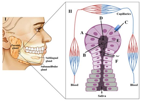
2. Physiology of Saliva

Saliva Composition and Production

In its broader sense, saliva composition includes the fluids produced by the different salivary glands and the gingival-crevicular secretions, along with cellular components, oral microbiota as well as food debris [7]. Saliva is a colourless liquid, slightly acidic, hypotonic and a mucoserous exocrine biological medium mainly composed of water (98%) with a production of approximately 0.5–1.5 L per day [5][6][8][5,6,8]. It is also possible to find different organic compounds including hormones, peptides, mucus, antibacterial compounds, enzymes, proteins, urea, uric acid, lactate and creatinine [2][4][8][9][2,4,8,9]. Ions such as Na+, K+, Ca2+, Cl and HCO3−, are also present in saliva [5][9][5,9]. The overall composition of saliva is affected by internal (related to the type of secretory glands and their stimulation) and external (personal habits including diet and lifestyle) factors [5][9][10][5,9,10]. A deeper understanding of the interplay of these factors on saliva production and composition can be found elsewhere [11][12][11,12].
The autonomic nervous system plays a key role in the production of saliva. This control is exerted by various nerves that regulate the viscosity, flow rate, volume and concentration of various constituents of saliva. The sublingual and minor mucus glands are regulated by stimulation of sympathetic nerves whereas the parotid and submandibular glands are stimulated by the parasympathetic innervation [4][5][8][13][4,5,8,13]. Saliva is mostly produced by three pairs of major salivary glands: i) parotid (which contribute for ~23% of a total saliva production), ii) submandibular (which contribute for ~65% of a total saliva production) and iii) sublingual (which contribute for a ~4% of a total saliva production (Figure 1) [4][5][8][9][4,5,8,9]. Additionally, numerous minor salivary glands (around 300–1000 units of von Ebner’s, mucous gland, labial and palatine glands, among other) distributed throughout the oral mucosa contribute with around ~8% of total saliva production [4][5][8][13][4,5,8,13]. Each type of salivary gland produces a specific secretion [8][10][8,10]. The parotid produces a serous fluid, the submandibular a sero-mucous, the sublingual secretes only mucous saliva and the minor glands produce viscous secretions [8][10][8,10]. The initial or primary saliva is an isotonic fluid produced by the acini, a cluster of cells (acinus) with a berry-type structure that are present in the salivary glands. These acini drain into the salivary ducts with small ‘striated’ ducts opening into wider intercalated and excretory ducts [10][14][10,14]. The acini are surrounded by blood capillaries that enable the exchange of substances from the circulation into the salivary glands [10]. This transport is done through three distinct mechanisms, namely selective transport using passive diffusion, ultrafiltration through pores or active (energy-dependent) transport against concentration gradients leading to the enrichment of the saliva composition [8][10][14][15][8,10,14,15] (Figure 1). During the passive diffusion, several compounds cross the cell membranes of capillaries and acini. Lipid-soluble compounds (e.g.,: unconjugated steroids) are close to unbound plasma concentrations, while hydrophilic compounds (e.g.,: conjugated steroids) reach only 1% of unbound plasma concentrations [3][8][14][3,8,14]. In turn, small polar molecules (having molecular weight below 1.9 kDa, as ions and some hormones) can be transported to saliva by ultrafiltration [8][14][8,14]. This transport is done through narrow junctions in the apical pole of the acini. Finally, for some electrolytes and larger molecules like peptides, the passage from capillaries to saliva will be dependent on an active transport mechanism [8][10][14][8,10,14]. Beyond these mechanisms of transport from the capillaries, elements such as bacteria, epithelial cells, erythrocytes, leukocytes, food debris or other contaminants may be also present in the saliva, causing significant variations to the final saliva composition [3][8][3,8]. This fact is particularly relevant in the context of the applicability of saliva as a diagnostic tool for different diseases. The connection between local (salivary glands) and systemic (blood) circulation sources provides a good option for looking into disease biomarkers or studying a physiological state. Moreover, saliva can be collected non-invasively, more affordable, accessible and painless way than blood, for instance [8][13][8,13].
Figure 1. (I)-The major salivary glands; (II)–Acini structure and the different mechanisms of transport of plasma compounds into a salivary gland. A–Entry of components by simple filtration; B–Entry of liposoluble compounds by passive diffusion C–active transport; D–Active pumping of Na+ ions and concomitant entry of H2O; E–component produced/secreted by the salivary glands; F–Pumping of Na+ ions into the blood producing hypotonic fluid; G–Liposoluble compounds; H–H2O, electrolytes (adapted from [16]).

3. Putative Salivary Biomarkers for Oral Diseases

As mentioned, ODs are a serious problem worldwide, affecting mostly the poor and marginalized groups in society with particularly higher incidence rate observed in the LMICs [1]. Strikingly, the most prevalent ODs tooth decay (caused by dental caries) and periodontal disease (gum inflammation) could be easily preventable with better oral care and access to medical services. It is therefore remarkable how such problems continue to be neglected. Cancers of the lips and oral cavity are also very prevalent, ranking in the top 15 of most common cancers in the world in 2018 [17]. Several other conditions can affect the oral cavity and its soft and hard tissues, but fortunately they have a lower global impact. The risk factors for the development of ODs are well-known, being the most relevant the abuse of sugar, alcohol and tobacco. In turn, these risk factors are also the main cause of other clinical conditions, as diabesity (diabetes and obesity co-occurrence), caries (excessive sugar consumption) and different forms of cancer (alcohol and tobacco abuse) [1]. Therefore, it is not surprising that great deal of efforts were made to identify the salivary biomarkers for ODs.

3.1. Gingivitis and Periodontal Disease

Gingivitis constitutes the initial stage of periodontal disease characterized by the inflammation of gums or gingiva. It is caused by the bacterial plaques build up on teeth and consequent inflammation of the surrounding tissues [18][24]. If not treated properly, gingivitis will progress to periodontal disease-causing tissue and bone degradation and eventual tooth loss. A non-targeted proteomic analysis of saliva by two-dimensional gel electrophoresis (2DE) and matrix-assisted laser desorption/ionization time-of-flight mass spectrometry (MALDI-TOF MS) identified the upregulation of immunoglobulin and keratins in gingivitis patients (n = 10) as compared to controls (n = 10) [19][25]. Another non-targeted study of saliva, found several proteins (among mucins, albumins, lactoferrins, histatins and amylases) altered in periodontitis [20][21][22][23][26,27,28,29]. The label free quantitative proteomic analysis of a periodontitis cohort by Bostanci et al. [24][30] revealed that lactoferrin, lacritin, sCD14, Mucin 5B and Mucin7 levels were reduced as compared to controls. This result suggests that the lower disease resistance offered by periodontitis patients is due to the reduced antimicrobial properties exhibited by their saliva. The matrix metalloproteinases (MMPs) are the proteases involved in the extracellular matrix remodelling and linked to periodontal inflammation and collagen degradation [25][31]. Particularly, MMP8 is considered as important biomarker for periodontitis. Furthermore, recent proteomic investigation also confirmed that MMP8 along with other cytokines (interleukin-1b–IL-1b, RANK/RANKL/OPG) were overexpressed in periodontitis [26][32]. In fact, Gursoy et al. [27][33] proposed a cumulative risk score using the salivary concentrations of Porphyromonas gingivalis, IL-1β, and MM8 as a novel and non-invasive model for the risk assessment of advanced periodontitis. The level of several other salivary cytokines, IL-6, IL-8, IL-17A, and tumour necrosis factor α (TNF- α) have been shown to be affected by periodontitis progress [28][34]. The identification of so many inflammatory markers in different studies involving periodontitis seems obvious given the progression of the gum inflammation from an initial gingivitis stage to periodontitis. In this case, however further studies are necessary to identify which markers are specific to the gum inflammation.

3.2. Dental Caries

Tooth decay, also known as dental caries, is one of the major ODs observed across all age groups worldwide. This multifactorial disease is triggered by the excessive consumption of fermentable carbohydrates (sugars, as glucose, fructose, sucrose and maltose), poor oral hygiene and inadequate fluoride exposure [29][45]. This combination eventually results in the formation and progression of a microbial biofilm producing acids which cause the demineralization of dental tissues and finally dental cavities [29][45]. Given that the prevalence of dental caries is positively correlated with the microbial load of Streptococcus mutans and Lactobacillus in the saliva [30][46], these two bacteria should be involved in the cascade leading to tooth decay. Porphyromonas gingivalis is another bacterium with a relevant role in ODs as it was implicated in the biofilm formation of bacterial plaque. Furthermore, it also plays an important role in the progression of periodontal disease as well as in the onset of different systemic pathologies, including rheumatoid arthritis, cardiovascular pathologies, and neurodegenerative pathologies (reviewed in [31][47]).

3.3. Oral Cancer

Oral cancer is the sixth most frequent malignant disease across the globe and oral squamous cell carcinoma (OSCC) is the most often reported subtype. The onset of oral cancer is often asymptomatic, but overall seems to be outcome of progressive premalignant conditions such as leukoplakia and oral lichen planus [32][33][50,51]. Metabolic reprogramming in oral cancer is not yet well understood and therefore, investigation of metabolic alterations is crucial for detecting novel diagnostic biomarkers and understand the disease progression. In a couple of studies saliva was used to unveil the metabolomic signature of oral cancer. Sugimoto et al. [34][52] carried out a salivary metabolomic analysis by capillary electrophoresis time-of-flight mass spectrometry (CE-TOF-MS) using a cohort of 156 individuals comprising of 69 patients with oral cancer and 87 controls. Overall, 57 metabolites were detected and out of these, 28 were found to be differentially expressed in oral cancer as compared to controls. Higher levels of salivary polyamines, piperidines and taurine were detected in oral cancer group and considered as promising screening biomarkers. In another study, Ishikawa et al. [32][50] reported that s-adenosylmethinine and pipecolate were upregulated in the saliva of oral cancer patients as compared to the control samples and possess a good diagnostic potential for the early detection of the disease [35][53]. In another study, the same authors proposed that the salivary metabolites indole-3-acetate and ethanolamine phosphate have the potential to discriminate OSCC from oral leukoplakia (OLP) [36][54]. Wei et al. [37][55] performed a salivary metabolomic study in three groups comprising oral cancer, oral leukoplakia and controls by UPLC coupled with Quadrupole TOF-MS. The study revealed that the metabolic set composed by valine, phenylalanine and lactic acid exhibits a moderate specificity and sensitivity to discriminate oral cancer from oral leukoplakia and controls [37][55]. Using the same approach, Wang et al. [38][56] performed a salivary metabolomic analysis using a cohort of 60 subjects (30 oral cancer patients and 30 controls subjects). The authors reported a pool of five salivary metabolites (propionylcholine, N-acetyl-L-phenylalanine, sphinganine, phytosphingosine, and S-carboxymethyl-L-cysteine) exhibiting good sensitivity, specificity and accuracy to discriminate oral cancer patients from controls. Furthermore, Lohavanichbutr et al. [39][57] applied 1H-NMR and LC-MS/MS to unveil putative salivary metabolic signatures for oral cancer and observed that glycine and proline were significantly altered in the malignant group when compared to the controls.
The earliest salivary proteomics analysis of OSCC pointed out the role of higher levels of several cytokines (IL-6, IL-8, IL-1 and TNF-α) in the proinflammatory and proangiogenic functions [40][58]. Related with this, more recently Aziz et al. reported augmented levels of IL-4, IL-10, IL-13 and IL-1RA in the saliva of patients with OSCC [41][59]. In another study, immunoglobulins were detected at higher levels in OSCC patients as compared to controls and found to be involved in angiogenesis [42][60]. A similar observation was reported for several cell surface receptor glycoproteins, namely CD44, CD59 and CEA, which were found overexpressed in patients with OSCC [43][61]. Different zinc finger proteins were also found augmented in OSCC patients, namely ZNF510 [44][62] or Cyfra 21–1 and CK 19 [43][44][45][61,62,63]. Hu et al. [41][59] performed a global proteomic analysis of salivary samples (OSCC = 64, control = 64) and proposed a panel of five proteins (M2BP, MRP14, CD59, catalase and profilin) which exhibit with high sensitivity (90%) and specificity (83%) to discriminate OSCC patients from controls. In a larger study, Yu et al. carried out proteomic analysis of a cohort comprising 131 OSCC patients, 103 low risk OPMDs (oral potentially malignant disorders) patients, 130 high risk OPMDs patients and 96 controls. The authors proposed four proteins–MMP1, KNG1, ANXA2 and HSPA5 that were not only able to discriminate the OSCC from controls, but also predict OPMDs progression into OSCC [46][64]. In a very recent study of OSCC salivary proteome, Sivdasan et al. reported CD44, S100A7 and S100P as novel potential biomarkers for the early detection of OSCC [47][65]. DNA methylation and conformational alterations on histones are potential molecular signatures for different cancers, including those affecting the salivary glands [48][66]. Under this context, Li et al. observed that the long non-coding RNA (lncRNA) RBM5-AS1, which promotes the proliferation, migration, and invasion of OSCC cells in vitro, was highly expressed in OSCC tumour tissues and cancer cell lines [49][67]. Furthermore, Park et al., reported that OSCC patients have lower levels of salivary miR-125a and miR-200a, strongly suggesting that specific salivary miRNAs can be used in oral cancer detection [50][68].

3.4. Oral Potentially Malignant Disorders (OPMD)

OPMDs are clinical manifestations that aggregate conditions as the oral lichen planus and oral leukoplakia (OLP), which possess risk of malignant transformation. Several prospective studies have predicted a 1% progression rate of OPMD into oral cancer [51][52][69,70]. Yang et al. [53][71] carried out 2DE and MALDI-TOF MS analysis of an OLP study cohort comprising 20 cases (patients = 10, control = 10) and reported an urinary up-regulated prokallikrein and reduced palate, lung and nasal epithelium carcinoma-associated protein (PLUNC) as novel biomarkers for oral lichen planus. In another study, Souza et al. [54][72] discovered a positive correlation of S1008, S1009 and haptoglobin with the inflammatory cytokines and oral lichen planus pathology. Furthermore, Camisasca et al. [55][73] performed a 2DE-MALDI-TOF analysis of a leukoplakia cohort consisting of 15 patients and 10 controls and identified keratin 10 as an important candidate protein biomarker for the disease pathology.

3.5. Burning Mouth Syndrome

Burning mouth syndrome is characterized by a chronic intraoral inflammation without any visible lesions in oral cavity. This syndrome is particularly prevalent in post-menopausal women and its etiology is poorly understood [56][74]. Ji et al. [57][75] reported a salivary global quantitative proteomic study to identify candidate proteins biomarkers for burning mouth syndrome in a cohort of 19 patients and 19 healthy control volunteers. The study identified three overexpressed proteins viz. α-enolase, IL-18 and kallikrein-13, as potential markers for the burning mouth syndrome.

3.6. Recurrent Aphthous Ulceration (RAS)

RAS is a common oral pathology recognized by ulcers in the mucosal lining of lip, tongue and soft palate. Sometimes, it also occurs at places as the isometric mucosa of the hard palate. Li et al. [58][76] identified dysregulated tryptophan and steroid hormone metabolism as a signature of the RAS in study cohort of 94 individuals (RAS = 45, Control = 49) by employing LC-MS/MS. This dysregulation of tryptophan metabolism and the hormonal imbalance are plausible causes for the depression, stress and reduced salivary immunity among RAS patients.

4. Salivary Volatomics

In the previous section, a significant number of studies identifying potential biomarkers for ODs were discussed. That included mostly higher molecular-weight constituents such as proteins. This section will focus specifically on the subset of potential biomarkers for ODs that are the volatile organic compounds (VOCs).
Saliva as most biofluids, also contains VOCs in its composition. Currently VOCs are being explored as potential biomarkers for ODs. Furthermore, many of them have been also detected in other biofluids [59][60][61][62][63][77,78,79,80,81], a fact that it is very important because it adds relevant information to our understanding of human metabolism in health and disease. This has obviously applications to a myriad of clinical conditions, as malignancies, infections, cardiovascular problems or genetic disorders (reviewed in Malathi, et al. [64][82]). This review will focus only the ODs.
According to the most recent data reviewed by Milanowski, Pomastowski, Ligor and Buszewski [60][78] almost 500 VOCs have been identified in the oral cavity. This includes data from reference studies as the human volatilome showing that 359 out of 1840 VOCs were identified in the saliva [61][79] and the 317 VOCs identified in a 10-days follow up salivary analysis [62][80]. In another study, 90 out of 166 VOCs were found common to saliva and axillary sweat samples [63][81]. Overall, salivary VOCs derive from different sources; a part of them results from the metabolic activity of different cells in the body (review in [60][78]) that eventually reach the serum, blood, gingival exudate, nasal cavity or the gastrointestinal reflux, among others. The acinar cells that compose the salivary glands for instance, are highly vascularized allowing the exchange of blood components including VOCs through different mechanisms as passive diffusion, ultrafiltration and active diffusion [16][65][66][16,88,89]. However, the oral microbiota activity as well as food debris, commercial products (toothpaste for instance) and environmental contaminants have a significant contribution to the VOCs identified in saliva. The oral cavity seems to be colonized by a huge number of bacteria (50 to 100 billion bacteria from 300 to 700 different species [63][67][68][81,90,91]). In fact, there are many salivary VOCs such as aliphatic amines, branched chain fatty acids, 2,3-butanedione, 2,3-pentanedione, pyrrole, indole, phenol, and volatile Sulphur-containing compounds (VSCs, as hydrogen sulphide, methyl mercaptan, dimethyl sulphide and dimethyl disulphide) that are produced by oral bacteria [60][62][63][68][78,80,81,91]. In turn, the hydrocarbons which are consistently the most abundant salivary VOCs reflect their possible origin in food, fragrances and cosmetics. Finally, many of the long-chain alkane derivatives such as hexane, octane and undecane are probably environmental contaminants (air pollutants). The same applies to aromatic compounds, as benzene, toluene, xylenes and styrene [60][78]. This metabolic characterization of the source of the VOCs identified in saliva (synthetized in Table 1) is crucial for diagnostic purposes. In the context of the ODs, it is of paramount importance to know the source of a given VOC so we can infer if certain clinical condition is present and retain information about its progression or response to treatment. The identification of microorganisms using their volatile metabolic activity has been already reported for several clinical conditions, including pulmonary bacterial infections caused by Escherichia coli, Pseudomonas aeruginosa, Staphylococcus aureus and Klebsiella pneumoniae [69][92] as well as for Candida fungus causing oral candidiasis [70][86] (Table 1). Unique VOCs signatures for bacteria species that often colonize our mouth, S. mutans, L. salivarius and P. acidifaciens ([71][93]) have been also described. These in vitro studies clearly point to the potential of salivary VOCs as putative biomarkers for ODs. In fact, several features have been associated with periodontal disease, as increased amounts of VSCs and presence of pyridines, which are absent in the saliva of healthy controls [60][78]. These are, nevertheless, VOCs that can arise from different pathways and sources and it is likely that many confounding factors are affecting the reported results. Also, dimethyl disulphide has been associated to halitosis and different metabolites such as indole and skatole or phenol and p-cresol have been shown to be specific of the bacterial fermentation of tryptophan and phenolic amino acids, respectively (Table 1, reviewed in [60][78]). There is an obvious interplay between periodontal disease and halitosis because patients affected by periodontal disease are at higher risk for halitosis detection than healthy individuals. In fact, it has been reported that the posterior portion of the tongue dorsum constitutes an important source of odorous compounds, possibly produced by P. gingivalis identified in these patients [72][94]. Nevertheless, careful analysis of such data should be taken because alternative sources such as diet, environmental contamination or even other endogenous contributions may drive variations in some of these VOCs. Blood borne halitosis for instance, is caused by malodourous compounds generated elsewhere in the organism, carried through the blood stream to the lungs where they diffuse across the pulmonary alveolar membrane to enter the breath. Regarding this, Torsten, Gomez-Moreno and Aguilar-Salvatierra [73][83] reported that the metabolism of at least nine medications can release dimethyl sulphide, carbon disulphide and several VSCs in breath causing a drug-related halitosis. Moreover, the metabolism of penicillamine raises the pH level favouring the proliferation of gram-negative bacteria in the oral cavity which in turn causes halitosis. Therefore, these forms of halitosis are not caused by any specific disease in the oral cavity. Anxiety is another condition that can favour changes in oral microbiota leading to significant variations in oral VSCs and consequently halitosis [74][84].
Table 1. Relevant salivary VOCs with potential for oral disease discrimination.

Condition

Putative Volatile Biomarker

Metabolic Context

Ref

Periodontal disease


pyridine and three methylpyridine isomers (picolines)

detected in patients but not in controls

[60]

[78]

hydrogen sulphide

oral bacteria infection

methyl mercaptan

oral bacteria infection

Halitosis


dimethyl disulphide

oral bacterial infection

[60]

[78]

dimethyl disulphide, carbon disulphide, VSCs

drug-related metabolism

[73]

[83]

VSCs

microbial degradation products of the sulphur-containing amino acids cysteine, cystine and methionine

[60]

[78]

VSCs

augmented levels detected upon anxiety challenge

[74]

[84]

VSCs, aliphatic amines, branched chain fatty acids, indole and phenol

oral bacteria metabolism

[59]

[77]

Putrescine, cadaverine, histamine, tyramine, indole, skatole, mercaptans and sulphides

microbial metabolism of proteinaceous substrates

[75]

[85]

2,3-butanedione; 2,3-pentanedione;

Phenol; pyrrole; indole and dimethyl disulphide

bacterial

metabolism of lipids and carbohydrates

[62]

[80]

indole and skatole

bacterial fermentation products of tryptophan

[60]

[78]

phenol and p-cresol

bacterial putrefaction metabolites of phenolic amino acids

Oral candidiasis


3-methyl-2-butanone and styrene

Candida albicans infection

[70]

[86]

p-xylene, 2-octanone, 2-heptanone and n-butyl acetate

Candida krusei infection

Oral cancer


1,4-dichlorobenzene; 1,2-decanediol; 2,5-di-tert-butylphenol and E-3-decen-2-ol

identified in head and neck cancer cohorts

[76]

[87]

Dietary origin


2-heptanone, benzaldehyde, dodecanal, 2-butyl-1-octanol, allyl isothiocyanate

examples of ketones, aldehydes, alcohols, esters and VSCs obtained from our diet

[62]

[80]

Oxidative stress


hexanal and nonanal

general markers for oxidative damage (endogenously produced from

membrane lipid oxidation)

[62]

[80]

Environmental contaminants (air pollutants)


long-chain alkane derivatives (hexane, octane and undecane); aromatic compounds (as benzene, toluene, xylenes and styrene)

common air pollutants found in saliva

[60]

[78]

Legend: VSCs–volatile sulphur compounds.
Cancer development and progression causes metabolic shifts that can be detected in different biofluids, including in saliva [34][52]. In this regard, volatomic fingerprints able to discriminate BC from healthy individuals have been already proposed [77][78][95,96]. Taware et al. applied the same principle to oral cancer and analysed the salivary volatile composition of a small cohort of 32 oral cancer patients and 27 controls. The authors reported that the volatiles 1,4-dichlorobenzene, 1,2-decanediol, 2,5-bis1,1-dimethylethylphenol and E-3-decen-2-ol were significantly associated with the malignant disease, possessing an excellent discriminatory potential [76][87].