The coronavirus disease 2019 (COVID-19) pandemic caused by severe acute respiratory syndrome coronavirus 2 (SARS-CoV-2) has had a widespread impact on health, including a substantial mortality among patients with various pre-existing health conditions. Patients with cardiovascular disease (CVD) are more susceptible to the development of severe COVID-19 infection. The incidence of mechanical complications of acute coronary syndrome (ACS) increased fivefold after the declaration of a state of emergency in Japan. Therefore, vaccination against SARS-CoV-2 is generally recommended in patients with CVD, as is vaccination against other infectious agents. The BNT162b2 mRNA COVID-19 vaccine has shown promising efficacy and safety, mainly in people without apparent pre-existing comorbidities. A nationwide mass vaccination study focused on the estimated vaccine effectiveness of patients with various comorbidities such as heart disease. No data, however, are available regarding the vaccine effectiveness in patients with CVD alone. So it's necessary to investigate the humoral response of patients with CVD to the BNT162b2 mRNA COVID-19 vaccine compared to that in healthcare workers (HCWs).
- coronavirus disease 2019 (COVID-19)
- vaccine
- cardiovascular disease
- immunogenicity
1. Analysis on Results
1.1. Baseline Characteristics of the Study Participants
| Patients ( | n | = 85) | HCWs ( |
|---|
| Variables | n | = 179) | ||||
|---|---|---|---|---|---|---|
| Univariable Analysis | Multivariable Analysis | |||||
| β Coefficient | 95% CI | p | Value | β Coefficient | 95% CI | |
| Age, y | 74 (68–77) | 49 (41–55) | ||||
| Male | 67 (79) | 58 (32) | ||||
| 8 (9) | ||||||
| Statins | ||||||
| 46 (54) | ||||||
| Antiplatelet drugs | ||||||
| 37 (44) | ||||||
| Anticoagulant drugs | ||||||
| 16 (19) | ||||||
| Intervals between the first vaccination and sampling, day | ||||||
| 14.7 ± 1.9 | ||||||
| 14.7 ± 1.7 | ||||||
| Intervals between the second vaccination and sampling, day | 14.9 ± 1.7 | 14.3 ± 1.6 | ||||
1.2. Seropositivity after Vaccination
| Variables | Univariable Analysis | Multivariable Analysis | ||||||||
|---|---|---|---|---|---|---|---|---|---|---|
| Odds Ratio | 95% CI | p | Value | Odds Ratio | 95% CI | p | Value | |||
| p | Value | |||||||||
| Patients with CVD (vs. HCWs) | 0.01 | 0.01 to 0.03 | <0.001 | 0.08 | 0.02 to 0.33 | <0.001 | ||||
| Patients with CVD (vs. HCWs) | −0.22 | −0.34 to −0.10 | <0.001 | −0.32 | −0.60 to −0.04 | 0.02 | ||||
| Age (per 1 year increment) | 0.86 | 0.82 to 0.89 | <0.001 | 0.95 | 0.90 to 0.99 | |||||
| Age (per 1 year increment) | 0.02 | −0.14 | −0.27 to −0.02 | 0.03 | 0.12 | −0.08 to 0.31 | 0.25 | Hypertension | 56 (66) | 16 (9) |
| Male | 0.19 | 0.10 to 0.34 | <0.001 | 0.78 | 0.30 to 2.00 | |||||
| Male | 0.61 | −0.22 | −0.34 to −0.10 | <0.001 | −0.17 | −0.30 to −0.03 | 0.02 | Dyslipidemia | 58 (68) | 5 (3) |
| Allergic disease | 5.33 | 2.58 to 11.0 | <0.001 | 1.16 | 0.39 to 3.45 | 0.79 | Diabetes | 26 (31) | 1 (1) | |
| Hypertension | <0.001 | 0.60 | 0.21 to 1.74Allergic disease | 8 (9) | 86 (48) | |||||
| 0.35 | ||||||||||
| Dyslipidemia | 0.05 | 0.03 to 0.10 | <0.001 | 0.88 | 0.26 to 2.95 | 0.84 | Diagnosis | |||
| Diabetes | 0.07 | 0.02 to 0.19 | <0.001 | 0.63 | 0.19 to 2.10 | 0.45 | Coronary artery disease | 53 (63) | NA | |
| Arrhythmia | 9 (11) | |||||||||
| Hypertensive heart disease | 10 (12) | |||||||||
| 0.09 | Cardiomyopathy | 6 (7) | ||||||||
| Aortic dissection or aneurysm | 4 (5) | |||||||||
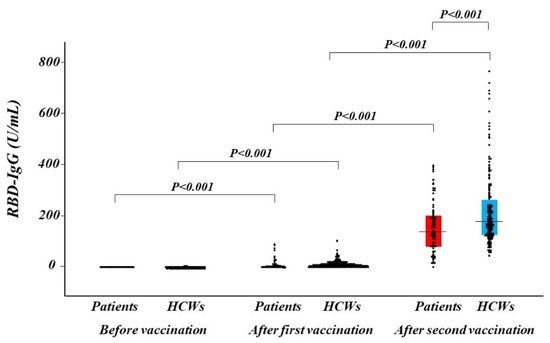
1.3. Antibody Titers after Vaccination
1.3. Antibody Titers after Vaccination
| Hypertension | ||||||
| −0.21 | −0.33 to −0.08 | 0.001 | −0.17 | −0.34 to 0.01 | 0.06 | |
| Dyslipidemia | −0.14 | −0.26 to −0.01 | 0.03 | 0.17 | −0.03 to 0.38 | 0.10 |
| Diabetes | −0.14 | −0.26 to −0.01 | 0.03 | −0.04 | −0.18 to 0.10 | 0.60 |
| Valvular disease | ||||||
| 3 (4) | ||||||
| 0.05 to 0.16 | ||||||
| Previous myocardial infarction | 26 (31) | |||||
| Previous coronary revascularization | 38 (45) | |||||
| Paroxysmal or persistent AF | 12 (14) | |||||
| Medications | ||||||
| RAAS inhibitors | 41 (48) | NA | ||||
| Beta-blockers | 34 (40) | |||||
| Diuretics | ||||||