Virtual Anthropology and Paleoneurology: Comparison
Please note this is a comparison between Version 2 by Catherine Yang and Version 1 by Antoine Balzeau.

Advances in neuroscience have made it possible to obtain increasing information on the anatomy of the brain, at ever-higher resolutions, with different imaging techniques, on ever-larger samples. At the same time, paleoanthropology has to deal with partial reflections on the shape of the brain, on fragmentary specimens and small samples in an attempt to approach the morphology of the brain of past human species. Paleoanthropology has much to gain from interacting more with the field of neuroimaging. Improving our understanding of the morphology of the endocast necessarily involves studying the external surface of the brain and the link it maintains with the internal surface of the skull. The contribution of neuroimaging will allow us to better define the relationship between brain and endocast. Models of intra- and inter-species variability in brain morphology inferred from large neuroimaging databases will help make the most of the rare endocasts of extinct species. Moreover, exchanges between these two disciplines will also be beneficial to our knowledge of the Homo sapiens brain. Documenting the anatomy among other human species and including the variation over time within our own species are approaches that offer us a new perspective through which to appreciate what really characterizes the brain of humanity today. 

  • Virtual Anthropology
  • Paleoneurology

1. Does the Endocast Reflects the Brain?

Paleoneurology is a fascinating topic, dealing with anatomical and biological aspects of past humans and, in addition, potential behavioral implications. The field is, of course, highly debated, for multiple reasons.
The main reason relates to the complex nature of the material that researchers analyze. Indeed, the soft tissues that constitute the brain never fossilize. Scientists only have to deal with the shallow imprints of the convolutions that the brain forms on the internal surface of the skull. This incomplete reflection of the brain is named the (brain) endocast. The brain presses on and leaves marks on the inner surface of the skull throughout a person’s life. This was true for the humans who lived a few million years ago, but also for all of us. The phenomenon is particularly intense during the period of accelerated growth of the brain, and therefore of the cranial box which surrounds it, during the first years of life. The whole process is intertwined, so that the shape of the adult skull is reminiscent of the moment of peak brain development. The behavior of the skull can be described as that of a morphological black box, retaining information that later makes it possible to reconstitute its original contents. Therefore, when a fossil skull is discovered, its inner surface is molded, either physically or virtually, using imaging methods, to reconstruct its endocast. This model represents the preserved imprints of the external surface of the brain. However, the correspondence between these limited records of convolutional [1][2] patterns and details of the surface of the brain remains to be demonstrated in modern humans. A few pioneer studies have considered this problem [1][2]. Moreover, it is necessary to develop new tools for the automatic and reliable determination of the endocranial sulci [3].
In the context of the PaleoBRAIN project, financed by the ANR, we are conducting a direct investigation of the correlation between the shape of the brain and that of the intracranial cast within a sample of modern humans using MRI (for Magnetic Resonance Imaging) acquisitions, including some with a specific sequence that allows the characterization of bone tissues. The comparison of morphometric data and anatomical traits between the brain and the endocast will be performed using state-of-the-art quantification methodologies. But our large dataset could probably also be used to refine the methodology dedicated to the sulcus detection in the endocast. Current methodologies use differential geometry to detect sulci as ravine or crest lines [1][2]. A key component in the design of such robust detectors is the amount of local smoothing performed before detection, which is usually tuned to the scale of the features to be detected. The T1-weighted MRI of our dataset can be used to define the ground truth using the sulci detected by the Brain VISA software. Subsequently, the optimal smoothing can be estimated using an inverse problem framework.
This project will also contribute to answering a key question about the evolution of the human brain. In many studies, the endocast is analyzed with distances characterized at maximal points of extension, maximal length or maximal width, or that correspond to intracranial points, such as endobregma or endolambda, for example [4][5], or with 3D methodologies that consider the surface as a whole [6][7]. These methodological approaches are justified by the complex nature of the material. Indeed, gyri and sulci are difficult to identify on the endocast (Figure 1). In this context, there is little information available about variations in the global size of the different lobes and their relationship with each other between hominin species.
Figure 1. Comparison between the position of the main sulci of the endocranial surface (in red) and the shape and position of the skull, including the course of the coronal suture (in blue) in Cro-Magnon 1, an Upper Paleolithic Homo sapiens; La Chapelle-aux-Saints 1, an Homo neanderthalensis; and Sambungmacan 3, an Homo erectus.
Moreover, H. neanderthalensis and fossil H. sapiens, which have the largest endocranial volume of all hominins, show different brain structures (Figure 1). These results illustrate that differences existed in the structure of the brain in addition to the well-known variation in size during human evolution. An important contribution to this topic will be to improve our ability to determine the location of the sulci and gyri on fossil hominin endocasts. To do so, a better knowledge of the anatomy and characteristics of hominids is necessary [8][9], together with a better knowledge of the brain–endocast relationship in living humans. Finally, it is fundamental to obtain a more generalized and simplified access to high-resolution endocranial data for fossil specimens. Indeed, this material is so complex that multiple appreciation by the few researchers dealing with paleoneurological information would certainly enhance our capacity for anatomical determination. It would also certainly help to minimize potential conflicting interpretations, which are very frequent in this small field of research.

2. What Can Be Deduced about a Species’ Folding Pattern from a Few Samples?

The very high intra-species variability of the cortical folding of Homo sapiens, illustrated by Figure 1, is a major difficulty for modern brain mapping. It should also warn us about the risk of over-interpretation inherent in the small number of samples available in paleoneurology. The idiosyncrasies of a specific brain are not necessarily representative of the folding pattern of its species. The amount of intra-species variability is species dependent. In great apes, it is less than in humans but still significant, especially in the frontal lobe. In baboons or macaques, it is almost non-existent. In species with a variable folding pattern, the match between the folds of an individual and its nomenclature can be difficult to establish and leads to confusion, especially when only an endocast is available [10][11]. In modern humans, the large sulci described in anatomical books are often split into pieces and reorganized into unusual folding patterns that are difficult to decipher (see Figure 2) [12]. Notably, these phenomena occur in the general population without developmental pathologies.
Figure 2. The hemispheres of five Homo sapiens and one chimp, with interruption of the central sulcus, which hosts sensorimotor areas (0.5% of occurrence). This kind of interruption is frequent in associative areas and leads to folding configurations that are difficult to decipher, which can be observed here in the frontal lobes.
The mysteries hidden behind the variability of cortical folding have led to the emergence of a multidisciplinary community that aims to understand these variations and their meaning. It associates biologists, who focus on the developmental phenomena that are at the origin of cortical folding (spatially heterogeneous neurogenesis, spatially heterogeneous chronology of synaptic development, etc.) [13], and physicists, who model the mechanical phenomena that result from these growth heterogeneities [14]. This new community also includes anatomists, who study the links between folding and the organization of cortical areas and fiber bundles [15], and computer scientists, who geometrically model the variability observed in the general population, and the specificities of developmental pathologies [16][17]. The progress made by this community could contribute to a better exploitation of the scarce data observed in the endocasts of the folding of extinct species. A better understanding of the rules driving cortical folding dynamics would provide insight into the architectural changes at the origin of the changes observed across species in endocasts. Endocasts are used as a proxy of the folding pattern, but the folding pattern is only a proxy of architecture, which is even more difficult to reverse-engineer. Current efforts for cracking the code behind folding patterns could contribute, for instance, to the discussion around the third frontal convolution when comparing sapiens, great apes, and extinct hominids. Joint modelling of folding variability and of functional variability will help to understand which features of the folding pattern can be used as landmarks of key cytoarchitectonic areas (see Figure 3) [18].
Figure 3. Machine learning can be used to model the variability of folding patterns. Here, the variability of the shape of the central sulcus is projected into a one-dimensional manifold representing the transition between a single knob and a double knob pattern. Functional mapping performed along this manifold shows that the two different folding patterns correspond to different localizations of functional areas along the central sulcus.

3. How Are Brain Asymmetries Quantified in the Hominin Fossil Record?

The number of brain structural asymmetries observable on endocranial casts and, consequently, in fossil specimens is limited due to several factors, which are of course related to the specificity of our material, which concerns only the external surface of the brain. By chance, features on the brain and endocast for which bilateral variation studies are possible are among the most consistent features available for cross-taxa studies on large samples. One important limiting point needs to be considered. Indeed, the difficulty in defining structural parameters and in establishing left-right homologies makes studies of brain asymmetries complex. Moreover, gross anatomical asymmetries of selected pairs of points may reflect combined asymmetries in brain subregions. The quantification of surface morphology, distance, or volume of discrete anatomical areas may not fully express real bilateral variation if their pattern of asymmetry is defined in reference to global anatomical brain areas.
Another limitation is that the methodologies employed in most previous studies of cerebral or endocranial asymmetries involved qualitative assessment or a simple index of bilateral traits and did not analyze departures from symmetry and different patterns of asymmetry (i.e., fluctuating and directional asymmetry, antisymmetry) in efficient and adapted ways [19]. It has indeed been shown that the brains of extant hominids demonstrated high levels of fluctuating asymmetry, allowing pronounced developmental plasticity and therefore making brains highly evolvable [20]. The quantification and analysis of the morphology—including the asymmetries—of the endocranial cavity need further development. Currently, the most advanced computational tools used in analysis of bilateral shape asymmetries rely on the standard framework of landmark-based morphometrics [21][22]. In this context, in addition to homologous landmarks between shapes for population studies, one must define homologous landmarks between the two sides of each shape under study. Analyses can then be carried out by using slight modifications of the linear distance-based [23] or superimposition [24][25][26][27] methods. However, we identified methodological problems underlying the theory and its application to the assessment of bilateral asymmetries [7][28][29]. Moreover, a limited set of landmarks is likely to be inadequate to capture the shape of intricate anatomical structures, or that of structures with few obvious salient features, such as brain endocasts. New methodological improvements are therefore necessary to better characterize and quantify bilateral asymmetries [30][31]. A specific methodology has been developed and tested on the endocast of the Cro-Magnon 1 fossil [32]. This approach is promising as it allows for an independent characterization of the asymmetries without referring to the potential global asymmetry of the object that is analyzed. New approaches based on machine learning could also be a source of inspiration. They allow us, for example, to establish the asymmetry of folding patterns without requiring the definition of homologous landmarks across subjects and hemispheres. For instance, the double-knob configuration of the central sulcus, depicted in Figure 3, is more frequent in the left hemisphere [33]. These new approaches could contribute to the old question of the language-related asymmetry of the third frontal convolution, which is difficult to tackle because of the large intraspecies variability of the related folding patterns [34].

4. The Complex Definition of Brain Features and of Their Application to the Fossil Record

A general problem concerns the lack of homogeneity in the definition of brain asymmetries and of the methods used to quantify them. For example, one of the most studied brain asymmetries on brain endocasts concern the petalias. LeMay [35][36] initially considered the antero-posterior projection of the frontal and occipital lobes, respectively. By contrast, later studies generalized the term ‘petalias’ to a wide range of anatomical traits. Some studies indeed referred to bilateral differences in the lateral extension of the posterior area of the frontal lobes [37], to other anatomical areas of the brain, and even to volumetric variations between hemispheres [38][39][40]. It is therefore difficult to compare data obtained on petalias if studies do not consider the same brain features. Nevertheless, it was largely accepted that this pattern of asymmetries appeared with early Homo [36][41] and is more common in right-handed individuals [35][36][42][43][44]. Based on an original methodology applied to the largest samples ever used, we demonstrated a shared specific pattern of protrusions of the frontal and occipital across all hominids, including extant African great apes, modern humans, and hominin fossils [31][45]. These asymmetries are a topic of debate in non-human primate brain studies [35][38][39][46][47][48] and paleoanthropology [49][37][50][51][52] because of their relationship with handedness and other specific aspects of human cognition. Similar results were obtained recently by an independent team [7]. H. sapiens appear to have more asymmetrical petalias than other extant great apes, but a shared pattern is observed, suggesting that a globally asymmetric brain is the ancestral condition. A recent study questioned this observation [53]. However, this is a good example of differences in the definition of the anatomical traits that are analyzed. These authors measured the bilateral variation in lateral extension of slices of the brain. This trait is not directly comparable to our analyses of the 3D position of the occipital poles [54] or to the 3D displacement between the left and right corresponding anatomical area. Another good illustration of the problem is Broca’s area, whose extension is defined differently according to authors [55]. This functional area is impossible to characterise on brain endocasts. However, we conducted a comparative study on the size, shape, and position of the third frontal convolution in great apes, H. sapiens, and hominin fossils [54]. The neuroanatomical asymmetries as quantified in our work show a pattern that is different from what was previously accepted based on qualitative data. Our main finding was a shared pattern of asymmetry in Broca’s area in all hominins and Pan paniscus, as well as an increase in the size of this area during human evolution. We also identified that Pan troglodytes and Pan paniscus have differences in their asymmetry patterns in the third frontal convolution. This topic is of great interest for future research. More generally, brain and endocranial studies have to rely on a clear definition of the anatomical features that are analyzed and an effort to use similar protocols will certainly enhance the reproducibility of our studies.

5. How to Grow a Hominin Brain?

The knowledge of ontogenetic patterns in fossil human species is scarce [56][57][58][59] and, to date, no information is available about the evolution of brain lateralization during growth and development. Both H. neanderthalensis and H. sapiens have enlarged brains compared with other hominins, but their respective organizations and morphologies are different, each of the two species having “grown” large brains through specific evolutionary processes. Much remains unknown about what these processes are, and how they are rooted in the hominin evolutionary tree. In the case of H. neanderthalensis, although some changes in gross cerebral morphology during childhood are documented, researchers have presented conflicting results concerning how their endocranial growth patterns relate to those of other primates. While the post-natal Neandertal ontogenetic trajectory is deemed closer to that of chimpanzees than to that of H. sapiens by some researchers, emphasizing a unique globularization phase in H. sapiens [59], others find that the mode of cerebral growth is largely similar in H. neanderthalensis and H. sapiens, emphasizing instead the characteristic morphologies of each species at birth, and refuting the idea of the derived nature of the post-natal cerebral growth trajectory in H. sapiens [60]. Nevertheless, these studies only consider the global shape of the internal surface of the skull. Additionally, available data addressing cerebral growth do not provide enough details, so that much of “how” the Neandertal brain grows remains unknown (e.g., do the contributions of the different lobes to total brain volume remain stable throughout infancy and childhood?).
We previously demonstrated that tThe two species have distinct brain organizations [61], but this important biological aspect has not yet been considered in the study of brain growth in H. neanderthalensis. The emergence of large databases on the brain development of sapiens, and to a lesser extent of extant non-human primates [62], could contribute to these debates.

6. Brain Endocast and Function

The question of the relationship between brain shape and function in hominins has been explored in previous studies [63]. According to their authors, they “show that Neanderthals had significantly larger visual systems than contemporary anatomically modern humans (indexed by orbital volume) and that when this, along with their greater body mass, is taken into account, Neanderthals have significantly smaller adjusted endocranial capacities than contemporary anatomically modern humans.” For the authors, these results had implications for interpreting variations in brain organization in terms of social cognition. Indeed, larger visual systems would have implied smaller adjacent anatomical areas, including the parietal areas related to social skills. Their final conclusion was that the extinction of H. neanderthalensis was due to weaker social cognition compared to modern humans. This study suffered from methodological limitations. The main problem was that they were improperly interpreting data mostly derived from the research of one of the authors of this paper [61]. These authors considered that our data for the external extension of the occipital lobe were directly related to the size of the visual cortex. However, such a direct interpretation was not demonstrated. Moreover, they did not measured any anatomical areas on the endocasts of H. neanderthalensis or of contemporary H. sapiens. All those approximations make any interpretation in terms of behaviors impossible.
This example should not prevent us from analyzing morphological variation among hominins species and exploring functional and behavioral implications. However, this needs to be undertaken on a solid anatomical framework, particularly in the context of interspecies comparisons, and with more caution for the evaluation of the potential link between brain anatomy and suspected function.

References

  1. Jean Dumoncel; Gérard Subsol; Stanley Durrleman; Anne Bertrand; Edwin de Jager; Anna C. Oettlé; Zarina Lockhat; Farhana E. Suleman; Amélie Beaudet; Are endocasts reliable proxies for brains? A 3D quantitative comparison of the extant human brain and endocast. Journal of Anatomy 2020, 238, 480-488, 10.1111/joa.13318.
  2. Marc Fournier; Benoît Combès; Neil Roberts; José Braga; Sylvain Prima; Mapping the distance between the brain and the inner surface of the skull and their global asymmetries. Medical Imaging 2011: Image Processing 2011, 79620Y, 79620Y-79620Y-7, 10.1117/12.876795.
  3. Edwin J. De Jager; Albert N. Van Schoor; Jakobus W. Hoffman; Anna C. Oettlé; Caroline Fonta; Muriel Mescam; Laurent Risser; Amélie Beaudet; Sulcal pattern variation in extant human endocasts. Journal of Anatomy 2019, 235, 803-810, 10.1111/joa.13030.
  4. Holloway Ralph L., Broadfield Douglas C., Yuan Michael S.. The Human Fossil Record; Holloway Ralph L., Broadfield Douglas C., Yuan Michael S., Eds.; John Wiley and Sons: london, 2005; pp. 315.
  5. Emiliano Bruner; Dominique Grimaud-Hervé; Xiujie Wu; José Manuel de la Cuétara; Ralph Holloway; A paleoneurological survey of Homo erectus endocranial metrics. Quaternary International 2015, 368, 80-87, 10.1016/j.quaint.2014.10.007.
  6. Simon Neubauer; Jean-Jacques Hublin; Philipp Gunz; The evolution of modern human brain shape. Science Advances 2018, 4, eaao5961, 10.1126/sciadv.aao5961.
  7. Simon Neubauer; Philipp Gunz; Nadia A. Scott; Jean-Jacques Hublin; Philipp Mitteroecker; Evolution of brain lateralization: A shared hominid pattern of endocranial asymmetry is much more variable in humans than in great apes. Science Advances 2020, 6, eaax9935, 10.1126/sciadv.aax9935.
  8. Dean Falk; Christoph P.E. Zollikofer; Marcia Ponce De León; Katerina Semendeferi; José Luis Alatorre Warren; William D. Hopkins; Identification of in vivo Sulci on the External Surface of Eight Adult Chimpanzee Brains: Implications for Interpreting Early Hominin Endocasts. Brain, Behavior and Evolution 2018, 91, 45-58, 10.1159/000487248.
  9. José Luis Alatorre Warren; Marcia S. Ponce de León; William D. Hopkins; Christoph P. E. Zollikofer; Evidence for independent brain and neurocranial reorganization during hominin evolution. Proceedings of the National Academy of Sciences 2019, 116, 22115-22121, 10.1073/pnas.1905071116.
  10. Dean Efalk; Interpreting sulci on hominin endocasts: old hypotheses and new findings. Frontiers in Human Neuroscience 2014, 8, 134, 10.3389/fnhum.2014.00134.
  11. Jean-François Mangin, Guillaume Auzias, Olivier Coulon, Zhong Sun, Denis Rivière, et al.. Sulci as Landmarks. Toga, Arthur W. Brain Mapping: an Encyclopedic Reference, pp."45 - 52",, 2015
  12. Jean-François Mangin; Yann Le Guen; Nicole Labra; Antoine Grigis; Vincent Frouin; Miguel Guevara; Clara Fischer; Denis Rivière; William D. Hopkins; Jean Régis; et al.Zhong Yi Sun “Plis de passage” Deserve a Role in Models of the Cortical Folding Process. Brain Topography 2019, 32, 1035-1048, 10.1007/s10548-019-00734-8.
  13. Cristina Llinares-Benadero; Víctor Borrell; Deconstructing cortical folding: genetic, cellular and mechanical determinants. Nature Reviews Neuroscience 2019, 20, 161-176, 10.1038/s41583-018-0112-2.
  14. Tuomas Tallinen; Jun Young Chung; François Rousseau; Nadine Girard; Julien Lefèvre; Jun Young Chung L. Mahadevan; On the growth and form of cortical convolutions. Nature Physics 2016, 12, 588-593, 10.1038/nphys3632.
  15. David C. Van Essen; A tension-based theory of morphogenesis and compact wiring in the central nervous system. Nature 1997, 385, 313-318, 10.1038/385313a0.
  16. J.-F. Mangin; J. Lebenberg; S. Lefranc; N. Labra; G. Auzias; M. Labit; M. Guevara; H. Mohlberg; P. Roca; J. Dubois; et al.F. LeroyG. Dehaene-LambertzA. CachiaT. DickscheidO. CoulonC. PouponD. RivièreK. AmuntsZ.Y. Sun Spatial normalization of brain images and beyond. Medical Image Analysis 2016, 33, 127-133, 10.1016/j.media.2016.06.008.
  17. Cachia, A.; et al; Towards Deciphering the Fetal Foundation of Normal Cognition and Cognitive Symptoms From Sulcation of the Cortex . Front. Neuroanat. 2021, 15, 68, 10.3389/fnana.2021.712862 .
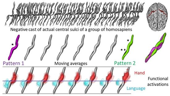
  18. Z. Y. Sun; P. Pinel; D. Rivière; A. Moreno; S. Dehaene; J.-F. Mangin; Linking morphological and functional variability in hand movement and silent reading. Brain Structure and Function 2015, 221, 3361-3371, 10.1007/s00429-015-1106-8.
  19. Palmer, A.R.; Strobeck, C. Fluctuating asymmetry analyses revisited. In Developmental Instability: Causes and Consequences; Polak, M., Ed.; Oxford University Press: Oxford, UK, 2003; pp. 279–319.
  20. Aida Gómez-Robles; William D. Hopkins; Chet C. Sherwood; Increased morphological asymmetry, evolvability and plasticity in human brain evolution. Proceedings of the Royal Society of London. Series B: Biological Sciences 2013, 280, 20130575, 10.1098/rspb.2013.0575.
  21. Timothy D. Weaver; Philipp Gunz; Using geometric morphometric visualizations of directional selection gradients to investigate morphological differentiation. Evolution 2018, 72, 838-850, 10.1111/evo.13460.
  22. Philipp Mitteroecker; Philipp Gunz; Advances in Geometric Morphometrics. Evolutionary Biology 2009, 36, 235-247, 10.1007/s11692-009-9055-x.
  23. Richtsmeier, J.T.; Cole, T.M.; Lele, S.R. An invariant approach to the study of fluctuating asymmetry: Developmental instability in a mouse model for Down syndrome. In Modern Morphometrics in Physical Anthropology; Springer: Berlin, Germany, 2005; pp. 187–212.
  24. J. T. Kent; Shape, Procrustes tangent projections and bilateral symmetry. Biometrika 2001, 88, 469-485, 10.1093/biomet/88.2.469.
  25. Christian Peter Klingenberg; Grant S. McIntyre; GEOMETRIC MORPHOMETRICS OF DEVELOPMENTAL INSTABILITY: ANALYZING PATTERNS OF FLUCTUATING ASYMMETRY WITH PROCRUSTES METHODS. Evolution 1998, 52, 1363-1375, 10.1111/j.1558-5646.1998.tb02018.x.
  26. Christian Peter Klingenberg; Marta Barluenga; Axel Meyer; SHAPE ANALYSIS OF SYMMETRIC STRUCTURES: QUANTIFYING VARIATION AMONG INDIVIDUALS AND ASYMMETRY. Evolution 2002, 56, 1909-1920, 10.1111/j.0014-3820.2002.tb00117.x.
  27. K. Mardia; Fl Bookstein; Ij Moreton; Statistical assessment of bilateral symmetry of shapes. Biometrika 2000, 87, 285-300, 10.1093/biomet/87.2.285.
  28. Combès, B.; Hennessy, R.; Waddington, J.L.; Roberts, N.; Prima, S. Automatic symmetry plane estimation of bilateral objects in point clouds. In Proceedings of the 2008 IEEE Conference on Computer Vision and Pattern Recognition-CVPR’2008, Anchorage, AK, USA, 24–26 June 200
  29. Combès, B.; Fournier, M.; Kennedy, D.N.; Braga, J.; Roberts, N.; Prima, S. EM-ICP strategies for joint mean shape and correspondences estimation: Applications to statistical analysis of shape and of asymmetry. In Proceedings of the 8th IEEE International Symposium on Biomedical Imaging: From Nano to Macro (ISBI’2011), Chicago, IL, USA, 30 March–2 April 2011; pp. 1257–1263.
  30. Emam ElHak Abdel Fatah; Natalie R. Shirley; Mohamed R. Mahfouz; Benjamin M. Auerbach; A three-dimensional analysis of bilateral directional asymmetry in the human clavicle. American Journal of Physical Anthropology 2012, 149, 547-559, 10.1002/ajpa.22156.
  31. Antoine Balzeau; Emmanuel Gilissen; Endocranial shape asymmetries in Pan paniscus, Pan troglodytes and Gorilla gorilla assessed via skull based landmark analysis. Journal of Human Evolution 2010, 59, 54-69, 10.1016/j.jhevol.2010.03.013.
  32. A. Balzeau; D. Grimaud-Hervé; F. Détroit; R. L. Holloway; B. Combès; S. Prima; First description of the Cro-Magnon 1 endocast and study of brain variation and evolution in anatomically modern Homo sapiens. Bulletins et mémoires de la société d'anthropologie de Paris 2012, 25, 1-18, 10.1007/s13219-012-0069-z.
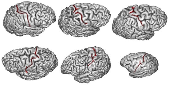
  33. Zhong Yi Sun; Stefan Klöppel; Denis Rivière; Matthieu Perrot; Richard Frackowiak; Hartwig Siebner; Jean-François Mangin; The effect of handedness on the shape of the central sulcus. NeuroImage 2011, 60, 332-339, 10.1016/j.neuroimage.2011.12.050.
  34. Trisanna Sprung-Much; Nicole Eichert; Erika Nolan; Michael Petrides; Broca’s area and the search for anatomical asymmetry: commentary and perspectives. Brain Structure and Function 2021, 2021, 1-9, 10.1007/s00429-021-02357-x.
  35. Marjorie Lemay; MORPHOLOGICAL CEREBRAL ASYMMETRIES OF MODERN MAN, FOSSIL MAN, AND NONHUMAN PRIMATE. Annals of the New York Academy of Sciences 1976, 280, 349-366, 10.1111/j.1749-6632.1976.tb25499.x.
  36. Marjorie LeMay; Asymmetries of the skull and handedness: Phrenology revisited. Journal of the Neurological Sciences 1977, 32, 243-253, 10.1016/0022-510x(77)90239-8.
  37. Ralph L. Holloway; Marie Christine De La Costelareymondie; Brain endocast asymmetry in pongids and hominids: Some preliminary findings on the paleontology of cerebral dominance. American Journal of Physical Anthropology 1982, 58, 101-110, 10.1002/ajpa.1330580111.
  38. William D Hopkins; Lori Marino; Asymmetries in cerebral width in nonhuman primate brains as revealed by magnetic resonance imaging (MRI). Neuropsychologia 2000, 38, 493-499, 10.1016/s0028-3932(99)00090-1.
  39. Dawn L. Pilcher; Elizabeth A. D. Hammock; William D. Hopkins; Cerebral volumetric asymmetries in non-human primates: A magnetic resonance imaging study. Laterality: Asymmetries of Body, Brain and Cognition 2001, 6, 165-179, 10.1080/713754406.
  40. William D. Hopkins; Jared P. Taglialatela; Adrien Meguerditchian; Talia Nir; Natalie M. Schenker; Chet C. Sherwood; Gray matter asymmetries in chimpanzees as revealed by voxel-based morphometry. NeuroImage 2008, 42, 491-497, 10.1016/j.neuroimage.2008.05.014.
  41. Ralph L. Holloway; Volumetric and asymmetry determinations on recent hominid endocasts: Spy I and II, Djebel Ihroud I, and the salèHomo erectus specimens, with some notes on neandertal brain size. American Journal of Physical Anthropology 1981, 55, 385-393, 10.1002/ajpa.1330550312.
  42. LeMay, M.; Billig, M.S.; Geschwind, N. Asymmetries of the brains and skulls of nonhuman primates. In Primate Brain Evolution, Methods and Concepts; Falk, D., Armstrong, E., Eds.; Plenum Press: New York, NY, USA, 1976; pp. 263–277.
  43. Albert M. Galaburda; Marjorie LeMay; Thomas L. Kemper; Norman Geschwind; Right-Left Asymmetries in the Brain. Science 1978, 199, 852-856, 10.1126/science.341314.
  44. Dean Falk; Charles Hildebolt; James Cheverud; Michael Vannier; R. Criss Helmkamp; Lyle Konigsberg; Cortical asymmetries in frontal lobes of Rhesus monkeys (Macaca mulatta). Brain Research 1990, 512, 40-45, 10.1016/0006-8993(90)91167-f.
  45. Antoine Balzeau; Emmanuel Gilissen; Dominique Grimaud-Hervé; Shared Pattern of Endocranial Shape Asymmetries among Great Apes, Anatomically Modern Humans, and Fossil Hominins. PLoS ONE 2012, 7, e29581, 10.1371/journal.pone.0029581.
  46. LeMay, M. Asymmetries of the brains and skulls of nonhuman primates. In Cerebral Lateralization in Nonhuman Species; Glick, S.D., Ed.; Academic Press: New York, NY, USA, 1985; pp. 233–245.
  47. Donald P. Cain; Juhn A. Wada; An Anatomical Asymmetry in the Baboon Brain. Brain, Behavior and Evolution 1979, 16, 222-226, 10.1159/000121838.
  48. James M. Cheverud; Dean Falk; Charles Hildebolt; Allen J. Moore; R. Criss Helmkamp; Michael Vannier; Heritability and Association of Cortical Petalias in Rhesus Macaques (Macaca mulatta). Brain, Behavior and Evolution 1990, 35, 368-372, 10.1159/000115881.
  49. Phillip V. Tobias; The brain of Homo habilis: A new level of organization in cerebral evolution. Journal of Human Evolution 1987, 16, 741-761, 10.1016/0047-2484(87)90022-4.
  50. William D. Hopkins; Kimberley Phillips; Amanda Bania; Sarah E. Calcutt; Molly Gardner; Jamie Russell; Jennifer Schaeffer; Elizabeth V. Lonsdorf; Stephen R. Ross; Steven J. Schapiro; et al. Hand preferences for coordinated bimanual actions in 777 great apes: Implications for the evolution of handedness in Hominins. Journal of Human Evolution 2011, 60, 605-611, 10.1016/j.jhevol.2010.12.008.
  51. Stephanie L. Bogart; Jean-François Mangin; Steven J. Schapiro; Lisa Reamer; Allyson J. Bennett; Peter J. Pierre; William D. Hopkins; Cortical sulci asymmetries in chimpanzees and macaques: A new look at an old idea. NeuroImage 2012, 61, 533-541, 10.1016/j.neuroimage.2012.03.082.
  52. Michael C. Corballis; Gjurgjica Badzakova-Trajkov; Isabelle S. Häberling; Right hand, left brain: genetic and evolutionary bases of cerebral asymmetries for language and manual action. WIREs Cognitive Science 2011, 3, 1-17, 10.1002/wcs.158.
  53. Li Xiang; Timothy Crow; Neil Roberts; Cerebral torque is human specific and unrelated to brain size. Brain Structure and Function 2019, 224, 1141-1150, 10.1007/s00429-018-01818-0.
  54. Antoine Balzeau; Emmanuel Gilissen; Ralph L. Holloway; Sylvain Prima; Dominique Grimaud-Hervé; Variations in size, shape and asymmetries of the third frontal convolution in hominids: Paleoneurological implications for hominin evolution and the origin of language. Journal of Human Evolution 2014, 76, 116-128, 10.1016/j.jhevol.2014.06.006.
  55. Simon S. Keller; Timothy Crow; Anne Foundas; Katrin Amunts; Neil Roberts; Broca’s area: Nomenclature, anatomy, typology and asymmetry. Brain and Language 2009, 109, 29-48, 10.1016/j.bandl.2008.11.005.
  56. Antoine Balzeau; Dominique Grimaud-Hervé; Teuku Jacob; Internal cranial features of the Mojokerto child fossil (East Java, Indonesia). Journal of Human Evolution 2005, 48, 535-553, 10.1016/j.jhevol.2005.01.002.
  57. Philipp Gunz; Simon Neubauer; Lubov Golovanova; Vladimir Doronichev; Bruno Maureille; Jean-Jacques Hublin; A uniquely modern human pattern of endocranial development. Insights from a new cranial reconstruction of the Neandertal newborn from Mezmaiskaya. Journal of Human Evolution 2012, 62, 300-313, 10.1016/j.jhevol.2011.11.013.
  58. Philipp Gunz; Simon Neubauer; Bruno Maureille; Jean-Jacques Hublin; Brain development after birth differs between Neanderthals and modern humans. Current Biology 2010, 20, R921-R922, 10.1016/j.cub.2010.10.018.
  59. Simon Neubauer; Philipp Gunz; Jean-Jacques Hublin; Endocranial shape changes during growth in chimpanzees and humans: A morphometric analysis of unique and shared aspects. Journal of Human Evolution 2010, 59, 555-566, 10.1016/j.jhevol.2010.06.011.
  60. Marcia S. Ponce de León; Thibaut Bienvenu; Takeru Akazawa; Christoph P.E. Zollikofer; Brain development is similar in Neanderthals and modern humans. Current Biology 2016, 26, R665-R666, 10.1016/j.cub.2016.06.022.
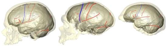
  61. Antoine Balzeau; Ralph L. Holloway; Dominique Grimaud-Hervé; Variations and asymmetries in regional brain surface in the genus Homo. Journal of Human Evolution 2012, 62, 696-706, 10.1016/j.jhevol.2012.03.007.
  62. Coulon, O.; Sein, J.; Auzias, G.; Nazarian, B.; Anton, J.L.; Rousseau, F.; Velly, L.; Girard, N. High temporal resolution longitudinal observation of fetal brain development. A baboon pilot study. In Proceedings of the 26th Annual Meeting of the Organization for Human Brain Mapping, Montreal, QC, Canada, 23 June–3 July 2020.
  63. Eiluned Pearce; Chris Stringer; R. I. M. Dunbar; New insights into differences in brain organization between Neanderthals and anatomically modern humans. Proceedings of the Royal Society of London. Series B: Biological Sciences 2013, 280, 20130168, 10.1098/rspb.2013.0168.
More