Building and functioning of the human brain requires the precise orchestration and execution of myriad molecular and cellular processes, across a multitude of cell types and over an extended period of time. Neural Stem Cells (NSCs) represent the heart of these processes, since they increase the pool of neural progenitors and are the founders of all the neural progeny which will constitute the adult human brain.
- Human neural stem cells
- Neurodevelopment
- Corticogenesis
- In vitro models
1. Human Neural Stem Cells: When and Where?
The immense complexity of the human brain is reflected in its cellular organization and in the cognitive and behavioral repertoire that defines us as human. The human brain is the product of an evolutionary and developmental history that resulted in its progressive enlargement and specialization. Our central nervous system (CNS) develops through a dynamic and prolonged process in which myriad cell types are generated by neural stem cells (NSCs) and assembled into an intricate synaptic circuitry. Deviations from the normal course of development can lead to a variety of pathologies, including neurological and psychiatric disorders that affect some of the most distinctly human aspects of cognition and behavior
During early CNS development, the neural tube is comprised of a pseudostratified layer of neuroepithelial cells lining the central cavity
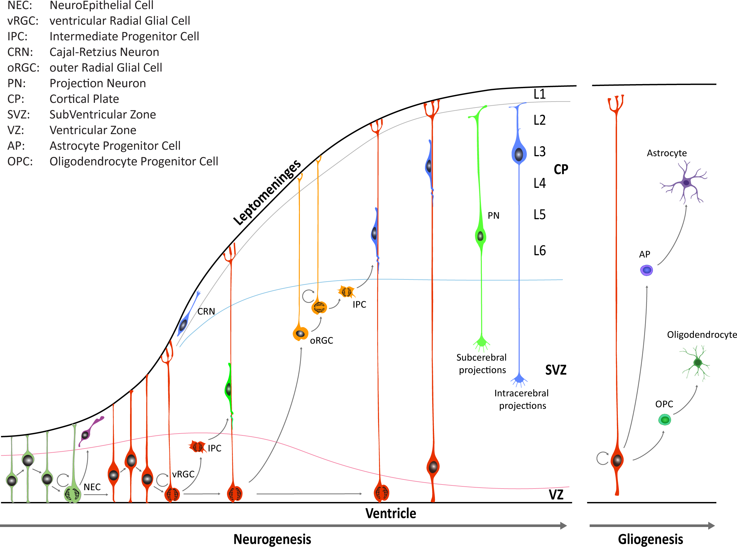
Figure 1. Schematic illustration of neocortical development. Neuroepithelial cells (NECs) undergo symmetric cell division to expand the initial pool and later transition into ventricular radial glia cells (vRGCs). vRGCs begin asymmetric cell division to generate another vRGC and a nascent projection neuron. Neurons then migrates radially from the ventricular zone (VZ) along the RGC basal processes into the cortical plate (CP). Early-born projection neurons (PNs) settle in the deep layers (Layers 5 and 6), and later-born neurons in upper layers. Additionally, some populations of RGC daughter cells convert themselves into intermediate progenitor cells (IPCs) or outer radial glial cells (oRGCs) in the subventricular zone (SVZ). After the neurogenic stages, gliogenesis occurs, generating astrocytes and oligodendrocytes.
Until the seventh post-conceptional week (pcw), depending on the region of the CNS, neuroepithelial cells undergo primarily symmetric divisions in order to expand the stem cell pool
[3]
[8]
[10]
During early neurogenesis, at the beginning of the second trimester, the vRGCs and IPCs give rise to neurons present in the deep layers. On the other hand, oRGCs give rise to later born IPCs, and differentiate predominantly into upper layer neurons (
[11]
Neural Stem Cells at a glance:
Neuroepithelial Cells
Radial Glia Cells (RGCs):
Intermediate Progenitor Cells (IPCs or Basal Progenitors)
Adult Neural Stem Cells
2.
In vitro systems of Neural Stem Cells
Our knowledge of NSCs has been revolutionized as optimized culture protocols have been put in place
[12]
[13]
[14]
post‐mortem
On the other hand, embryonic neuroepithelial cells possess a great self‐renewing potential and wide multilineage differentiation. Thanks to these unique properties, neuroepithelial cells represent an ideal candidate for
in vitro
[22]
[23]
post‐mortem
[23]
[23]
[24]
[24]
Human pluripotent stem cells (hPSCs), which include both ESCs and iPSCs, represent an extraordinary
ex vivo
in vitro
[12]
[25]
[26]
in vitro
[26]
[27]
[27]
[28]
in vivo.
Acknowledgments
References
- Alejandro L. Diaz; Joseph G. Gleeson; The Molecular and Genetic Mechanisms of Neocortex Development. Clinics in Perinatology 2009, 36, 503-512, 10.1016/j.clp.2009.06.008.
- Jan H. Lui; David V. Hansen; Arnold R. Kriegstein; Development and Evolution of the Human Neocortex. Cell 2011, 146, 18-36, 10.1016/j.cell.2011.06.030.
- John C. Silbereis; Sirisha Pochareddy; Ying Zhu; Mingfeng Li; Nenad Sestan; The Cellular and Molecular Landscapes of the Developing Human Central Nervous System.. Neuron 2016, 89, 248-68, 10.1016/j.neuron.2015.12.008.
- André M.M. Sousa; Kyle A. Meyer; Gabriel Santpere; Forrest O. Gulden; Nenad Sestan; Evolution of the Human Nervous System Function, Structure, and Development. Cell 2017, 170, 226-247, 10.1016/j.cell.2017.06.036.
- Arnold R. Kriegstein; Arturo Alvarez-Buylla; The glial nature of embryonic and adult neural stem cells.. Annual Review of Neuroscience 2009, 32, 149-84, 10.1146/annurev.neuro.051508.135600.
- Joshua J. Breunig; Tarik F Haydar; Pasko Rakic; Neural Stem Cells: Historical Perspective and Future Prospects. Neuron 2011, 70, 614-625, 10.1016/j.neuron.2011.05.005.
- Simone A Fietz; Iva Kelava; Johannes Vogt; Michaela Wilsch-Bräuninger; Denise Stenzel; Jennifer L. Fish; Denis Corbeil; Axel Riehn; Wolfgang Distler; Robert Nitsch; et al.Wieland B. Huttner OSVZ progenitors of human and ferret neocortex are epithelial-like and expand by integrin signaling. Nature Neuroscience 2010, 13, 690-699, 10.1038/nn.2553.
- David V. Hansen; Jan H. Lui; Philip R. L. Parker; Arnold R. Kriegstein; Neurogenic radial glia in the outer subventricular zone of human neocortex. Nature 2010, 464, 554-561, 10.1038/nature08845.
- Isabel Reillo; Víctor Borrell; Germinal Zones in the Developing Cerebral Cortex of Ferret: Ontogeny, Cell Cycle Kinetics, and Diversity of Progenitors. Cerebral Cortex 2011, 22, 2039-2054, 10.1093/cercor/bhr284.
- Tomasz J. Nowakowski; Alex A. Pollen; Carmen Sandoval-Espinosa; Arnold R. Kriegstein; Transformation of the Radial Glia Scaffold Demarcates Two Stages of Human Cerebral Cortex Development.. Neuron 2016, 91, 1219-1227, 10.1016/j.neuron.2016.09.005.
- Bradley J. Molyneaux; Paola Arlotta; João R. L. Menezes; Jeffrey D. Macklis; Neuronal subtype specification in the cerebral cortex. Nature Reviews Neuroscience 2007, 8, 427-437, 10.1038/nrn2151.
- Luciano Conti; Elena Cattaneo; Neural stem cell systems: physiological players or in vitro entities?. Nature Reviews Neuroscience 2010, 11, 176-187, 10.1038/nrn2761.
- B. Reynolds; S Weiss; Generation of neurons and astrocytes from isolated cells of the adult mammalian central nervous system. Science 1992, 255, 1707-1710, 10.1126/science.1553558.
- Luciano Conti; Steven M. Pollard; Thorsten Gorba; Erika Reitano; Mauro Toselli; Gerardo Biella; Yirui Sun; Sveva Sanzone; Qi-Long Ying; Elena Cattaneo; et al.Austin Smith Niche-Independent Symmetrical Self-Renewal of a Mammalian Tissue Stem Cell. PLoS Biology 2005, 3, e283, 10.1371/journal.pbio.0030283.
- Steven M. Pollard; Luciano Conti; Yirui Sun; Donato Goffredo; Austin Smith; Adherent Neural Stem (NS) Cells from Fetal and Adult Forebrain. Cerebral Cortex 2006, 16, i112-i120, 10.1093/cercor/bhj167.
- D Goffredo; Luciano Conti; F Di Febo; G Biella; A Tosoni; G Vago; I Biunno; A Moiana; D Bolognini; M Toselli; et al.Elena Cattaneo Setting the conditions for efficient, robust and reproducible generation of functionally active neurons from adult subventricular zone-derived neural stem cells. Cell Death & Differentiation 2008, 15, 1847-1856, 10.1038/cdd.2008.118.
- Ilaria Albieri; Marco Onorati; Giovanna Calabrese; Alessia Moiana; Daniele Biasci; Aurora Badaloni; Stefano Camnasio; Dimitrios Spiliotopoulos; Zoltan Ivics; Elena Cattaneo; et al.Giangiacomo G. Consalez A DNA transposon-based approach to functional screening in neural stem cells. Journal of Biotechnology 2010, 150, 11-21, 10.1016/j.jbiotec.2010.07.027.
- Marco Onorati; Stefano Camnasio; Maurizio Binetti; Christian B. Jung; Alessandra Moretti; Elena Cattaneo; Neuropotent self-renewing neural stem (NS) cells derived from mouse induced pluripotent stem (iPS) cells. Molecular and Cellular Neuroscience 2010, 43, 287-295, 10.1016/j.mcn.2009.12.002.
- Marco Onorati; Maurizio Binetti; Luciano Conti; Stefano Camnasio; Giovanna Calabrese; Ilaria Albieri; Francesca Di Febo; Mauro Toselli; Gerardo Biella; Ben Martynoga; et al.François GuillemotGiangiacomo G. ConsalezElena Cattaneo Preservation of positional identity in fetus-derived neural stem (NS) cells from different mouse central nervous system compartments. Cellular and Molecular Life Sciences 2010, 68, 1769-1783, 10.1007/s00018-010-0548-7.
- Yirui Sun; Steven M. Pollard; Luciano Conti; Mauro Toselli; Gerardo Biella; Georgina Parkin; Lionel Willatt; Anna Falk; Elena Cattaneo; Austin Smith; et al. Long-term tripotent differentiation capacity of human neural stem (NS) cells in adherent culture. Molecular and Cellular Neuroscience 2008, 38, 245-258, 10.1016/j.mcn.2008.02.014.
- Lilian Hook; Joaquim Vives; Norma Fulton; Mathew Leveridge; Sarah Lingard; Martin D. Bootman; Anna Falk; Steven M. Pollard; Timothy E. Allsopp; Dennise Dalma-Weiszhausz; et al.Ann TsukamotoNobuko UchidaThorsten Gorba Non-immortalized human neural stem (NS) cells as a scalable platform for cellular assays. Neurochemistry International 2011, 59, 432-444, 10.1016/j.neuint.2011.06.024.
- Jignesh Tailor; Raja Kittappa; Ketty Leto; Monte Gates; Melodie Borel; Ole Paulsen; Sonia Spitzer; Ragnhildur Thora Karadottir; Ferdinando Rossi; Anna Falk; et al.Austin Smith Stem cells expanded from the human embryonic hindbrain stably retain regional specification and high neurogenic potency.. The Journal of Neuroscience 2013, 33, 12407-22, 10.1523/JNEUROSCI.0130-13.2013.
- Marco Onorati; Zhen Li; Fuchen Liu; André M.M. Sousa; Naoki Nakagawa; Mingfeng Li; Maria Teresa Dell’Anno; Forrest O. Gulden; Sirisha Pochareddy; Andrew T.N. Tebbenkamp; et al.Wenqi HanMihovil PletikosTianliuyun GaoYing ZhuCandace BichselLuis VarelaKlara Szigeti-BuckSteven LisgoYalan ZhangAnze TestenXiao-Bing GaoJernej MlakarMara PopovicMarie FlamandStephen M. StrittmatterLeonard K. KaczmarekE. S. AntonTamas HorvathBrett D. LindenbachNenad Sestan Zika Virus Disrupts Phospho-TBK1 Localization and Mitosis in Human Neuroepithelial Stem Cells and Radial Glia.. Cell Reports 2016, 16, 2576-2592, 10.1016/j.celrep.2016.08.038.
- Maria Teresa Dell’Anno; Xingxing Wang; Marco Onorati; Mingfeng Li; Francesca Talpo; Yuichi Sekine; Shaojie Ma; Fuchen Liu; William B. J. Cafferty; Nenad Sestan; et al.Stephen M. Strittmatter Human neuroepithelial stem cell regional specificity enables spinal cord repair through a relay circuit.. Nature Communications 2018, 9, 3419, 10.1038/s41467-018-05844-8.
- Yechiel Elkabetz; Georgia Panagiotakos; George Al Shamy; Nicholas D. Socci; Viviane Tabar; Lorenz Studer; Human ES cell-derived neural rosettes reveal a functionally distinct early neural stem cell stage. Genes & Development 2008, 22, 152-165, 10.1101/gad.1616208.
- Philipp Koch; Thoralf Opitz; Julius A. Steinbeck; Julia Ladewig; Oliver Brüstle; A rosette-type, self-renewing human ES cell-derived neural stem cell with potential for in vitro instruction and synaptic integration. Proceedings of the National Academy of Sciences 2009, 106, 3225-3230, 10.1073/pnas.0808387106.
- Wenlin Li; Woong Sun; Y. Zhang; Wanguo Wei; Rajesh Ambasudhan; Peng Xia; Maria Talantova; Tongxiang Lin; Janghwan Kim; Xiaolei Wang; et al.Woon Ryoung KimStuart A. LiptonKang ZhangSheng Ding Rapid induction and long-term self-renewal of primitive neural precursors from human embryonic stem cells by small molecule inhibitors. Proceedings of the National Academy of Sciences 2011, 108, 8299-8304, 10.1073/pnas.1014041108.
- Reuven Edri; Yakey Yaffe; Michael J. Ziller; Naresh Mutukula; Rotem Volkman; Eyal David; Jasmine Jacob-Hirsch; Hagar Malcov; Carmit Levy; Gideon Rechavi; et al.Irit Gat-ViksAlexander MeissnerYechiel Elkabetz Analysing human neural stem cell ontogeny by consecutive isolation of Notch active neural progenitors. Nature Communications 2015, 6, 6500, 10.1038/ncomms7500.