Lactoferrin: Comparison
Please note this is a comparison between Version 2 by Catherine Yang and Version 4 by Catherine Yang.

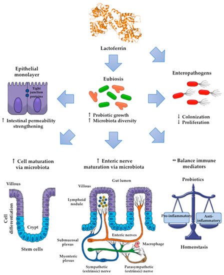
Lactoferrin (Lf) is an iron-binding milk glycoprotein that promotes the growth of selected probiotic strains. The effect of Lf on the growth and diversification of intestinal microbiota may have an impact on several issues, including (i) strengthening the permeability of the epithelial cell monolayer, (ii) favoring the microbial antagonism that discourages the colonization and proliferation of enteric pathogens, (iii) enhancing the growth and maturation of cell-monolayer components and gut nerve fibers, and (iv) providing signals to balance the anti- and pro-inflammatory responses resulting in gut homeostasis.

  • lactoferrin
  • lactoferricin
  • milk

1. Introduction

Bovine and human milk contains a wide array of bioactive components, including the iron-binding glycoprotein known as lactoferrin (Lf). This protein displays antimicrobial, anti-inflammatory, and immunomodulatory activities that contribute to the maintenance of homeostasis and to the control of life-threatening diseases in the intestine of consumers, mainly in neonates [1]. As its milk counterpart, Lf present in mucosal secretions and neutrophil secondary granules displays a wide array of functions including antimicrobial activity, as well as modulatory actions on immune response, cell proliferation, and iron metabolism, among some others [2]. Lf is a mammalian monomeric molecule of approximately 80 kDa that is organized into N- and C-terminal lobes whose divergent structural features determine, in part, its exceptional multifunctional characteristics [3]. Milk is an enriched source of Lf; in addition, it becomes a source of lactoferricin (Lfcin), which is a peptide derived from the Lf N-terminus by gastric pepsin digestion that also show antimicrobial activity against pathogens [4].

2. Lactoferrin: Effects on Probiotic Growth

2.1. Bulk Milk

An analysis of milk fractions separated by ultrafiltration demonstrated that, in human milk, the bifidogenic activity of the nonprotein fraction (containing glycans, among other components) was greater than that of the protein fraction for B. Pennsylvanicus, B. bifidum, and B. longum [5]. By contrast, the nonprotein and protein fractions of cow milk had similar growth-promoting effects on B. infantis, B. bifidum, and B. breve [5]. These findings may reflect the divergent composition of glycans and proteins in human and cow milk. Unlike cow milk, human milk contains lacto-N-biose (LNB), which is used as a substrate by bifidobacteria capable of using complex human milk oligosaccharides, such as B. infantis [6]. An analysis of fractioned samples indicated that bulk milk displayed a higher degree of probiotic activity than milk protein fractions tested individually [7]. The bifidogenic properties of milk were related to the content of bioactive milk proteins, although the activity of casein was unclear [8][5][9]. Both Lf and α-lactalbumin isolated from cow milk displayed stronger growth-promoting effects on B. infantis and B. breve than on B. bifidum strains [5]. For B. infantis, the bifidogenic activity of a mixture containing Lf, lactoperoxidase, and lysozyme was higher than the activities of the single components tested separately [7]. Bifidogenic activity for B. infantis and B. breve was observed for Lf from mature milk but not Lf from colostrum [10]. Lf present in milk is a glycosylated protein that contains β-N-glycans used for bifidobacterial growth [11][12][9]. These results suggest a role for the bifidogenic activity of Lf as a “provider” of N-glycans that increase the expression of genes required for their utilization [11]. In turn, β-glycans induce the gene expression and translation of proteins (ATP-binding cassette (ABC) and phosphodiesterase proteins) involved in the transport of sugars and their concomitant incorporation into the bifid shunt fermentative pathway [13]. These findings suggest that the individual activity of each protein is synergized to enhance the growth of probiotics and is dependent on milk stage maturation, which, in turn, impacts the extent of glycosylation of Lf.

2.2. Apo and HoloLactoferrin

The antimicrobial action of iron-depleted bLf can also be seen against protozoan like Entamoeba histolytica trophozoites [14]. These findings indicate that iron-free Lf either has an antimicrobial effect against pathogenic and probiotic bacteria or displays a selective growth-promoting activity on some probiotic strains.
Additional evidence showed that apoLf (both bovine and human) inhibited the growth of B. infantis, B. bifidum and L. acidophilus, whereas 66% iron-saturated bLf (holo66bLf) inhibited the growth of bifidobacteria but not lactobacilli [15]. Conversely, both holo98bLf and holo98hLf inhibited the growth of lactobacilli but not bifidobacteria. In single-culture assays, the growth of foodborne pathogens was decreased by apobLf, apohLf, and holo66bLf but was unaltered by holo98bLf. In coculture assays, apobLf and holo66bLf selectively retarded E. coli O157:H7 growth without affecting B. infantis growth [15]. An analysis of metal-bound Lf forms demonstrated that apobLf, Cu-bLf, and Fe-bLf enhanced the growth of bifidobacterial strains [16]. Moreover, Cu-bLf and Zn-bLf had stronger inhibitory action than apobLf on the growth of pathogenic strains of E. coli and S. aureus [16]. The bactericidal action of apobLf and Cu-bLf was strong against E. coli and was weak against B. breve, whereas the inhibitory or bactericidal action of holobLf was weak or absent [16].
These results support that, unlike the iron-saturated form, iron-depleted bLf limits the growth of multidrug-resistant pathogens without affecting the proliferation of some probiotics and contributes to the natural innate mechanism for maintaining the microbiota balance and gut homeostasis.
Other experimental settings documented that holobLf but not apobLf enhanced the growth of L. acidophilus. Moreover, both holobLf and apobLf stimulated the growth of B. breve, B. infantis, and B. bifidum, although they did not have an effect on B. longum [17]. Under culture conditions of iron deprivation, the growth of B. breve was enhanced by holohLf and was inhibited by apohLf [18]. The effects of iron deprivation or supplementation on growth promotion appear to be specific to each probiotic strain and Lf source. A possible mechanism is that Lf in the iron-loaded form provides iron as an essential factor for the proper function of the enzymes involved in iron reduction, DNA replication, and repair and for ABC glycan transporters (cell membrane permease) in some bifidobacterial strains [19][20]. The effects of Lf on growth modulation by supplying or depleting iron for probiotic growth are influenced by additional environmental conditions as described below.

2.3. Probiotic Culture Conditions

The inhibitory and stimulatory effects of Lf on the growth of probiotics have been tested under various conditions of aerobiosis, anaerobiosis, time, temperature, and iron depletion in cultures [21][10][18][22]. Experimental results have indicated that the inhibitory concentration-dependent effect of bLf (iron less than 15 mg/100 g of protein) was similar under aerobic or anaerobic conditions [22]. Under aerobic conditions, bLf had a greater inhibitory effect on the growth of pathogenic strains than on probiotic strains but displayed strong growth-promoting effects on L. rhamnosus ATCC 7469 and L. acidophilus BCRC 14065 in a dose-independent manner [22]. Assays examining the effect of temperature indicated that bLf showed inconsistent probiotic activity at 37 °C; at 22 °C, the growth of probiotic strains (B. breve and lactobacilli strains) was selectively retarded, but the addition of bLf resumed probiotic proliferation in a dose-dependent manner [21]. These findings provide insights into the development of conditions for the selective growth of probiotic strains by controlling the temperature and bLf dosage. Other experimental in vitro settings in cultures of Lactococcus (L.) lactis subsp. cremoris JCN20076 evidenced that growth promotion activity was found higher with heat-treated bLf at 65 °C for 30 min followed by heat-treated bLf at 80 °C for 5 min while unheated bLf showed less activity on L. lactis subsp. cremoris growth [23]. According to these findings, the impact of heating on the potentiation of growth promotion activity of bLf is a critical advantage given that the manufacturing of infant formulas entails a thermal process of pasteurization.
In iron-free cultures, the growth-promoting effects of holohLf on B. brevis resulted from the direct extraction of iron-bound Lf and eventual uptake by probiotic bacteria [18]. By contrast, in other assays, the probiotic activity of bovine and human Lf was not influenced by the degree of iron saturation [10]. These discoveries suggest that environmental factors may affect the extent of iron saturation of Lf and, in turn, its ability to enhance or decrease the growth of probiotics that express Lf-binding proteins, as described in the following section.

2.4. Probiotic Lactoferrin-Binding Proteins

Several experiments have accounted for the impact of the interaction of Lf with probiotic surface components on Lf activity. A Western blot analysis of the protein fractions of sonicates from B. bifidum indicated the presence of bLf-binding proteins in both the membrane (69 kDa) and cytosolic fractions (20, 35, 50, and 66 kDa) [24]. These experiments used biotinylated apobLf and streptavidin-avidin-labeled horseradish peroxidase. Other assays have demonstrated the growth-promoting activity of both apo- and iron-loaded bLf forms in probiotic strains expressing membrane Lf-binding proteins, including L. acidophilus (21, 41, and 67 kDa), B. breve, and B. bifidum (both 69 kDa), and B. infantis (67 kDa) but not B. longum [17]. The probiotic activity of bLf isolated from mature milk was coincident with a strong degree of binding of B. bifidum and B. breve. The underlying mechanisms are apparently independent of bLf-iron saturation extent, although probiotic growth might result from the binding of Lf with probiotic proteins to be transported to the inner milieu for the concomitant cleavage and transport of Lf-linked glycans used as energy sources, as depicted in Figure 1.
Figure 1. Simplified hypothetical mechanisms that may account for the in vitro growth-promoting effects of lactoferrin (Lf) on probiotics (bifidobacteria and lactobacilli). (a) Lf glycans undergo enzymatic hydrolysis to produce both intact oligosaccharides and the products of cleavage (disaccharides and monosaccharides). Intact and hydrolyzed glycans are transported via cell membrane permeases to the cytosol. In the cytosol, glycans are terminally hydrolyzed to yield monosaccharides used by cytosolic enzymes as substrates of fermentation via the Embden–Meyerhof–Parnas or bifid shunt pathways to produce energy for cell growth. (b) The cationic surface of Lf interacts electrostatically with acidic components of the cell wall that are negatively charged, such as teichoic acids. This interaction may favor the internalization of Lf in the periplasmic space and its eventual binding to cell membrane proteins for translocation to the cytosol. In the presence of ATP, Lf dissociates to the N-lobe and C-lobe. The latter may interact with DNA to modulate genes involved in the mechanisms of DNA replication for cell growth. (c) Diferric Lf reaches the periplasmic space, where iron in ferric form is released and then translocated via a permease to cytosolic compartments. Ferric ions are considered an essential cofactor of enzymes involved in crucial processes of metabolism and DNA replication for growth. RasMol™ 1994 by Roger Sayle© was used as the software for modeling and coloring the tertiary protein structures.
In bifidobacteria, surface proteins involved in the hydrolysis and uptake of glycans (intact human milk oligosaccharides, lacto-N-biose (LNB), and free monosaccharides) are ultimately funneled to the fermentative bifid shunt pathway [6][13]. Bifidobacteria express membrane proteins related to metabolism and proteins involved in energy production and conversion [25]. Other experimental assays demonstrated that bLf from colostrum could bind B. bifidum and B. breve despite lacking growth-promoting activity. These findings should indicate that the bifidogenic properties of bLf are independent of the receptor binding capacity [10]. Visualization by confocal laser-scanning microscopy using biotinylated Lf and fluorescein-conjugated avidin showed Lf binding to the pole of Bifidobacterium cells [26][27]. A Lf-binding protein with a molecular mass of 67 kDa was found in the membrane and cytosolic fractions of bifidobacterial cells, including B. longum [26][27]. An analysis of each Lf lobe indicated that the N-lobe was the presumable site of interaction of full-length Lf with bifidobacterial proteins; by contrast, the C-lobe lacked the binding activity to bifidobacterial proteins despite promoting bifidobacterial growth [28]. A potential mechanism underlying these findings may involve ionic interactions of cationic Lf with anionic surface proteins that enable Lf uptake by bifidobacteria and subsequent potential growth-promoting activity. Human milk Lf forms a molecular complex with ATP via the C-lobe that leads to its dissociation into monomeric forms that interact with macromolecules such as DNA [29]. Human milk Lf includes enzymes such as malto-oligosaccharidase, ATPase, DNase, RNase, and phosphatase [30]. Lf acts as a DNA transcriptional factor and as an ATP-binding protein that results in the dissociation of monomeric forms that interact with DNA [31]. Future studies may provide substantive evidence that supports the probiotic effect of Lf by acting as a DNA transcriptional factor.
By contrast, hydrophobicity and autoaggregation are physicochemical parameters that reflect the ability of bacteria to interact with components of the surrounding environment [32]. In this regard, assays of bifidobacterial cells in liquid media demonstrated that autoaggregation was reduced by bLf, bovine transferrin, and ovotransferrin. Surface hydrophobicity was altered by bovine transferrin but not by bLf or Tf [33]. These results may reflect the wide array of the extent of aggregation and surface hydrophobicity, which affects the plasticity of the adaptation of bifidobacteria in the intestinal milieu [34][35].

2.5. Lactoferrin Hydrolysates and Lf-Derived Peptides

Although their actual mechanism of action is not fully known, the activity of Lf hydrolysates and Lf peptides on the growth of lactic acid bacteria is iron-independent, since these derivatives are unable to bind iron. Culture assays in bifidobacteria and lactobacilli strains showed that bacterial growth was either inhibited or unaltered by the hydrolysate of apobLf obtained by treatment with pepsin [36]. Moreover, compared with the full-length protein, the apobLf hydrolysate displayed strong inhibitory action on the growth of foodborne pathogens and MRSA [36][37]. Combining culture supernatants from apobLf-resistant probiotic strains with apobLf hydrolysate resulted in synergistic inhibitory action against foodborne pathogens and MRSA [36][37]. These results may reflect the role of the synergistic antimicrobial effect of bLf hydrolysate and probiotic strains resistant to bLf hydrolysate in the intestinal milieu. Stronger antimicrobial activity of apoLf hydrolysate than the full-length Lf protein may rely on its ability to enhance the entrance of the secreted antimicrobial compounds released by the probiotic strains in the culture supernatant within the MRSA bacteria [36][37]. In contrast to this inhibitory activity, bLf hydrolysates exhibited growth-promoting properties on bifidobacterial strains [38]. The bifidogenic activity on B. breve was greater with apobLf (pepsin) hydrolysate than with full-length Lf, while on B. infantis, bifidogenic activity was only found for apobLf hydrolysate. Assays on B. breve and B. longum subsp. infantis cultures indicated that a synthetic peptide from bLfcin showed stronger bifidogenic activity than natural bLf hydrolysate, whereas no bifidogenic activity was detected on an amino acid mixture containing the same amino acid sequences as the synthetic active peptide [38]. According to these effects, bLf hydrolysates and synthetic bLf peptides present in maternal milk or even in infant formula may provide intestinal benefits in infants, in addition to the benefits given by the whole Lf protein.
In human milk subjected to pepsin proteolysis, peptide derivatives from the polymeric immunoglobulin receptor (pIgR) and hLf enhanced the growth of B. bifidum. Treatment with proteolytic enzymes caused no loss of the bifidogenic activity of these peptides. A synthetic peptide derivative C(1)AV GGG CIAL(10) with a sulfide bridge in C(1) and C(7) displayed the same activity as native peptides [38]. These interesting results may explain the ability of degradation of pIgR and Lf to render active peptides refractory to enzymatic hydrolysis, promote the growth of bifidogenic cells, and inhibit the proliferation of pathogenic bacteria in the large intestine of infants [39]. In addition, the products of proteolysis of Lf by lactic acid bacteria peptidases have been characterized. Compared to other milk-derived peptides that underwent full hydrolysis, the 25-residue peptide Lfcin was more resistant to proteolytic degradation by these peptidases during incubation with Streptococcus thermophilus and L. delbrueckii subsp. bulgaricus strains used in the yogurt-making industry [40]. The biological activity was observed in peptide sequences located in the C-lobe of Lf. Synthetic N-L-N-R (C-lobe residues 563–566) enhanced the growth of L. acidophilus used to produce fermented milk while displaying strong antibacterial activity against Pseudomonas spp. and E. coli strains that reduce milk quality [41]. Based on these results, some hypothetical mechanisms that may account for the effects of Lf on in vitro probiotic growth are depicted in Figure 1.

3. Lactoferrin: Modulatory Effects on the Gut Microbiota

3.1. Human neonates

Importantly, it has been demonstrated that hLf benefits the nascent gut health and immune development and functioning in preterm and neonate infants. These effects are due to Lf favors the decrease of its permeability and increase of its maturation [42]. The impact of Lf on the colonization of intestinal microbiota has also been addressed in neonate populations, since ongoing intestinal maturation puts them at risk for life-threatening diseases, including necrotizing enterocolitis [43][44]. Buccigrossi et al. (2007) found that both hLf and bLf exert a mucosal trophic effect on enterocytes (Caco2 cells) that is related to its concentration; at high Lf concentrations, it was promoted a more rapid proliferation of these cells, whereas at low Lf concentration it was induced their differentiation [45].
The role of Lf on the colonization of microbiota has been addressed during the early stages of intestinal maturation in neonates fed maternal milk and an infant formula diet [43]. In early trials, a relationship between the bLf contained in an infant formula diet and the fecal microbiota in neonates was not seen [46][47]. In current studies, the abundance of fecal bifidobacteria and lactobacilli was significantly associated with the levels of Lf in feces from breastfed newborns [48]. These discoveries suggest that hLf levels in neonates are beneficial for contributing to the establishment of the gut microbiota.
Regarding the microbiota composition and its relation with Lf intake, Mastromarino et al. (2014) measured the content of Lf and the microbiota of breast milk and of feces of infants at birth and one month after delivery [48]. Interestingly, in preterm infants, higher concentrations of fecal Lf at birth and 30 days after delivery were observed than in full-term infants; also, the amount of fecal bifidobacteria and lactobacilli were significantly associated with the concentration of fecal Lf. These results suggested that Lf promotes a bifidogenic microflora in the gut in neonate and preterm infants. High levels of fecal Lf in in the first days of life contribute to a strong early host-microbe interaction that could be important for the composition of the neonatal gut microbiota and the development of these microorganisms, in addition to the antimicrobial activity of this milk glycoprotein. This interaction is critical for having a healthy immune system and a correct metabolic program in newborns.
A clinical trial in VLBW (<1500 g) babies tested the effect of a rrhLf, administered at 150 mg/kg/day every 12 h by a nasogastric route from day one to 28 of life. Proteobacteria and Firmicutes were the major Phyla in feces from babies treated or untreated (placebo) with rhLf. It should be noted that the fecal abundance of the pathogenic species Enterobacter, Klebsiella, and Staphylococcus was decreased, while Citrobacter abundance was increased, but it was not associated with infections [49]. In breastfed babies, the abundance of fecal bifidobacteria was predominant, and that of the facultative anaerobes was poor; by contrast, in babies fed formula containing bLf (10 or 100 mg/ml), obligate anaerobes (Clostridium and Bacteroides) were predominantly seen in regard to bifidobacteria [50]. A delicate balance of microbiota members is prone to the disturbance in preterm infants [51]; thus, findings indicate that the effect of bLf dosage on promoting anaerobic growth must be kept in mind to prevent potential risks in dysbiosis in newborns.
In a retrospective cohort study in preterm babies fed bLf in combination with LGG, the incidence of severe necrotizing colitis was significantly reduced, and the resolution of this disease was improved. Notably, bLf had no collateral effects, but a severe case of sepsis by LGG was found [52]. These results indicate that due to the extreme fragility of very low birth weight neonates, stringent precautions should be taken to avoid the risk of severe cases of neonatal sepsis.
Currently, several recommendations of these therapies using Lf and probiotics against NEC and other gut diseases have been addressed. For example, the therapy with Lf must be as early in the life as possible, more than 100 mg/day is the recommended dosage, Lf is apparently more effective in preterm than in term infants, and the efficacy versus Gram-negative bacteria could be limited. Important gaps in the knowledge exist concerning dosages, schedule, duration of treatment, most effective probiotic strains, and interactions of probiotics with human and bovine milk [53][54]. As stated below, experimental findings in preterm piglets support these claims [55][56].

3.2. Piglet Model

Given their physiological and anatomical resemblance to humans, piglets have been used in experimental studies as a model for the neonatal gastrointestinal tract to provide insights about the presumable mechanisms underlying the role of Lf and its derivatives on intestinal homeostasis in neonates [43].
A recombinant Lf fusion peptide consisting of Lfcin and lactoferrampin (Lfampin) expressed in Pichia pastoris has been shown to exhibit potent probiotic effects on bifidobacteria and lactobacilli throughout the gastrointestinal tract in weaned piglets [57]. Additional trials in healthy full-term piglets demonstrated that bLf, in combination with probiotics increased the richness of microbiota in the small and large intestine. Interestingly, bLf reversed the effects of probiotics on the increase or decrease of ferric or ferrous iron transport system abundance, respectively [58]. Thus, the iron-binding ability of bLf on ferric ions seems to affect the role of probiotics on microbiota modulation.
Piglet trials documented the substantive effects of bLf or recombinant hLf on stimulating the abundance of a wide array of microbiota members and on improving body weight gain [59][60]. Recombinant Lfcin-Lfampin expressed in Photorhabdus luminescens also enhanced the growth of bifidobacteria and lactobacilli and body mass gain [61]. Glycan degradation results in the formation of SFCA, which in turn are a source of energy for the epithelial cells necessary for ongoing gut maturation [51]. Thus, underlying mechanisms seem to be associated with the ability of Lf or its derivatives to favor the diversity of microbiota members with a pivotal role in breakdown glycans.
The microbiome has a critical role in intestinal maturation by providing signals for the development of innervations of the enteric nervous system connected in turn with the central nervous system (CNS); conversely, CNS and enteric nerves modulate intestinal maturation via microbiome signaling pathways [62]. Notably, the effect of bLf on the abundance and diversity of microbiota in piglets seems to affect the intestinal expression of neurotransmitters such as ileal vasoactive intestinal peptide released by enteric nerve fibers [59] and the expression of parameters associated with the maturation of enteric nerves such as brain-derived neurotrophic factor (BDNF) and ubiquitin carboxy-terminal hydrolase 1 [ubiquitin thiol esterase (UCHL1)] [59][63]. These results suggest a presumable mechanism for the role of bLf on the interplay between the gut-brain axis and the microbiome, resulting in the maturation of enteric nerve fibers.
The impact of bLf on intestinal maturation has also been demonstrated with the increased activity of brush-border enzymes, such as jejunal lactase, as found in piglets fed formula containing probiotics and bioactive milk components, including bLf [59]. The intestinal alkaline phosphatase activity was enhanced in piglets fed a sow milk replacement containing bLf [63]. Benefits on epithelial architecture have been evidenced in the small intestine by larger crypt area, depth and width and thinner lamina propria, as found in piglets fed bLf, recombinant hLf or Lfcin-Lfampim from P. luminescens [63][61][64]. bLf also enhanced jejune crypt proliferation, depth, area, and the crypt mRNA expression of β-catenin mRNA, as documented in colostrum-deprived piglets fed formula containing bLf [65]. The upregulation of bLf on intestinal growth may result from the elicitation of the β-catenin-Wnt signaling pathway [65]. β-Catenin mRNA encodes a cytosolic protein expressed at crypts that is regarded as a key effector of Wnt signaling; the latter drives the self-renewal and proliferation of stem cells and their concomitant differentiation to other cell components of the epithelial monolayer [66]. The findings provide evidence that supports the role of Lf in growth and maturation in the neonatal intestine.
Maternal bLf supplementation increased the pregnancy rate, litter size, and survival, and the levels of IgA antibodies in the serum of gilts and their litter [67]. These findings indicate that bLf consumption provided benefits on survival and immunity during pregnancy and lactation. In piglets fed daily for the first seven or 14 days of life with a formula containing bLf (367 or 1300 mg/kg body weight), the serum IgG antibodies increased; however, bLf did not affect the cellularity of the lymphoid populations (B cells and T cells), NKCs and neutrophils. In supernatants from lipopolysaccharide (LPS)-primed mesenteric lymph node cell cultures treated with bLf at 367 mg/kg of body mass, the IL-6 and IL-10 levels were enhanced, whereas the tumor necrosis factor (TNF)-α level was unaffected [68]. The consumption of transgenic milk containing hLf had significant effects on the decrease in circulating neutrophils and the increase in lymphocytes without affecting cytokine expression [64], whereas in piglets fed transgenic milk containing recombinant human lysozyme and rhLf, the number of peripheral blood cells was increased without affecting the expression of TNF-α, IL-6, TGF-β and TLR4 [64]. Accordingly, these results indicate that natural or recombinant Lf products display a tendency to either not affect or downmodulate the markers of inflammation.

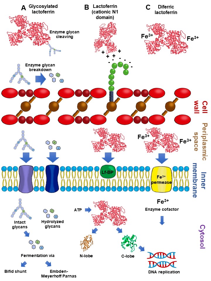
By contrast, piglets fed the chimera Lfcin-Lfampin showed enhanced serum antioxidant enzymes such as glutathione peroxidase and peroxidase, as well as the effectors of the adaptive immunity branch including IgA, IgG, and IgM antibodies and the components of the innate response involved in protection from the deleterious effects of inflammation [61]. This finding agrees with the observation that rhLf-cow milk increased TLR-2 mRNA expression in the ileum and the levels of colonic IgG and nuclear factor-κB (NF-κB) p65, concomitant with the increase in spleen IL-2, -4, and -5 and plasmatic IgG, IgA and IL-12, and IL-10 [69]. Although the assessment of parameters was systemic, the compartmental modulatory action on inflammatory and immune components underlies the protective role of recombinant Lf derivatives in the intestinal milieu. According to the above results, some presumable mechanisms that account for the impact of Lf on intestinal microbiota growth are depicted in Figure 2.

Figure 2. The impact of Lf on the promotion of the growth and diversity of intestinal microbiota may entail (i) strengthening of the permeability of the epithelial cell monolayer; (ii) favoring of the microbial antagonism that discourages the colonization and proliferation of enteric pathogens, enhancing the growth and maturation of (iii) cell-monolayer components and (iv) gut nerve fibers; and (v) providing signals to balance the anti- and proinflammatory responses resulting in homeostasis.

References

  1. Giansanti, F.; Panella, G.; Leboffe, L.; Antonini, G. Lactoferrin from Milk: Nutraceutical and Pharmacological Properties. Pharmaceuticals 2016, 9, 61.
  2. Nazir, S.N.M.; Yasmeen, A.; Usman, S. Review study on lactoferrin: A multifunctional protein. Sky J. Food Sci. 2017, 6, 014–020.
  3. Albar, A.H.; Almehdar, H.A.; Uversky, V.N.; Redwan, E.M. Structural heterogeneity and multifunctionality of lactoferrin. Curr. Protein Pept. Sci. 2014, 15, 778–797.
  4. Park, Y.W.; Nam, M.S. Bioactive Peptides in Milk and Dairy Products: A Review. Korean J. Food Sci. Anim. Resour. 2015, 35, 831–840.
  5. Petschow, B.W.; Talbott, R.D. Response of bifidobacterium species to growth promoters in human and cow milk. Pediatric Res. 1991, 29, 208–213.
  6. Sela, D.A.; Mills, D.A. Nursing our microbiota: Molecular linkages between bifidobacteria and milk oligosaccharides. Trends Microbiol. 2010, 18, 298–307.
  7. Loh, S., Jr.; Maznah, I. The effect of different milks and milk proteins on the growth of Bifidobacterium infantis ATCC 27920 in vitro. Malays. J. Nutr. 1999, 5, 61–70.
  8. Petschow, B.W.; Talbott, R.D. Growth promotion of Bifidobacterium species by whey and casein fractions from human and bovine milk. J. Clin. Microbiol. 1990, 28, 287–292.
  9. Kodama, A. The biotying of Bifidobacterium isolated from healthy infants in Wakayama and Osaka district; Bifidobacterium growth promoting activities of human casein and lactoferrin. Pediatr Int. 1983, 25, 486.
  10. Petschow, B.W.; Talbott, R.D.; Batema, R.P. Ability of lactoferrin to promote the growth of Bifidobacterium spp. in vitro is independent of receptor binding capacity and iron saturation level. J. Med. Microbiol. 1999, 48, 541–549.
  11. Garrido, D.; Nwosu, C.; Ruiz-Moyano, S.; Aldredge, D.; German, J.B.; Lebrilla, C.B.; Mills, D.A. Endo-beta-N-acetylglucosaminidases from infant gut-associated bifidobacteria release complex N-glycans from human milk glycoproteins. Mol. Cell. Proteom. 2012, 11, 775–785.
  12. Karav, S.; German, J.B.; Rouquie, C.; Le Parc, A.; Barile, D. Studying Lactoferrin N-Glycosylation. Int. J. Mol. Sci. 2017, 18, 870.
  13. Zhao, J.; Cheung, P.C. Comparative proteome analysis of Bifidobacterium longum subsp. infantis grown on beta-glucans from different sources and a model for their utilization. J. Agric. Food Chem. 2013, 61, 4360–4370.
  14. Leon-Sicairos, N.; Lopez-Soto, F.; Reyes-Lopez, M.; Godinez-Vargas, D.; Ordaz-Pichardo, C.; de la Garza, M. Amoebicidal activity of milk, apo-lactoferrin, sIgA and lysozyme. Clin. Med. Res. 2006, 4, 106–113.
  15. Griffiths, E.A.; Duffy, L.C.; Schanbacher, F.L.; Dryja, D.; Leavens, A.; Neiswander, R.L.; Qiao, H.; DiRienzo, D.; Ogra, P. In vitro growth responses of bifidobacteria and enteropathogens to bovine and human lactoferrin. Dig. Dis. Sci. 2003, 48, 1324–1332.
  16. Saito, H.; Miyakawa, H.; Ishibashi, N.; Yoshitaka, T.; Hayasawa, H.; Shimamura, S. Effect of Iron-Free and Metal-Bound Forms of Lactoferrin on the Growth of Bifidobacteria.; E. coli and S. aureus. Biosci. Microflora 1996, 15, 1–7.
  17. Kim, W.S.; Ohashi, M.; Tanaka, T.; Kumura, H.; Kim, G.Y.; Kwon, I.K.; Goh, J.S.; Shimazaki, K. Growth-promoting effects of lactoferrin on L. acidophilus and Bifidobacterium spp. Biometals 2004, 17, 279–283.
  18. Miller-Catchpole, R.; Kot, E.; Haloftis, G.; Furmanov, S.; Bezkorovainy, A. Lactoferrin can supply iron for the growth of Bifidobacterium breve. Nutr Res. 1997, 17, 205–213.
  19. Cronin, M.; Zomer, A.; Fitzgerald, G.F.; van Sinderen, D. Identification of iron-regulated genes of Bifidobacterium breve UCC2003 as a basis for controlled gene expression. Bioengineered 2012, 3, 157–167.
  20. Lanigan, N.; Bottacini, F.; Casey, P.G.; O’Connell Motherway, M.; van Sinderen, D. Genome-Wide Search for Genes Required for Bifidobacterial Growth under Iron-Limitation. Front. Microbiol. 2017, 8, 964.
  21. Chen, P.W.; Liu, Z.S.; Kuo, T.C.; Hsieh, M.C.; Li, Z.W. Prebiotic effects of bovine lactoferrin on specific probiotic bacteria. Biometals 2017, 30, 237–248.
  22. Chen, P.W.; Ku, Y.W.; Chu, F.Y. Influence of bovine lactoferrin on the growth of selected probiotic bacteria under aerobic conditions. Biometals 2014, 27, 905–914.
  23. Kim, W.S. Effects of Heat-treated Bovine Lactoferrin on the Growth of Lactococcus lactis subsp. cremoris JCM 20076. J. Milk Sci. Biotechnol. 2019, 37, 129–135.
  24. Kim, W.S.; Tanaka, T.; Kumura, H.; Shimazaki, K. Lactoferrin-binding proteins in Bifidobacterium bifidum. Biochem. Cell Biol. 2002, 80, 91–94.
  25. Wasko, A.; Polak-Berecka, M.; Kalita, M. Protein profiles from intact cells as a tool in Bifidobacterium characteristics. Pol. J. Microbiol. 2012, 61, 305–310.
  26. Rahman, M.M.; Kim, W.S.; Ito, T.; Kumura, H.; Shimazaki, K. Examination of bovine lactoferrin binding to bifidobacteria. Appl. Biochem. Microbiol. 2008, 44, 529–532.
  27. Rahman, M.M.; Kim, W.S.; Ito, T.; Kumura, H.; Shimazaki, K. Growth promotion and cell binding ability of bovine lactoferrin to Bifidobacterium longum. Anaerobe 2009, 15, 133–137.
  28. Rahman, M.; Kim, W.S.; Kumura, H.; Shimazaki, K. Bovine lactoferrin region responsible for binding to bifidobacterial cell surface proteins. Biotechnol. Lett. 2009, 31, 863–868.
  29. Semenov, D.V.; Kanyshkova, T.G.; Buneva, V.N.; Nevinsky, G.A. Human milk lactoferrin binds ATP and dissociates into monomers. Biochem. Mol. Biol. Int. 1999, 47, 177–184.
  30. Kanyshkova, T.G.; Babina, S.E.; Semenov, D.V.; Isaeva, N.; Vlassov, A.V.; Neustroev, K.N.; Kul’minskaya, A.A.; Buneva, V.N.; Nevinsky, G.A. Multiple enzymic activities of human milk lactoferrin. Eur. J. Biochem. 2003, 270, 3353–3361.
  31. He, J.; Furmanski, P. Sequence specificity and transcriptional activation in the binding of lactoferrin to DNA. Nature 1995, 373, 721–724.
  32. Vinderola, C.G.; Medici, M.; Perdigon, G. Relationship between interaction sites in the gut, hydrophobicity, mucosal immunomodulating capacities and cell wall protein profiles in indigenous and exogenous bacteria. J. Appl. Microbiol. 2004, 96, 230–243.
  33. Rahman, M.; Kim, W.S.; Kumura, H.; Shimazaki, K. In vitro effects of bovine lactoferrin on autoaggregation ability and surface hydrophobicity of bifidobacteria. Anaerobe 2008, 14, 73–77.
  34. Andriantsoanirina, V.; Teolis, A.C.; Xin, L.X.; Butel, M.J.; Aires, J. Bifidobacterium longum and Bifidobacterium breve isolates from preterm and full term neonates: Comparison of cell surface properties. Anaerobe 2014, 28, 212–215.
  35. Canzi, E.; Guglielmetti, S.; Mora, D.; Tamagnini, I.; Parini, C. Conditions affecting cell surface properties of human intestinal bifidobacteria. Antonie Van Leeuwenhoek 2005, 88, 207–219.
  36. Chen, P.W.; Jheng, T.T.; Shyu, C.L.; Mao, F.C. Antimicrobial potential for the combination of bovine lactoferrin or its hydrolysate with lactoferrin-resistant probiotics against foodborne pathogens. J. Dairy Sci. 2013, 96, 1438–1446.
  37. Chen, P.W.; Jheng, T.T.; Shyu, C.L.; Mao, F.C. Synergistic antibacterial efficacies of the combination of bovine lactoferrin or its hydrolysate with probiotic secretion in curbing the growth of meticillin-resistant Staphylococcus aureus. J. Med. Microbiol. 2013, 62, 1845–1851.
  38. Oda, H.; Wakabayashi, H.; Yamauchi, K.; Sato, T.; Xiao, J.Z.; Abe, F.; Iwatsuki, K. Isolation of a bifidogenic peptide from the pepsin hydrolysate of bovine lactoferrin. Appl. Environ. Microbiol. 2013, 79, 1843–1849.
  39. Liepke, C.; Adermann, K.; Raida, M.; Magert, H.J.; Forssmann, W.G.; Zucht, H.D. Human milk provides peptides highly stimulating the growth of bifidobacteria. Eur. J. Biochem. 2002, 269, 712–718.
  40. Paul, M.; Somkuti, G.A. Hydrolytic breakdown of lactoferricin by lactic acid bacteria. J. Ind. Microbiol. Biotechnol. 2010, 37, 173–178.
  41. Kim, W.S.; Ohashi, M.; Shimazaki, K. Inhibitory Effects of Synthetic Peptides Containing Bovine Lactoferrin C-lobe Sequence on Bacterial Growth. Korean J. Food Sci. Anim. Resour. 2016, 36, 452–457.
  42. Goldman, A.S. Modulation of the gastrointestinal tract of infants by human milk. Interfaces and interactions. An evolutionary perspective. J. Nutr. 2000, 130, 426S–431S.
  43. Donovan, S.M. The Role of Lactoferrin in Gastrointestinal and Immune Development and Function: A Preclinical Perspective. J. Pediatrics 2016, 173, S16–S28.
  44. Schuller, S.S.; Kramer, B.W.; Villamor, E.; Spittler, A.; Berger, A.; Levy, O. Immunomodulation to Prevent or Treat Neonatal Sepsis: Past, Present, and Future. Front. Pediatrics 2018, 6, 199.
  45. Buccigrossi, V.; de Marco, G.; Bruzzese, E.; Ombrato, L.; Bracale, I.; Polito, G.; Guarino, A. Lactoferrin induces concentration-dependent functional modulation of intestinal proliferation and differentiation. Pediatr Res. 2007, 61, 410–414.
  46. Balmer, S.E.; Scott, P.H.; Wharton, B.A. Diet and fecal flora in the newborn: Lactoferrin. Arch. Dis. Child. 1989, 64, 1685–1690.
  47. Wharton, B.A.; Balmer, S.E.; Scott, P.H. Fecal flora in the newborn. Effect of lactoferrin and related nutrients. Adv. Exp. Med. Biol. 1994, 357, 91–98.
  48. Mastromarino, P.; Capobianco, D.; Campagna, G.; Laforgia, N.; Drimaco, P.; Dileone, A.; Baldassarre, M.E. Correlation between lactoferrin and beneficial microbiota in breast milk and infant’s feces. Biometals 2014, 27, 1077–1086.
  49. Sherman, M.P.; Sherman, J.; Arcinue, R.; Niklas, V. Randomized Control Trial of Human Recombinant Lactoferrin: A Substudy Reveals Effects on the Fecal Microbiome of Very Low Birth Weight Infants. J. Pediatrics 2016, 173, S37–S42.
  50. Roberts, A.K.; Chierici, R.; Sawatzki, G.; Hill, M.J.; Volpato, S.; Vigi, V. Supplementation of an adapted formula with bovine lactoferrin: 1. Effect on the infant fecal flora. Acta paediatrica 1992, 81, 119–124.
  51. Unger, S.; Stintzi, A.; Shah, P.; Mack, D.; O’Connor, D.L. Gut microbiota of the very-low-birth-weight infant. Pediatric Res. 2015, 77, 205–213.
  52. Meyer, M.P.; Alexander, T. Reduction in necrotizing enterocolitis and improved outcomes in preterm infants following routine supplementation with Lactobacillus GG in combination with bovine lactoferrin. J. Neonatal-Perinat. Med. 2017, 10, 249–255.
  53. Manzoni, P. Clinical Benefits of Lactoferrin for Infants and Children. J. Pediatr 2016, 173, S43–S52.
  54. Manzoni, P.; Mostert, M.; Stronati, M. Lactoferrin for prevention of neonatal infections. Curr. Opin. Infect. Dis 2011, 24, 177–182.
  55. Nguyen, D.N.; Li, Y.; Sangild, P.T.; Bering, S.B.; Chatterton, D.E. Effects of bovine lactoferrin on the immature porcine intestine. Br. J. Nutr. 2014, 111, 321–331.
  56. Nguyen, D.N.; Jiang, P.; Stensballe, A.; Bendixen, E.; Sangild, P.T.; Chatterton, D.E. Bovine lactoferrin regulates cell survival, apoptosis and inflammation in intestinal epithelial cells and preterm pig intestine. J. Proteom. 2016, 139, 95–102.
  57. Tang, X.S.; Shao, H.; Li, T.J.; Tang, Z.R.; Huang, R.L.; Wang, S.P.; Kong, X.F.; Wu, X.; Yin, Y.L. Dietary supplementation with bovine lactoferrampin-lactoferricin produced by Pichia pastoris fed-batch fermentation affects intestinal microflora in weaned piglets. Appl. Biochem. Biotechnol. 2012, 168, 887–898.
  58. Grzywacz, K.; Butcher, J.; Romain, G.; Li, J.; Stintzi, A. The impact of probiotics and lactoferrin supplementation on piglet gastrointestinal microbial communities. Biometals 2019, 32, 533–543.
  59. Berding, K.; Wang, M.; Monaco, M.H.; Alexander, L.S.; Mudd, A.T.; Chichlowski, M.; Waworuntu, R.V.; Berg, B.M.; Miller, M.J.; Dilger, R.N.; et al. Prebiotics and Bioactive Milk Fractions Affect Gut Development, Microbiota, and Neurotransmitter Expression in Piglets. J. Pediatr. Gastroenterol. Nutr. 2016, 63, 688–697.
  60. Hu, W.; Zhao, J.; Wang, J.; Yu, T.; Wang, J.; Li, N. Transgenic milk containing recombinant human lactoferrin modulates the intestinal flora in piglets. Biochem. Cell Biol. 2012, 90, 485–496.
  61. Tang, Z.; Yin, Y.; Zhang, Y.; Huang, R.; Sun, Z.; Li, T.; Chu, W.; Kong, X.; Li, L.; Geng, M.; et al. Effects of dietary supplementation with an expressed fusion peptide bovine lactoferricin-lactoferrampin on performance, immune function and intestinal mucosal morphology in piglets weaned at age 21 d. Br. J. Nutr. 2009, 101, 998–1005.
  62. Sherman, M.P.; Zaghouani, H.; Niklas, V. Gut microbiota, the immune system, and diet influence the neonatal gut-brain axis. Pediatric Res. 2015, 77, 127–135.
  63. Yang, C.; Zhu, X.; Liu, N.; Chen, Y.; Gan, H.; Troy, F.A.; Wang, B. Lactoferrin up-regulates intestinal gene expression of brain-derived neurotrophic factors BDNF, UCHL1 and alkaline phosphatase activity to alleviate early weaning diarrhea in postnatal piglets. J. Nutr. Biochem. 2014, 25, 834–842.
  64. Cooper, C.A.; Nelson, K.M.; Maga, E.A.; Murray, J.D. Consumption of transgenic cows’ milk containing human lactoferrin results in beneficial changes in the gastrointestinal tract and systemic health of young pigs. Transgenic Res. 2013, 22, 571–578.
  65. Reznikov, E.A.; Comstock, S.S.; Yi, C.; Contractor, N.; Donovan, S.M. Dietary bovine lactoferrin increases intestinal cell proliferation in neonatal piglets. J. Nutr. 2014, 144, 1401–1408.
  66. Qi, Z.; Chen, Y.G. Regulation of intestinal stem cell fate specification. Sci. China Life Sci. 2015, 58, 570–578.
  67. Jahan, M.; Kracht, S.; Ho, Y.; Haque, Z.; Bhattachatyya, B.N.; Wynn, P.C.; Wang, B. Dietary lactoferrin supplementation to gilts during gestation and lactation improves pig production and immunity. PloS ONE 2017, 12, e0185817.
  68. Comstock, S.S.; Reznikov, E.A.; Contractor, N.; Donovan, S.M. Dietary bovine lactoferrin alters mucosal and systemic immune cell responses in neonatal piglets. J. Nutr. 2014, 144, 525–532.
  69. Li, Q.; Hu, W.; Zhao, J.; Wang, J.; Dai, Y.; Zhao, Y.; Meng, Q.; Li, N. Supplementation transgenic cow’s milk containing recombinant human lactoferrin enhances systematic and intestinal immune responses in piglets. Mol. Biol. Rep. 2014, 41, 2119–2128.
More