Macrophage Migration Inhibitory Factor in Alzheimer′s Disease: Comparison
Please note this is a comparison between Version 2 by Peter Tang and Version 1 by Ferdinando Nicoletti.

The most common form of dementia is accounted for by Alzheimer′s disease (AD) that includes between 50% and 75% of the cases of dementia with a doubling of its prevalence every five years after the age of 65 years. Macrophage migration inhibitory factor (MIF) is a pleiotropic cytokine produced by several cells of the innate and adaptive immune system, as well as non-immune cells. Dismantling the exact role of MIF and its receptors in AD may offer novel diagnostic and therapeutic opportunities in AD. 

  • Alzheimer′s disease
  • macrophage migration inhibitory factor
  • neuroinflammation

1. Introduction

The term “dementia” refers to a cognitive decline that interferes with everyday life. The course of the disease is usually irreversible, and severely affects a patient′s social surroundings. Dementia represents an increasing public health, social, and financial problem due to population aging [1].
The most common form of dementia is accounted for by Alzheimer′s disease (AD) that includes between 50% and 75% of the cases of dementia with a doubling of its prevalence every five years after the age of 65 years [2][3]. AD is a neurodegenerative disease afflicting more than 45 million people worldwide with approximately 1% of those in their 60s and up to 8% of those above the age of 85. Although AD is prevalently sporadic, mutations in the three genes–amyloid precursor protein (APP), presenilin 1 (PSEN1), and presenilin 2 (PSEN2)–cause a rare (<0.5%) familial form of AD (fAD) that develops at an earlier age (between 30 and 50 years) than the sporadic case [2][3]. The remaining forms of the disease are due to vascular dementia (VaD), mixed Alzheimer′s and VaD, dementia with Lewy bodies, and frontotemporal dementia [2][3].
The progressive increase of the elderly population has been consequently accompanied by an increase in the prevalence of AD that is now favoring the adequate awareness of the relevance and the medical and social burden of AD [3].
Epidemiological and genetic studies, along with preclinical and clinical observations, have offered relevant insights able to dismantle the heterogenous nature of “typical” late onset AD. It is generally accepted that AD depends on a complex interaction of genetic and environmental factors, with the former taking account for ~70% of AD risk [3]. In particular, it is known that three variants, ε2, ε3, and ε4 of the APOE gene account for the highest risk of developing sporadic AD: in fact, ε4-carries have an odds ratio (OR) for AD of ~3 in heterozygotes and of ~12 in homozygotes, as compared to non-ε4 carriers [3]. Twenty additional genetic risk factors have been identified and are related to inflammation, cholesterol metabolism, and endosomal-vesicle recycling pathways [3]. The use of next generation sequencing has also revealed a number of other low frequency genes that confer relatively higher risk for AD, shedding light on a potential pathogenetic mechanism. Whilst each of these risk genes can, per se, confer a small increase in the OR, their combination in a polygenic risk score can almost double case prediction from chance [3].
AD usually appears in a subacute manner in the preclinical phase characterized by reduced memory, cognition, and multiple personality changes. The course of the disease is usually progressive over a different period of time ranging from mild cognitive impairment to overt dementia that leads to complete cognitive impairment and physical disability and death, due to immobility [4][5] (Figure 1).
Figure 1. Clinical course of Alzheimer′s disease (AD) from asymptomatic to symptomatic stage. MCI: Mild Cognitive Impairment.

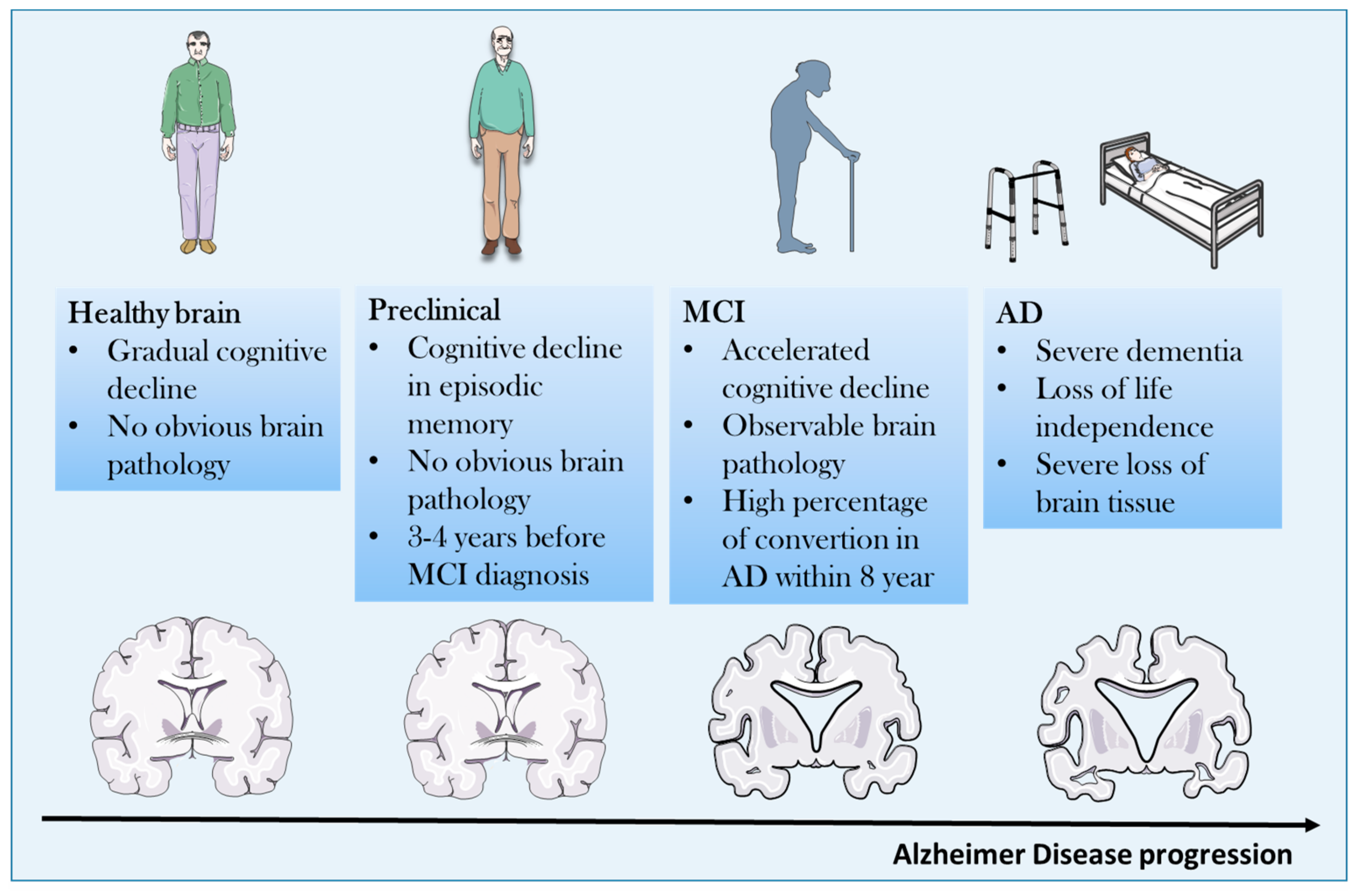
2. Pathogenetic Concepts

There are at least four major pathogenetic players in the development of AD [2]. Given the heterogeneity of the disease, each of them may contribute differently to the induction, onset, and progression of the disease in individual cases. These hypotheses include the amyloid cascade hypothesis, the tau hypothesis, the cholinergic hypothesis, and the excitotoxicity hypothesis. Although each of them has also been evaluated as a potential therapeutic target, the clinical outcomes have been disappointing [1] for several possible reasons. For example, intriguing proposals of the reasons beyond the failure of specific targeting of the amyloid hypothesis and the tau hypothesis in the clinical setting are elegantly presented in a recent review [6].
There are also other well promoting factors in the pathogenesis of AD including hyperinsulinemia and diabetes, as well as the occurrence of accompanying neuroinflammation [2]. It has also recently been proposed that a common pathogenetic pathway connecting diabetes, subchronic inflammation, and AD may rely on the activation of the common pathogenetic pathway consisting of PTN-MK-RPTPβ/ζ Axis [7]. Strengthening the causal link between deregulated glycaemic homeostasis, including insulin secretion and resistance and AD, is the recent identification of a newly indicated form of diabetes named “type 3” diabetes that is characterized as a brain-insulin resistant state linked to AD [8]. Although the precise cellular type that develops insulin resistance in the brain has not yet been identified, recent studies suggest that cortical neurons develop resistance to insulin after prolonged exposure to high insulin concentrations. This occurs through inhibition of phosphorylation of Akt, p70S6K, and GSK-3β [9]. As MIF inhibits Akt phosphorylation its high expressions during brain pathologies may exacerbate insulin resistance in neurons [10].
We have previously discussed that the Apoε4 (Apoε4) allele of the apolipoprotein (apo) gene, has also been identified as the primary genetic risk factor for AD [1][2].

3. Neuroinflammation and AD

That upregulated immunoinflammatory events may play an important pathogenetic role in the development of AD is supported by several preclinical and clinical observations and this area of research has gained much interest during the last decade.

3.1. Innate Immune System and AD

The better understanding of the functioning of the innate immune system, with the emerging role played from interaction of danger associated molecular pathway (DAMP) with pathogen recognition receptors (PRR) on cells of the innate immune system has allowed us to gain important insight into immune pathogenetic concepts of AD. It is proposed that during the development of AD misfolded and aggregated proteins might act as DAMP that binds pathogen PRR including class A scavenger receptor A1, CD36, CD14, α6β1 integrin, CD47, and toll like receptors expressed by microglial cells and astrocytes. This would in turn determine the release of inflammatory mediators such as NO and ROS, as well as proinflammatory cytokines, including TNF-α and IL-1β [11][12][13][14].

3.2. Proinflammaory Cytokines and AD

It has therefore been proposed that pro-inflammatory cytokines may contribute to AD pathogenesis through multiple pathways, including the induction of indoleamine 2,3-dioxygenase that increases the levels of the quinolinic acid, a neurotoxic factor, and in turn promotes tau hyperphosphorylation [11][12][13][14]. The role of anti-inflammatory cytokines, primarily belonging to the Th2 and Th3 cell subsets in the process of AD, is also worth mentioning as they might exert protective effects against AD by counteracting the effects of pro-inflammatory cytokines [11][12][13][14]. For example, transforming growth factor (TGF)-β that is a prototypical anti-inflammatory cytokine produced by Th3 cells is capable of ameliorating Aβ-induced cytotoxicity, in vivo and in vitro, and deficiency of TGF-β1 promotes both Aβ accumulation and NFTs formation [11][12][13][14]. This pathogenetic hypothesis is also consistent with the epidemiologic evidence indicating an affirmative influence of non-steroidal anti-inflammatory drugs (NSAIDs) on delaying the progression of AD and have suggested that a blockade of the ongoing inflammatory processes may delay the progression AD [1].
It is important to note that despite local recruitment of brain microglia to the sites of amyloid deposition, they ultimately fail in preventing the formation of β-amyloid plaques [11][12][13][14].
Several studies have reported that pro-inflammatory cytokines are augmented in patients with AD, and genetic polymorphisms for these cytokines have also been reported but often remain unconfirmed in patients with the same or different ethnicity [15].
Accordingly, though with some contradictory findings, prototypical pro-inflammatory cytokines of the innate immune system including IL-1β, TNF-α, IL-6, IL-12, and IL-23 have been found augmented in plaques and or CSF of AD patients and animal models of the disease. In transgenic mouse models of AD, inflammatory cytokines correlate with amyloid load [15].
In addition, an antibody directed against IL-12/IL23 ameliorates the course of the disease in a mouse model of AD [16].

3.3. Proinflammatory Cytokines as Biomarkers During AD Development and Progression

Recent studies have also shown that immune pro-inflammatory cytokines, such as macrophage migration inhibitory factor (MIF) and YKL-40, TNF receptors and sTREM2, are associated with tau pathology and brain aging, thus suggesting that these molecules may be useful diagnostic markers and therapeutic targets [17].
Another study showed that the CSF markers -IL-15, MCP-1, VEGFR-1, sICAM1, sVCAM-1, and VEGF-D- are independently associated with the CSF tau and p-tau181 levels [18]. Together with the observation of Bacher and colleagues [19] that MIF is augmented in AD, these data may suggest that measuring blood levels of MIF may represent a diagnostic biomarker that may be useful both for diagnosis and therapeutic monitoring of the disease, at least in a well-defined subset of patients that are characterized by larger production of MIF, and that may also be considered for tailored therapeutic approaches with specific MIF inhibitors. However, longitudinal or perspective studies on the possible use of MIF as diagnostic biomarkers are so far missing.
The pathogenetic relevance of neuroinflammation in AD models is highlighted from a systematic review that reports beneficial effects on the course of experimental rodent models of AD with several immunomodulatory agents including IL-1 receptor antagonist and TNF inhibitors [20].
As an important note to the neuroinflammatory hypothesis of AD, it is worth noting that the inability of plaque clearance by CNS phagocytes, along with upregulated inflammation, seems instrumental to disease progression. Accordingly, several genes associated with sporadic AD are involved in glial clearance of misfolded proteins. Also, external factors, i.e., systemic inflammation and obesity, promote disease development and evolution [21][22].
Simultaneously, and along this line of research, it is interesting to observe that peripheral phagocytes are able to effectively clear plaques and therapeutic strategies aiming at favoring recruitment of these cells into the CNS are actively being pursued. It has been suggested that Aβ immunotherapy could clear cerebral Aβ accumulations by activating phagocytes, and recent evidences generated in preclinical AD models propose that targeting the TGF-β-Smad 2/3 signaling is able to modulate blood-to-brain trafficking of these cells [23][24]. Also, the chemokine receptor Cx3cr1 pathway seems to control the chemotaxis of phagocytes toward AD affected neurons [23][24].

4. MIF: An Emerging Player in the Neuroinflammatory Hypothesis of AD

4.1. Biology, Physiology and Physiopathology of MIF

MIF is a pleiotropic cytokine produced by several cells of the innate and adaptive immune system, as well as non-immune cells including myocardial cells. MIF has been discovered at the end of the 60 and its name is due to its ability to inhibit the migration of macrophages. MIF has also attracted much attention because of its unique interaction with corticosteroids [25].
MIF has been shown to overcome the inhibitory effects of glucocorticoids on the production of pro-inflammatory cytokines by monocytes in vitro and to counteract steroid effects against lethal endotoxemia in vivo. MIF also counteracts glucocorticoid inhibition of T-cell in vitro by restoring the expression of IL-2 and interferon (IFN)-γ and may therefore play a major role in determining resistance to steroids. Through its ability to interact with steroids, MIF is also endowed with hormone-like properties and it has been shown to modulate the HPA axis [26].
Recent studies have highlighted the mode of action and signaling transmission of MIF, as well as its complex role played in regulation of immune responses.
MIF transduces its biological signals by binding to the CD74 receptor or the co-receptors, CXCR2, CXCR4, and CXCR7. In turn, a variety of signaling cascades, including the MAPK, PI3K/AKT, and NF-kB pathways are activated. By doing so, MIF activates pro-inflammatory events including the secretion of IL-6 and TNF-α and the activation of the inflammasome [27][28][29].
For this reason, MIF has been implicated in the pathogenesis of several autoimmune diseases including type 1 diabetes, multiple sclerosis, autoimmune hepatitis, and rheumatoid arthritis [30][31][32][33][34].
MIF may also contribute to immunoinflammatory events during development of Duchenne′s syndrome [35] and seems to play a pathogenetic role in major depressive disorders [36]. More recent studies also indicate that MIF is implicated in certain forms of cancer phenotypes such as melanoma, glioblastoma, prostate cancers, and neuroblastoma [37][38][39][40][41][42][43].
However, and in a manner similar to other cytokines with a primarily pro-inflammatory profile, MIF might also display anti-inflammatory activities that appear to be primarily mediated by its ability to activate AMPK and inhibit the JNK pathway. These effects may be taken into account by the capacity of MIF to reduce the development of an immunoinflammatory condition such as cardiac ischemia/reperfusion [44][45].

4.2. The Emerging Role of MIF Homologue, D-dopachrome Tautomerase (D-DT; MIF-2)

Adding interest to the role of MIF, a second structurally related MIF family member, D-DT, was recently characterized [46]. D-DT or MIF-2 was recognized to be a structural and functional homolog of MIF, which could exert overlapping effects, further raising the complexity of canonical MIF signaling pathways [46].
D-DT often, but not always, exerts synergistic and overlapping effects of MIF. Both MIF and D-DT play a key role in progressive forms of MS in male patients and D-DT has also been found to exert oncogenic effects [31][37].

5. MIF in Neurodegenerative Diseases

MIF in PD and ALS

The role of MIF in neurodegenerative diseases has also received much attention (Table 1). In agreement with its pleiotropic biological functions, emerging evidence indicates that MIF may play a complex role in neurodegenerative disorders with potential beneficial effects in Parkinson′s Diseases (PD) and amyotrophic lateral sclerosis (ALS).
Table 1. Evidences for the involvement of the Macrophage Migration Inhibitory Factor (MIF) in neurodegenerative disorders.

Disease

Preclinical Data

We will present here findings of experimental, human genetic, and clinical studies that have studied MIF in rodent models of AD and AD patients (Table 2), and will highlight emerging therapeutic opportunities for tailored modulation of the activity of MIF that may potentially be applied to AD patients. Dismantling the exact role of MIF and its receptors in AD may offer novel diagnostic and therapeutic opportunities in AD This focused review may also propel further interest on additional studies of the yet unexplored role of D-DT in AD.
Table 2. Evidences for the involvement of the Macrophage Migration Inhibitory Factor (MIF) in Alzheimer′s Disease (AD).

Preclinical Data

Human Data

References

Human Data

References

Parkinson′s Diseases (PD)

MIF reduces apoptosis and induces autophagy in an in vitro model of PD (SH-SY5Y cells exposed to MPP+)


[47]

ISO-1 reduces Aβ-induced toxicity in vitro


[19][53]

MIF is upregulated in mouse model of PD (induced by i.p. injection of MPTP)


[47]


↑ serum levels


↑ MIF in CNS of AD and MCI patients

[19][53]

[48

MIF colocalizes with Aβ plaques in APP23 transgenic mice

]


[19][53]

Amyotrophic Lateral Sclerosis (ALS)

MIF inhibits mutant SOD1 misfolding in motor neuron-like cells


↑ MIF in plasma of AD and MCI patients

[49][50]


[54]

Endogenous MIF knockdown in SOD1 mutant mice accelerates disease


[51][52]

MIF overexpression in the spinal cord improves ALS in SOD1 mutant mice


[51][52]

MPP+: 1-methyl-4-phenylpyridinium; MPTP: 1-Methyl-4-phenyl-1,2,3,6-tetrahydropyridin; i.p.: Intra-peritoneal; SOD1: superoxide Dismutase 1.
In particular, we have first shown that PD patients have elevated blood levels of MIF that do not correlate with severity of the disease [48]. On the basis of some beneficial effects observed with exogenously administered MIF in models of PD, we postulated that this increase might have reflected a compensatory attempt to counteract pathogenetic pathways in the course of the disease, and that endogenous MIF might have played a beneficial role in PD [48].

6. MIF and AD

In spite of these beneficial neuroprotective effects of MIF in PD and ALS, conflicting data have been generated as to whether MIF plays a harmful or beneficial role in AD. Until a few years ago, there was general agreement that MIF plays a key role as pathogenetic cytokine in AD pathogenesis and progression. Two recent papers have, however, questioned this view and propose that endogenous MIF may be beneficial in AD and that its augmented levels found in post mortem brains, CSF, and periphery of AD patients witness a compensatory attempt at counteracting insufficient biological function of MIF in the brain due to glycation, oxidation, and binding to plaques.

MIF deficiency attenuates tau hyperphosphorylation in astrocytes from APP/PS1 transgenic mice


[

55]

MIF overexpression prevents Aβ toxicity in SH-SY5Y cells


[56]

APP23/MIF+/− mice show significant memory impairment


[56]

References

  1. Briggs, R.; Kennelly, S.P.; O’Neill, D. Drug treatments in Alzheimer’s disease. Clin. Med. 2016, 16, 247–253.
  2. Adlimoghaddam, A.; Neuendorff, M.; Roy, B.; Albensi, B.C. A review of clinical treatment considerations of donepezil in severe Alzheimer′s disease. CNS Neurosci. Ther. 2018, 24, 876–888.
  3. Lane, C.A.; Hardy, J.; Schott, J.M. Alzheimer′s disease. Eur. J. Neurol. 2018, 25, 59–70.
  4. Dubois, B.; Hampel, H.; Feldman, H.H.; Scheltens, P.; Aisen, P.; Andrieu, S.; Bakardjian, H.; Benali, H.; Bertram, L.; Blennow, K.; et al. Preclinical Alzheimer’s disease: Definition, natural history, and diagnostic criteria. Alzheimer’s Dement. 2016, 12, 292–323.
  5. Melis, R.J.; Haaksma, M.L.; Muniz-Terrera, G. Understanding and predicting the longitudinal course of dementia. Curr. Opin. Psychiatry 2019, 32, 123–129.
  6. Sigurdsson, E.M. Alzheimer′s therapy development: A few points to consider. Prog. Mol. Biol. Transl. Sci. 2019, 168, 205–217.
  7. Herradon, G.; Ramos-Alvarez, M.P.; Gramage, E. Connecting Metainflammation and Neuroinflammation Through the PTN-MK-RPTPβ/ζ Axis: Relevance in Therapeutic Development. Front. Pharmacol. 2019, 10, 377.
  8. Shau, K. ‘Type 3′ diabetes: A brain insulin-resistant state linked to Alzheimer′s disease. Pr. Diabetes 2017, 34, 187–188.
  9. Kim, B.; Sullivan, K.A.; Backus, C.; Feldman, E.L. Cortical Neurons Develop Insulin Resistance and Blunted Akt Signaling: A Potential Mechanism Contributing to Enhanced Ischemic Injury in Diabetes. Antioxidants Redox Signal. 2011, 14, 1829–1839.
  10. Stošić-Grujičić, S.; Saksida, T.; Miljković, Đ.; Stojanović, I. MIF and insulin: Lifetime companions from common genesis to common pathogenesis. Cytokine 2020, 125, 154792.
  11. Heneka, M.T.; Carson, M.J.; El Khoury, J.; Landreth, G.E.; Brosseron, F.; Feinstein, U.L.; Jacobs, A.H.; Wyss-Coray, T.; Vitorica, J.; Ransohoff, R.M.; et al. Neuroinflammation in Alzheimer’s disease. Lancet Neurol. 2015, 14, 388–405.
  12. Webers, A.; Heneka, M.T.; Gleeson, P.A. The role of innate immune responses and neuroinflammation in amyloid accumulation and progression of Alzheimer’s disease. Immunol. Cell Boil. 2019, 12301.
  13. Minter, M.R.; Taylor, J.M.; Crack, P.J. The contribution of neuroinflammation to amyloid toxicity in Alzheimer′s disease. J. Neurochem. 2016, 136, 457–474.
  14. Su, F.; Bai, F.; Zhang, Z. Inflammatory Cytokines and Alzheimer’s Disease: A Review from the Perspective of Genetic Polymorphisms. Neurosci. Bull. 2016, 32, 469–480.
  15. Patel, N.S.; Paris, D.; Mathura, V.; Quadros, A.N.; Crawford, F.C.; Mullan, M.J. Inflammatory cytokine levels correlate with amyloid load in transgenic mouse models of Alzheimer′s disease. J. Neuroinflammation 2005, 2.
  16. Berg, J.V.; Prokop, S.; Miller, K.R.; Obst, J.; E Kälin, R.; Lopategui-Cabezas, I.; Wegner, A.; Mair, F.; Schipke, C.G.; Peters, O.; et al. Inhibition of IL-12/IL-23 signaling reduces Alzheimer’s disease–like pathology and cognitive decline. Nat. Med. 2012, 18, 1812–1819.
  17. Brosseron, F.; Kolbe, C.-C.; Santarelli, F.; Carvalho, S.; Antonell, A.; Castro-Gomez, S.; Tacik, P.; Namasivayam, A.A.; Mangone, G.; Schneider, R.; et al. AETIONOMY study group Multicenter Alzheimer′s and Parkinson′s disease immune biomarker verification study. Alzheimers. Dement. 2019.
  18. Popp, J.; Oikonomidi, A.; Tautvydaitė, D.; Dayon, L.; Bacher, M.; Migliavacca, E.; Henry, H.; Kirkland, R.; Severin, I.; Wojcik, J.; et al. Markers of neuroinflammation associated with Alzheimer’s disease pathology in older adults. Brain, Behav. Immun. 2017, 62, 203–211.
  19. Bacher, M.; Deuster, O.; Aljabari, B.; Egensperger, R.; Neff, F.; Jessen, F.; Popp, J.; Noelker, C.; Reese, J.P.; Al-Abed, Y.; et al. The Role of Macrophage Migration Inhibitory Factor in Alzheimer’s Disease. Mol. Med. 2010, 16, 116–121.
  20. Peplow, P.V.; Martinez, B. Amelioration of Alzheimer’s disease pathology and cognitive deficits by immunomodulatory agents in animal models of Alzheimer’s disease. Neural Regen. Res. 2019, 14, 1158–1176.
  21. McManus, R.M.; Heneka, M.T. Role of neuroinflammation in neurodegeneration: New insights. Alzheimer’s Res. Ther. 2017, 9, 14.
  22. Wang, M.-M.; Miao, D.; Cao, X.-P.; Tan, L.; Tan, L. Innate immune activation in Alzheimer’s disease. Ann. Transl. Med. 2018, 6, 177.
  23. Rezai-Zadeh, K.; Gate, D.; Gowing, G.; Town, T. How to get from here to there: Macrophage recruitment in Alzheimer’s disease. Curr. Alzheimer Res. 2011, 8, 156–163.
  24. Joaquín Merino, J.; Muñetón-Gómez, V.; Alvárez, M.-I.; Toledano-Díaz, A. Effects of CX3CR1 and Fractalkine Chemokines in Amyloid Beta Clearance and p-Tau Accumulation in Alzheimer’s Disease (AD) Rodent Models: Is Fractalkine a Systemic Biomarker for AD? Curr. Alzheimer Res. 2016, 13, 403–412.
  25. Fingerle-Rowson, G.; Koch, P.; Bikoff, R.; Lin, X.; Metz, C.N.; Dhabhar, F.S.; Meinhardt, A.; Bucala, R. Regulation of Macrophage Migration Inhibitory Factor Expression by Glucocorticoids in Vivo. Am. J. Pathol. 2003, 162, 47–56.
  26. Calandra, T.; Bucala, R. Macrophage Migration Inhibitory Factor (MIF): A Glucocorticoid Counter-Regulator within the Immune System. Crit. Rev. Immunol. 1997, 17, 77–88.
  27. Jankauskas, S.S.; Wong, D.W.; Bucala, R.; Djudjaj, S.; Boor, P. Evolving complexity of MIF signaling. Cell. Signal. 2019, 57, 76–88.
  28. Shin, M.S.; Kang, Y.; Wahl, E.R.; Park, H.-J.; Lazova, R.; Leng, L.; Mamula, M.; Krishnaswamy, S.; Bucala, R.; Kang, I. Macrophage Migration Inhibitory Factor Regulates U1 Small Nuclear RNP Immune Complex-Mediated Activation of the NLRP3 Inflammasome. Arthritis Rheumatol. 2019, 71, 109–120.
  29. Lang, T.; Lee, J.P.W.; Elgass, K.; Pinar, A.A.; Tate, M.D.; Aitken, E.H.; Fan, H.; Creed, S.J.; Deen, N.S.; Traore, D.A.K.; et al. Macrophage migration inhibitory factor is required for NLRP3 inflammasome activation. Nat. Commun. 2018, 9, 2223.
  30. Cavalli, E.; Mazzon, E.; Basile, M.S.; Mangano, K.; Marco, D.; Bramanti, P.; Nicoletti, F.; Fagone, P.; Petralia, M.C.; Di Marco, R. Upregulated Expression of Macrophage Migration Inhibitory Factor, Its Analogue D-Dopachrome Tautomerase, and the CD44 Receptor in Peripheral CD4 T Cells from Clinically Isolated Syndrome Patients with Rapid Conversion to Clinical Defined Multiple Sclerosis. Medicina 2019, 55, 667.
  31. Benedek, G.; Meza-Romero, R.; Jordan, K.; Zhang, Y.; Nguyen, H.; Kent, G.; Li, J.; Siu, E.; Frazer, J.; Piecychna, M.; et al. MIF and D-DT are potential disease severity modifiers in male MS subjects. Proc. Natl. Acad. Sci. USA 2017, 114, E8421–E8429.
  32. Fagone, P.; Mazzon, E.; Cavalli, E.; Bramanti, A.; Petralia, M.C.; Mangano, K.; Al-Abed, Y.; Bramati, P.; Nicoletti, F. Contribution of the macrophage migration inhibitory factor superfamily of cytokines in the pathogenesis of preclinical and human multiple sclerosis: In silico and in vivo evidences. J. Neuroimmunol. 2018, 322, 46–56.
  33. Bilsborrow, J.B.; Doherty, E.; Tilstam, P.V.; Bucala, R. Macrophage migration inhibitory factor (MIF) as a therapeutic target for rheumatoid arthritis and systemic lupus erythematosus. Expert Opin. Ther. Targets 2019, 23, 733–744.
  34. Günther, S.; Fagone, P.; Jalce, G.; Atanasov, A.G.; Guignabert, C.; Nicoletti, F. Role of MIF and D-DT in immune-inflammatory, autoimmune, and chronic respiratory diseases: From pathogenic factors to therapeutic targets. Drug Discov. Today 2019, 24, 428–439.
  35. Lombardo, S.D.; Mazzon, E.; Mangano, K.; Basile, M.S.; Cavalli, E.; Mammana, S.; Fagone, P.; Nicoletti, F.; Petralia, M.C. Transcriptomic Analysis Reveals Involvement of the Macrophage Migration Inhibitory Factor Gene Network in Duchenne Muscular Dystrophy. Genes 2019, 10, 939.
  36. Petralia, M.C.; Mazzon, E.; Fagone, P.; Basile, M.S.; Lenzo, V.; Quattropani, M.C.; Bendtzen, K.; Nicoletti, F. Pathogenic contribution of the Macrophage migration inhibitory factor family to major depressive disorder and emerging tailored therapeutic approaches. J. Affect. Disord. 2020, 263, 15–24.
  37. O’Reilly, C.; Doroudian, M.; Mawhinney, L.; Donnelly, S.C. Targeting MIF in Cancer: Therapeutic Strategies, Current Developments, and Future Opportunities. Med. Res. Rev. 2016, 36, 440–460.
  38. Penticuff, J.C.; Woolbright, B.L.; Sielecki, T.M.; Weir, S.J.; Taylor, J.A. MIF family proteins in genitourinary cancer: Tumorigenic roles and therapeutic potential. Nat. Rev. Urol. 2019, 16, 318–328.
  39. Soumoy, L.; Kindt, N.; Ghanem, G.; Saussez, S.; Journe, F. Role of Macrophage Migration Inhibitory Factor (MIF) in Melanoma. Cancers 2019, 11, 529.
  40. Lechien, J.R.; Nassri, A.; Kindt, N.; Brown, D.N.; Journe, F.; Saussez, S. Role of macrophage migration inhibitory factor in head and neck cancer and novel therapeutic targets: A systematic review. Head Neck 2017, 39, 2573–2584.
  41. Mangano, K.; Mazzon, E.; Basile, M.S.; Di Marco, R.; Bramanti, P.; Mammana, S.; Petralia, M.C.; Fagone, P.; Nicoletti, F. Pathogenic role for macrophage migration inhibitory factor in glioblastoma and its targeting with specific inhibitors as novel tailored therapeutic approach. Oncotarget 2018, 9, 17951–17970.
  42. Presti, M.; Mazzon, E.; Basile, M.S.; Petralia, M.C.; Bramanti, A.; Colletti, G.; Bramanti, P.; Nicoletti, F.; Fagone, P. Overexpression of macrophage migration inhibitory factor and functionally-related genes, D-DT, CD74, CD44, CXCR2 and CXCR4, in glioblastoma. Oncol. Lett. 2018, 16, 2881–2886.
  43. Cavalli, E.; Mazzon, E.; Mammana, S.; Basile, M.S.; Lombardo, S.D.; Mangano, K.; Bramanti, P.; Nicoletti, F.; Fagone, P.; Petralia, M.C. Overexpression of Macrophage Migration Inhibitory Factor and Its Homologue D-Dopachrome Tautomerase as Negative Prognostic Factor in Neuroblastoma. Brain Sci. 2019, 9, 284.
  44. Qi, D.; Hu, X.; Wu, X.; Merk, M.; Leng, L.; Bucala, R.; Young, L.H. Cardiac macrophage migration inhibitory factor inhibits JNK pathway activation and injury during ischemia/reperfusion. J. Clin. Investig. 2009, 119, 3807–3816.
  45. Tilstam, P.V.; Qi, D.; Leng, L.; Young, L.; Bucala, R. MIF family cytokines in cardiovascular diseases and prospects for precision-based therapeutics. Expert Opin. Ther. Targets 2017, 21, 671–683.
  46. Merk, M.; Mitchell, R.A.; Endres, S.; Bucala, R. D-dopachrome tautomerase (D-DT or MIF-2): Doubling the MIF cytokine family. Cytokine 2012, 59, 10–17.
  47. Li, S.; Nie, K.; Zhang, Q.; Guo, M.; Qiu, Y.; Li, Y.; Gao, Y.; Wang, L. Macrophage Migration Inhibitory Factor Mediates Neuroprotective Effects by Regulating Inflammation, Apoptosis and Autophagy in Parkinson’s Disease. Neuroscience 2019, 416, 50–62.
  48. Nicoletti, A.; Fagone, P.; Donzuso, G.; Mangano, K.; Dibilio, V.; Caponnetto, S.; Bendtzen, K.; Zappia, M.; Nicoletti, F. Parkinson’s disease is associated with increased serum levels of macrophage migration inhibitory factor. Cytokine 2011, 55, 165–167.
  49. Israelson, A.; Ditsworth, D.; Sun, S.; Song, S.; Liang, J.; Hruska-Plochan, M.; McAlonis-Downes, M.; Abu-Hamad, S.; Zoltsman, G.; Shani, T.; et al. Macrophage migration inhibitory factor as a chaperone inhibiting accumulation of misfolded SOD1. Neuron 2015, 86, 218–232.
  50. Shvil, N.; Banerjee, V.; Zoltsman, G.; Shani, T.; Kahn, J.; Abu-Hamad, S.; Papo, N.; Engel, S.; Bernhagen, J.; Israelson, A. MIF inhibits the formation and toxicity of misfolded SOD1 amyloid aggregates: Implications for familial ALS. Cell Death Dis. 2018, 9, 107.
  51. Leyton-Jaimes, M.F.; Benaim, C.; Abu-Hamad, S.; Kahn, J.; Guetta, A.; Bucala, R.; Israelson, A. Endogenous macrophage migration inhibitory factor reduces the accumulation and toxicity of misfolded SOD1 in a mouse model of ALS. Proc. Natl. Acad. Sci. USA 2016, 113, 10198–10203.
  52. Leyton-Jaimes, M.F.; Kahn, J.; Israelson, A. AAV2/9-mediated overexpression of MIF inhibits SOD1 misfolding, delays disease onset, and extends survival in mouse models of ALS. Proc. Natl. Acad. Sci. USA 2019, 116, 14755–14760.
  53. Oyama, R.; Yamamoto, H.; Titani, K. Glutamine synthetase, hemoglobin α-chain, and macrophage migration inhibitory factor binding to amyloid β-protein: Their identification in rat brain by a novel affinity chromatography and in Alzheimer’s disease brain by immunoprecipitation. Biochim. et Biophys. Acta (BBA)-Protein Struct. Mol. Enzym. 2000, 1479, 91–102.
  54. Lee, K.S.; Chung, J.H.; Lee, K.H.; Shin, M.-J.; Oh, B.H.; Hong, C.H. Bioplex analysis of plasma cytokines in Alzheimer’s disease and mild cognitive impairment. Immunol. Lett. 2008, 121, 105–109.
  55. Li, S.-Q.; Yü, Y.; Han, J.-Z.; Wang, D.; Liu, J.; Qian, F.; Fan, G.-H.; Bucala, R.; Ye, R.D. Deficiency of macrophage migration inhibitory factor attenuates tau hyperphosphorylation in mouse models of Alzheimer’s disease. J. Neuroinflammation 2015, 12, 177.
  56. Zhang, S.; Zhao, J.; Zhang, Y.; Zhang, Y.; Cai, F.; Wang, L.; Song, W. Upregulation of MIF as a defense mechanism and a biomarker of Alzheimer’s disease. Alzheimer’s Res. Ther. 2019, 11, 54.
More
Video Production Service