Liquid Biopsy-Based Biosensors: Comparison
Please note this is a comparison between Version 2 by Jessie Wu and Version 1 by Parvaneh Sardarabadi.

Globally, non-small cell lung cancer (NSCLC) is the leading cause of cancer deaths. The 5-year survival rate has remained at 16% for the past forty years despite advancements in chemotherapy and targeted therapies. Minimal residual disease (MRD) is described as the existence of either isolated tumour cells or circulating tumour cells in biological liquid of patients after removal of the primary tumour without any clinical signs of cancer. Recently, liquid biopsy has been promising as a non-invasive method of disease monitoring and treatment guidelines as an MRD marker. Liquid biopsy could be used to detect and assessment of earlier stages of NSCLC, post-treatment MRD, resistance to targeted therapies, immune checkpoint inhibitors (ICIs) and tumour mutational burden. MRD surveillance has been proposed as a potential marker for lung cancer relapse. Principally biosensors provide the quantitative analysis of various materials by converting biological functions into quantifiable signals. Biosensors are usually operated to detect antibodies, enzymes, DNA, RNA, EVs, and whole cells. Here we present a category of biosensors based on the signal transduction method for identifying biosensor-based biomarkers in liquid biopsy specimens to monitor lung cancer treatment

  • bio-sensor
  • liquid biopsy

1. Liquid Biopsy as an Alternative Technique for Tissue Biopsy

Liquid biopsy is considered as a new technique for detecting tumour cells and identifying any tumour-derived products, which contains cell-free nucleic acids, circulating tumour cells and extracellular vesicles, specifically exosomes (Figure 1).
Figure 1. Liquid biopsy in lung cancer. Liquid biopsy can be considered for plenty of clinical targets. Liquid biopsy biomarkers are categorised into three main groups: circulating free DNA (cfDNA), CTCs and EVs. cDNA can be used for detecting a wide variety of mutations, such as insertions, deletions and amplification. Capturing and identifying CTCs in whole blood are cooperative for checking proteins and RNA expression and analysing various DNA abnormalities.

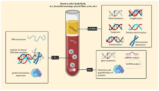
1.1. Circulating Cell-Free DNA (cfDNA) and Circulating Tumour DNA (ctDNA)

cfDNA (or ctDNA) monitoring is an optimistic technique for detecting MRD in tumours such as breast [61][1] and colon [62][2]. Natural cells release cfDNA into the biological fluids made by the ordinary activity of the cells, like apoptosis. Usually, the concentration of cfDNA is 5–10 ng/mL in body fluidics (with the range of 180–200 base pairs) [63][3]. However, the level of necrotic tumour cells (ctDNA) in cancerous tissue significantly increases [64][4]. cfDNA can present the heterogeneity of cancerous tissue regardless of any alteration during treatment or at different stages of cancer [65,66][5][6]. Preoperative ctDNA positivity in NSCLC advanced stage is a significant predictor of recurrence-free survival (RFS) and OS [63][3]. ctDNA identification after surgery increases the probability of early relapse detection and could play a critical role in stratifying patients for treatment [67][7]. Abbosh et al. demonstrated that phylogenetic ctDNA profiling tracks the subclonal nature of lung cancer relapse and metastasis. A unique procedure for ctDNA-driven therapeutic investigations proved a direct correlation between ctDNA concentration and computed tomography (CT) scan result of tumour mass [68][8].

1.2. Circulating Tumor Cells (CTCs)

CTCs or seeds of metastasis are isolated single migratory of cells or multi clusters of tumour cells which are measurable in the bloodstream of solid tumour cases. Similar to ctDNA, the lifetime of CTCs is 1~2.4 h, so instability of CTCs is a weakness of using this biomarker for investigation. However, the clinical application of CTCs has FDA approval (CellSearch® platform) [69][9]. CTCs are analysed by quite common techniques like qPCR, NGS, cytogenetic analyses like fluorescence in situ hybridisation (FISH). Moreover, several methods are considered for isolation and detection of CTCs, such as antigen isolation based (EpCAM) on the surface of the CTCs, size-based and circulating tumour cell deformation. In personalised medicine, analyses of CTCs have the potential for monitoring patients treated with immunotherapy and chemotherapy and may become a new biomarker for early prognosis and MRD in NSCLC. Epithelial CTCs in the bloodstream of NSCLC cases are rarely discovered and aligned with a poor prognosis to checkpoint inhibitors [70][10]. Some studies demonstrated a similarity of approximately 93% between expression of PD-L1 on CTCs and PD-L1 expression in matched-patient tumours [71][11]. Patients with CTCs PD-L1+ had a shorter survival time than patients with CTCs PD-L1 [72][12]. A day after surgery, CTCs numbers decrease to the lowest level, except for patients with confirmed recurrence, which increases after three days. These findings indicate that CTCs as a biomarker of biopsy fluid could be useful for monitoring MRD and responding to immunotherapy in early stage lung cancer after surgery [73][13].

1.3. Extracellular Vesicles (EVs)

EVs are bilayer biological vesicles found in extracellular fluids, including blood, urine and CSF. EVs are very complex in size, origin, content and function. In this regard, they are divided into different fractions and populations. The main classification of EVs could be divided into three main fractions based primarily on vesicle size (in diameter) and origin. These populations of EVs are exosomes, microvesicles (MVs) and apoptotic bodies. Exosomes are the smallest fraction or population among EVs. In contrast, MVs and apoptotic bodies are large in size and different in origin. Briefly, exosomes (30–150 nm in diameter) are originated from the multi vesicular endosomes (MVE) through exocytosis. MVs (150–1000 nm in diameter) are originated from cell cytoplasmic membrane through direct shedding of vesicles. The apoptotic bodies with heterogenous-sized vesicles (more than 1000 nm in diameter) are the cellular debris resulted from an apoptotic pathway. Besides the other vesicles’ populations, exosomes are more noticeable. Extensive research has been carried out for their biological and physiological functions in the human body, especially in specific conditions like cancers [74][14]. The vast potentials of exosomes and biological roles made them a new platform for different applications in biomedical sciences. Exosomes have a complex molecular content in their lumen, including RNAs, proteins, carbohydrates, lipids and metabolites. These exosomal contents belong to the cell that produces and secretes the exosomes. In this regard, exosomes harbour their parent-cell molecules that are different among various cells-secreted exosomes. The latter is the unique characteristic of exosomes (and MVs) that made them a source for biomarker discovery.
In the recent two decades, hundreds of biomarkers, so-called exosomal biomarkers, have been discovered and tested based on their sensitivity and specificity. These exosomal biomarkers are mRNAs, lncRNAs, miRNAs, proteins, carbohydrates and lipids. Due to the exosome’s vesicular nature, they are an excellent source for biomarker analysis versus non-exosomal biomarkers. Furthermore, exosomal biomarkers are resistant to harsh conditions and enzymatic digestion. So, they could be easily detected and analysed in different ways, including biosensors-based technology. However, technical aspects of exosomes purification and enrichment are challengeable from this point of view. Ultracentrifuge as a popular technique for exosome separation needs specialisation along with costly equipment. Low-cost techniques such as ELISA, Western blotting and flow cytometry require high concentrations of purified exosomes. As a consequence, purification, the isolation and detection of biomarkers on exosomes require innovative approaches.

2. Type of Biosensors for Detection of Lung Cancer Biomarkers in Liquid Biopsy

Biosensors, in the context of liquid biopsy MRD, based on bioreceptors, are classified into antibody, aptamer, DNA, enzyme, whole-cell and phage biosensors as well as based on the transducers are organised into electrochemical biosensors, electro-kinetic biosensors, optical biosensors and magnetic biosensors (Table 1). However, due to their remarkable properties, such as a biosensor’s limit of detection (LoD), wide linear dynamic range and excellent repeatability, they have gained popularity in recent years.
Table 1. Comparison of different biosensors used to diagnose MRD liquid biopsy.
Transduce Principle Advantages Disadvantages Application in Lung Cancer MRD
Electrochemical sensor Convert the biochemical interaction to electrical signals
-
User-friendly
-
Portability
-
Cost-effective
-
Easily miniaturised
-
Rapid
-
High-specificity
-
High-reproducibility
-
Low detection limit
-
Need a small sample volume
-
Label-free
-
Real-time detection (piezoelectric)
-
Can be performed in turbid samples
-
Low sensitivity
-
False result from nonspecific bonding
-
Low shelf life
-
Cells or biomarkers are no longer intact.
-
Sensitive to temperature or environment change implications
-
Easily miniaturised, making them ideal for personalised medicine in the MRD detection context.
-
Electrochemical aptasensors can be reused; therefore, a biosensor can be used for serial monitoring at different times. Aptasensors have lower sensitivity than other electrochemical methods.
-
Capable of detecting ctDNA at extremely low quantities.
-
Able to detect specific ctDNA mutations in untreated serum or blood
-
Sequencing results are provided faster than standard PCR and DNA techniques.
Magnetic biosensor Applying paramagnetic particles to detect biological interactions by monitoring magnetic property changes
-
Low background noise
-
Detection of multiple biomarkers at the same time
-
High sensitivity
-
Can be performed in turbid samples
-
Stability
-
No change in the nature of magnetics in response to chemical reagents
-
Costly
-
Need to multiple washing steps
-
Time-consuming
-
For quantitative MRD testing, magnetic nanoparticles used with other bio-sensing platforms such as LFIA
-
Magnetic tags apply to the isolation and characterisation of EVs and CTC, so no sample preparation is required in an integrated biosensor.
-
Application as an immunoassay-based liquid biopsy or DNA-based liquid biopsy or POC devices
-
NMR signal detection needs only a few sample purifications steps, making it ideal for developing liquid biopsy for low-quality fluid samples or those with excessive protein contamination.
Surface-enhanced Raman spectroscopy (SERS) Using molecules adsorbing on rough metal surfaces to generate Raman scattering
-
High spatial resolution
-
Non-invasive label-free
-
High sensitivity
-
Multiple detections
-
Quantification
-
Unique spectroscopic fingerprint
-
Low background noise
-
Complex structure
-
Requires a nanoprobe
-
-Time-consuming
-
Expensive equipment to read-out
-
Requires a trained technician
-
Non-stability in long-term storage
-
SERS tags have extraordinary multiplexing capacity.
-
Real-time monitoring of therapy response
Surface plasmon resonance biosensors (SPR) Alteration of SPR angle due to increasing of refractive index during binding of biomolecules on the sensor surface
-
High sensitivity
-
Cost effectivity
-
Real-time detection
-
Reliable
-
High sensitivity
-
Enzyme-free
-
Label-free
-
Stability
-
Complicated optics
-
Costly
-
Low sensitivity
-
Monitoring of immune checkpoint inhibitors (ICI)
Fluorescence-based biosensing An analytical signal of a photoluminescence emission mechanism
-
High photostability (quantum dots)
-
High sensitivity
-
Imagining technique
-
Requires a trained technician
-
Background fluorescence noise
-
Sample losses during the labelling and purification process
-
Costly
-
Monitoring of ICIs such as PDL1
-
QDs are used as energy donors in FRET for the CTC identification as a prognostic factor
-
Due to high sensitivity, reliability and reproducibility, fluorescence techniques are favourable for detecting MRD biomarkers than other optical methods.
Lateral flow immunoassay (LFIA) Optical properties
-
Rapid
-
Portable
-
User-friendly
-
Cost-effective
-
Additional processing or external equipment is not required
-
No need for skilled personnel
-
Long shelf life
-
No need for additional equipment
-
Qualitative or semi-quantitative read-out
-
Low sensitivity
-
LFIA test strips are handy for detecting EVs, especially when a rapid procedure is needed.
-
They have the potential to become a quantitative test.
-
For quantitative MRD testing, gold or latex nanoparticles are replaced with magnetic particles or quantum dots (QDots-based lateral flow test strip).
Chemiluminescence Electrochemistry and visual luminescence measurements
-
Low background noise
-
Simple instrumentation
-
High sensitivity
-
Broad dynamic range
-
No need for the external light source
-
Background noise
-
Reagent low stability
-
Time-consuming
-
Sample vanishing during the labelling and purification process
-
A shorter DNA region is required. Compared to other routine methods such as PCR-based tests, DNA with a short length (~18 nucleotides) is detectable.
Currently, biosensors based on nanomaterials (nanobiosensors) are widely established in the development of biosensors due to their great sensitivity. Biosensors employ a variety of nanomaterials, including gold nanoparticles (AuNPs), metal oxides (MOs), nanowires (NWs), nanorods (NRs), carbon nanotubes-based biosensors (CNTs), quantum dot (QDs), graphene and its derivatives and nanocomposites (dendrimers) with the range size of 1 to 100 nm [75,76,77][15][16][17]. The popularity of gold nanoparticles (AuNPs) is because of the fast response and boost of analyte’s uncovering signals even in limited concentration [78][18]. Immobilisation of different biomolecules on the surface of carbon nanotubes (CNTs) makes them applicable; scaffolds having the ability to combine with a transducer signal. Layering structure of GR sheet makes two divisions of CNTs, single-walled carbon nanotubes (SWCNTs) and multi-walled carbon nanotubes (MWCNTs) which are practical for different applications [79][19].

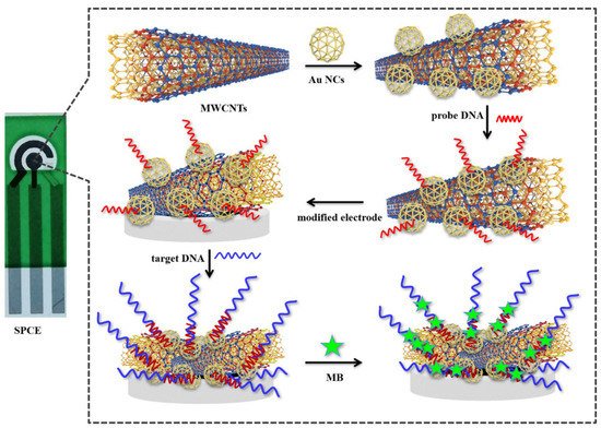
2.1. Electrochemical Sensor

Cells and DNA/RNA as a target of liquid biopsy were sensed with label-free electrical biosensors. To enhance signal detection, lncRNA biomarker MALAT1 has been assayed by a DNA-based electrochemical biosensor as a target for lung cancer monitoring (Figure 2). The biosensor was developed based on a gold nanocage coupled with an amidated multi-walled carbon nanotube (Au NCs/MWCNT-NH2)-decorated screen-printed carbon electrode (SPCE). The result demonstrated that the LoD of the biosensor is 42.8 fM with a wide linear range of 10−7–10−14 M [40][20]. The existence of cytokeratin 19 fragment (CYFRA 21-1) in serum is considerably matched up with NSCLC and may be able to predict tumour relapse [80][21].
Figure 2. Schematic representation of the SPCE electrochemical DNA biosensor to detect lncRNA biomarker MALAT1. Reprinted from ref. [40][20].
Sandwich-type immunosensor, employing immunosensor amino-functionalised carbon nanotube (MWCNT-NH2) within high efficiency in electrical conductivity and electron transfer and suitable biocompatibility, makes this nanotube an agreeable detector for abnormal expression of CYFRA21-1 in serum [81][22]. Assessment of CYFRA 21-1 has been identified as a potential prognostic factor in NSCLC. Studies showed that serial measurement of CYFRA 21-1 in patients with the advanced NSCLC-undergoing treatment and the impact of immune-checkpoint inhibitors (ICPIs) could be used to assess the outcome of early treatment without the need for frequent and expensive imaging procedures. It is a valuable marker to detect recurrence during monitoring after surgical resection [82][23]. Yoon et al. established the functionalised graphene oxide nanosheets chip for isolation of CTCs from early stage lung cancer patients’ moreover adjustable for advanced metastatic cancer patients. Graphene oxide nanosheets modified by phospholipid–polyethylene–glycol-amine (PL–PEG–NH2) and tetrabutylammonium (TBA) hydroxide were adsorbed onto the patterned gold surface and then chemically conjugated with NeutrAvidinis, which is the connection between crosslinker on graphene oxide nanosheets and EpCAM antibodies [83][24].
The electrochemical cyto-sensors (PDA NPs nanocomposites interface on the functionalised PGE) with high biocompatibility and sensitivity have been designed by Bolat et al. for label-free electrochemical detection of A-549 lung cancer cells in a wide linear range (1.0 × 10²–1.0 × 10⁵ cells mL−1) with a low detection limit (25 cells mL−1). In this regard utilising electrochemical cyto-sensors for bio-detection of cancer cells in liquid biopsy makes them a fast and straightforward sensor for bio-recognition elements to investigate the capture and detection of lung cancer cells in early stages [84][25]. Fusion of a self-assembled monolayer (SAM) of AuNP and determined aptamer on gold microelectrodes for target cells trapping fabricated a biosensor with high quantification stability operable for early lung cancer detection. The S–Au bonds between thiol-terminated aptamers and AuNP layers showed considerable selectivity assay for A549 lung CTCs cells capturing in the whole blood samples [85][26].
Commercialisation and mass production of user-friendly biosensors for liquid biopsy has been expanded. Electric field-induced release and measurement (EFIRM) liquid biopsy can identify ctDNA in patients with early stage NSCLC. An electrochemical control system with 96 parallel channels drives the EFIRM platform. This electrochemical control system is linked to a gold electrode array; the size of a standard microtiter plate. In an effort, EFIRM biosensors, which are effective, accurate, fast, user-friendly and inexpensive, are developed for non-invasive saliva-based EGFR gene mutation detection [86][27]. The EFIRM-based method was also used in the detection of the prognostic biomarker in liquid biopsy such as ultra-short circulating tumour DNA (usctDNA) in plasma and saliva [87][28] and p.L858R EGFR, exon19 del EGFR, p.E545K PIK3CA ctDNA in plasma [88][29] and exosomes [43][30]. The saliva-based EFIRM platform shows good concordance to plasma-based platforms (ddPCR and NGS) for longitudinally monitoring the combination of EGFR and PIK3CA ctDNA and may be a valuable platform for liquid biopsy MRD in NSCLC patients. EZLife Bio has developed a device based on the EFIRM technology that takes only 3 h to run EFIRM assays, which is substantially faster than NGS-based liquid biopsy methods. In many investigations, the pH value of the intracellular fluid environment of cancer is ≤7.1 [89][31]. The correlation between pH and cancer can be indicated as a biochemical marker for diagnosing and monitoring NCSLC cancer. Detection of CTCs in the blood either with deterministic lateral displacement (DLD) or without DLD devices by using the potential difference between the reference electrode (Ag/AgCl) and indicator electrode (ZnO) in a microfluidics-based pH sensor is reported by Ganesh Kumar Mani et al. [90][32].

2.2. Electro-Kinetic Biosensors

The pressure-driven fluid flow transport by advection enforces both a current and a potential at the charged micro-and nanochannel ends. This leads to an electrokinetic phenomenon that can be used as a sensor/transducer principle. The generated electricity is well-regulated as a sensor power supply, so this effect is useable as a passive sensor/transducer principle. In this way, the adsorption of biological material like biomarkers and physical changes like pressure/flow or pH value is detectable [91][33]. Recently, a multiplexed electro-kinetic sensor was used to monitor several small extracellular vesicles (sEVs) and tumour cell surface markers in sEVs isolated from pleural effusion (PE) fluid of NSCLC patients [92][34]. The detection method is based on the electro-kinetic principle, where the change in the streaming current is monitored as the surface markers of the sEVs interact with the affinity reagents immobilised on the inner surface of a silica microcapillary. This study obtained samples from patients with diverse genetic drivers of their cancers, including EGFR, ALK and KRAS. They were also given different treatment regimens, including PD-L1 immune checkpoint inhibitors, EGFR- and ALK-TKI. In NSCLC patients treated with EGFR-TKIs, PD-L1 expression is a poor prognostic factor. The biosensor measured the PD-L1 expression in sEVs. It showed that an electro-kinetic biosensor could detect various and simultaneous quantities of low concentration biomarkers in pleural fluid and predict or monitor therapy response. The electro-kinetic results were consistent with those obtained using conventional methods such as Western blot (WB) and proximity extension assay (PEA). Although the outcomes of this biosensor seemed acceptable, it is possible to enhance the signal of proteins by modifying the size and charge of the particles in the electro-kinetic sensor [93][35]. A microfluidic cathodic photoelectrochemical immunosensor for measuring CYFRA 21-1 has been developed. The process is based on the p–n junction of AgI/Bi2Ga4O9, with dissolved O2 acting as an electron acceptor to create the superoxide anion radical (O2) as the working microelectrode. When combined with a novel SOD@hMnO2 as the co-catalyst signal amplification label, O2 can be catalysed by SOD via a disproportionation reaction to produce O2 and H2O2; then, hMnO2 can trigger the decomposition of H2O2 to produce O2 and H2O2. The linearity of the microfluidic cathodic PEC biosensor chip ranged from 0.1 pg mL−1 to 100 ng mL−1, with a detection limit of 0.026 pg mL−1 [46][36].

2.3. Magnetic Biosensor

Whenever the magnetic field is enforced to the sensor, the resistance of giant magnetoresistance (GMR) sensor changes; as a result, a biomolecule which is labelled with magnets, generates a signal. High sensitivity and rapid response induced by a low magnetic field are the superiority of GMR material for biosensors. Furthermore, the stability of magnetic probes for labelling compared to other labels like fluorescent tags for long-time usage, external manageability, and no background noise are some advantages of magnetic biosensors [94][37]. For signal amplification, magnetic particles were employed in electrochemical biosensors [30][38].
Nair and colleagues measured the tissue inhibitor of metalloproteinase-1 (TIMP1) biomarker with the help of the immunoassay GMR sensor [95][39]. In this study, plasma TIMP1 levels and relevant clinical and imaging characteristics were adequate for diagnosing lung cancer in smokers. In a 2011 research, Pesta and colleagues demonstrated the prognostic effect of TIMP1 mRNA. Higher TIMP1 mRNA levels are associated with an adverse prognosis of patients [96][40]. The expression of TIMP-1 may serve as a prognostic biomarker as well as a therapeutic target in NSCLC patients [97][41]. It is assumed that TIMP-1 is a possible tissue biomarker for lymph invasion and distant metastasis of lung adenocarcinoma by promoting cancer metastasis [98][42].

2.4. Optical Biosensors

Optical biosensors are a compressed analytical device that takes advantage of the principle of optical measurements (absorbance, fluorescence, chemiluminescence and others). The yields of optical biosensors are signals which present the concentration of the analyte. Direct, real-time and label-free detection of substances are some of the optical biosensors’ benefits, along with high specificity and sensitivity. Generally, reference sensors are not required during the measurement since the comparison signal can be generated with the same light source as the scanning sensor [99][43].

2.4.1. Surface Enhanced Raman Spectroscopy (SERS)

SERS-based biosensors are divided into direct (label-free) detection and indirect detection (needing SERS tags). Label-free presents molecular fingerprint information of biomolecules. The drawback of label-free detection is the complexity and even weakness of signals from some substances; also, they are prone to signals from interfering molecules in the matrix. In indirect SERS sensing, unique SERS signals (SERS tags) are labelled on the intensifying substrates. SERS read-out is recognised after bounding targets and ligands or the reaction of SERS tags to environmental properties [100][44].
Exosomal PD-L1 in the blood is a good predictor of clinical response to anti-PD-1/PD-L1 therapy. Pang and colleagues applied the Anti-PD-L1 antibody modified Au@Ag@MBA (as the SERS tag) for exosomal PD-L1 labelling and quantification [101][45]. One of the most intriguing aspects of this biosensor was separating exosomes based on Fe3O4@TiO2 nanoparticles without antibodies or aptamers, which can enrich and separate exosomes from solution in 5 min with a 96.5 percent capture efficiency. In addition, TiO2 can specifically bind with the phosphate groups on the lipid bilayer of exosomes. This investigation showed that SERS-based biosensors could be utilised to monitor immunotherapy in the blood [102][46].
In another study, an ultrasensitive sensor based on an Ag nanorod array SERS substrate by assembling special hairpin-shaped molecular beacons (MBs) was developed for the detection of multiple lung cancer-related miRNA biomarkers [103][47]. Molecular beacons a hairpin-shaped 25 nucleotides DNA or RNA molecules with a fluorophore and a quencher. They do not fluoresce in the absence of their target complementary molecules. Instead, a spontaneous configuration change on MBs occurs due to hybridisation with their particular targets, and the dye and quencher separate from each other, resulting in fluorescence emission.

2.4.2. Surface Plasmon Resonance Biosensors (SPR)

Different interactions simultaneously with high throughput and sensitivity are characteristics of plasmon surface resonance imaging (SPRi). Spatially label-free optical detection and analysis of bio interaction in multiplex format make this technique an excellent detector for identifying many parameters simultaneously. The SPR technique could be used to identify the characteristics of NSCLC-related exosomes. Fan et al. recently developed a novel SPRi-based biosensing technique for detecting NSCLC-derived exosomes via multiple recognition sites with LoD of 104 particles/μL. The number of plasma exosomes identified with Anti CD63 differed across patients in a study of four healthy samples, four treated samples and four patients. Exosome levels were lower in healthy participants (approximately less than 10 RU) and higher in patients (about 25 RU). For patients undergoing therapy, intermediate values of the two groups were recorded. These findings suggest that immune-base sensing assay can be employed to assess treatment effectiveness by assessing the abundance of exosomes in plasma [50][48].
Some biosensors detect cargo within exosomes, such as circulating tumour RNAs (ctRNAs). Recourse to exosomes in blood and urine samples for early identification and monitoring treatment for various cancers such as prostate [104][49] and AML [105][50] has been reported.
Detection of MRD biomarkers in leukaemia has revealed that using several markers improves the accuracy and reliability of the MRD approach considerably [106][51]. As a result, seemingly using multiplex approaches in the diagnosis of MRD is desirable. Exosomal miRNAs have been proven to be useful as promising diagnostic biomarkers in patients with NSCLC. Since their remarkable stability and abundance, exosomes are better adapted for liquid biopsy than CTCs and cfDNA.
In the light of this, exosomal miRNAs have been considered as possible biomarkers for recurrent lung cancer [107][52]. However, information is scarce in this field, and specific conflicts must be resolved [108][53]. An SPRi-based biosensor was designed to investigate the simultaneous detection of multiplex 24 NSCLC-associated exosomal miRNAs in a clinical sample employing Au-on-Ag heterostructure and DNA tetrahedral framework (DTF) [49][54]. This biosensor has a detection range of 2 fM to 20 nM and a detection limit of 1.68 fM. In addition, DTF immobilisation on the gold array chip minimises nonspecific adsorption, resulting in fewer false-positive results due to interference factors in the input sample.
Liu et al. developed a highly sensitive, compact SPR biosensor to capture exosomes and characterise exosomal proteins. At concentrations as low as 2 × 10¹⁰ exosomes/mL, a compact SPR biosensor successfully detected EGFR expression levels in A549 exosomes, two times higher than the ELISA method (4 × 10¹⁰ exosomes/mL). Furthermore, there were no significant differences in exosomal EGFR expression in the clinical phase between the serum sample of the healthy and cancer patients [52][55], so this must be considered.

References

  1. Cirmena, G.; Garuti, A.; De Mariano, M.; Coco, S.; Ferrando, L.; Isnaldi, E.; Barbero, V.; Fregatti, P.; Del Mastro, L.; Ferrando, F.; et al. Circulating Tumor DNA Using Tagged Targeted Deep Sequencing to Assess Minimal Residual Disease in Breast Cancer Patients Undergoing Neoadjuvant Chemotherapy. J. Oncol. 2020, 2020, 8132507.
  2. Dizdar, L.; Fluegen, G.; van Dalum, G.; Honisch, E.; Neves, R.P.; Niederacher, D.; Neubauer, H.; Fehm, T.; Rehders, A.; Krieg, A.; et al. Detection of Circulating Tumor Cells in Colorectal Cancer Patients Using the GILUPI CellCollector: Results from a Prospective, Single-Center Study. Mol. Oncol. 2019, 13, 1548–1558.
  3. Chen, E.; Cario, C.L.; Leong, L.; Lopez, K.; Márquez, C.P.; Chu, C.; Li, P.S.; Oropeza, E.; Tenggara, I.; Cowan, J.; et al. Cell-free DNA Concentration and Fragment Size as a Biomarker for Prostate Cancer. Sci. Rep. 2021, 11, 5040.
  4. Cheng, F.; Su, L.; Qian, C. Circulating Tumor DNA: A Promising Biomarker in the Liquid Biopsy of Cancer. Oncotarget 2015, 7, 48832–48841.
  5. Russano, M.; Napolitano, A.; Ribelli, G.; Iuliani, M.; Simonetti, S.; Citarella, F.; Pantano, F.; Dell’Aquila, E.; Anesi, C.; Silvestris, N.; et al. Liquid Biopsy and Tumor Heterogeneity in Metastatic Solid Tumors: The Potentiality of Blood Samples. J. Exp. Clin. Cancer Res. 2020, 39, 95.
  6. Cucchiara, F.; Del Re, M.; Valleggi, S.; Romei, C.; Petrini, I.; Lucchesi, M.; Crucitta, S.; Rofi, E.; De Liperi, A.; Chella, A.; et al. Integrating Liquid Biopsy and Radiomics to Monitor Clonal Heterogeneity of EGFR-Positive Non-Small Cell Lung Cancer. Front. Oncol. 2020, 10, 2664.
  7. Peng, M.; Huang, Q.; Yin, W.; Tan, S.; Chen, C.; Liu, W.; Tang, J.; Wang, X.; Zhang, B.; Zou, M.; et al. Circulating Tumor DNA as a Prognostic Biomarker in Localized Non-small Cell Lung Cancer. Front. Oncol. 2020, 10, 561598.
  8. Abbosh, C.; Birkbak, N.J.; Wilson, G.A.; Jamal-Hanjani, M.; Constantin, T.; Salari, R.; Le Quesne, J.; Moore, D.A.; Veeriah, S.; Rosenthal, R.; et al. Phylogenetic Ctdna Analysis Depicts Early-Stage Lung Cancer Evolution. Nature 2017, 545, 446–451.
  9. Chen, L.; Bode, A.M.; Dong, Z. Circulating Tumor Cells: Moving Biological Insights into Detection. Theranostics 2017, 7, 2606–2619.
  10. Tamminga, M.; de Wit, S.; Hiltermann, T.J.N.; Timens, W.; Schuuring, E.; Terstappen, L.; Groen, H.J. Circulating Tumor Cells in Advanced Non-Small Cell Lung Cancer Patients are Associated with Worse Tumor Response to Checkpoint Inhibitors. J. Immunother. 2019, 7, 173.
  11. Cheng, Y.; Wang, T.; Lv, X.; Li, R.; Yuan, L.; Shen, J.; Li, Y.; Yan, T.; Liu, B.; Wang, L. Detection of PD-L1 Expression and Its Clinical Significance in Circulating Tumor Cells from Patients with Non-Small-Cell Lung Cancer. Cancer Manag. Res. 2020, 12, 2069–2078.
  12. Kong, D.; Zhang, W.; Yang, Z.; Li, G.; Cheng, S.; Zhang, K.; Feng, L. Correlation between PD-L1 Expression ON CTCs and Prognosis of Patients with Cancer: A Systematic Review and Meta-Analysis. OncoImmunology 2021, 10, 1938476.
  13. Wu, C.-Y.; Lee, C.-L.; Fu, J.-Y.; Yang, C.-T.; Wen, C.-T.; Liu, Y.-H.; Liu, H.-P.; Hsieh, J.C.-H.; Wu, C.-F. Circulating Tumor Cells as a Tool of Minimal Residual Disease Can Predict Lung Cancer Recurrence: A Longitudinal, Prospective Trial. Diagnostics 2020, 10, 144.
  14. Li, M.-Y.; Liu, L.-Z.; Dong, M. Progress on Pivotal Role and Application of Exosome in Lung Cancer Carcinogenesis, Diagnosis, Therapy and Prognosis. Mol. Cancer 2021, 20, 22.
  15. Su, H.; Li, S.; Jin, Y.; Xian, Z.; Yang, D.; Zhou, W.; Mangaran, F.; Leung, F.; Sithamparanathan, G.; Kerman, K. Nanomaterial-Based Biosensors for Biological Detections. Adv. Heal. Care Technol. 2017, 3, 19–29.
  16. Aykaç, A.; Gergeroglu, H.; Beşli, B.; Akkaş, E.; Yavaş, A.; Güler, S.; Güneş, F.; Erol, M. An Overview on Recent Progress of Metal Oxide/Graphene/CNTs-Based Nanobiosensors. Nanoscale Res. Lett. 2021, 16, 65.
  17. Naresh, V.; Lee, N. A Review on Biosensors and Recent Development of Nanostructured Materials-Enabled Biosensors. Sensors 2021, 21, 1109.
  18. Yeh, Y.-C.; Creran, B.; Rotello, V.M. Gold nanoparticles: Preparation, Properties, and Applications in Bionanotechnology. Nanoscale 2011, 4, 1871–1880.
  19. Tîlmaciu, C.-M.; Morris, M.C. Carbon Nanotube Biosensors. Front. Chem. 2015, 3, 59.
  20. Chen, M.; Wu, D.; Tu, S.; Yang, C.; Chen, D.; Xu, Y. A Novel Biosensor for the Ultrasensitive Detection of the Lncrna Biomarker MALAT1 in Non-Small Cell Lung Cancer. Sci. Rep. 2021, 11, 3666.
  21. Muley, T.; He, Y.; Rolny, V.; Wehnl, B.; Escherich, A.; Warth, A.; Stolp, C.; Schneider, M.A.; Meister, M.; Herth, F.J.; et al. Potential for the Blood-Based Biomarkers Cytokeratin 19 Fragment (CYFRA 21-1) and Human Epididymal Protein 4 (HE4) to Detect Recurrence during Monitoring after Surgical Resection of Adenocarcinoma of the Lung. Lung Cancer 2019, 130, 194–200.
  22. Yola, M.L.; Atar, N.; Özcan, N. A Novel Electrochemical Lung Cancer Biomarker Cytokeratin 19 Fragment Antigen 21-1 Immunosensor Based on Si3N4/Mos2 Incorporated Mwcnts and Core–Shell Type Magnetic Nanoparticles. Nanoscale 2021, 13, 4660–4669.
  23. Dall’Olio, F.G.; Abbati, F.; Facchinetti, F.; Massucci, M.; Melotti, B.; Squadrilli, A.; Buti, S.; Formica, F.; Tiseo, M.; Ardizzoni, A. CEA and CYFRA 21-1 as Prognostic Biomarker and as a Tool for Treatment Monitoring in Advanced NSCLC Treated with Immune Checkpoint Inhibitors. Adv. Med. Oncol. 2020, 12, 1–13.
  24. Yoon, H.J.; Kim, T.H.; Zhang, Z.; Azizi, E.; Pham, T.M.; Paoletti, C.; Lin, J.; Ramnath, N.; Wicha, M.S.; Hayes, D.F.; et al. Sensitive Capture of Circulating Tumour Cells by Functionalized Graphene Oxide Nanosheets. Nat. Nanotechnol. 2013, 8, 735–741.
  25. Bolat, G.; Vural, O.A.; Yaman, Y.T.; Abaci, S. Polydopamine Nanoparticles-Assisted Impedimetric Sensor Towards Label-Free Lung Cancer Cell Detection. Mater. Sci. Eng. C 2021, 119, 111549.
  26. Nguyen, N.-V.; Jen, C.-P. Selective Detection of Human Lung Adenocarcinoma Cells Based on the Aptamer-Conjugated Self-Assembled Monolayer of Gold Nanoparticles. Micromachines 2019, 10, 195.
  27. Wei, F.; Lin, C.-C.; Joon, A.; Feng, Z.; Troche, G.; Lira, M.E.; Chia, D.; Mao, M.; Ho, C.L.; Su, W.C.; et al. Noninvasive Saliva-Based EGFR Gene Mutation Detection in Patients with Lung Cancer. Am. J. Respir. Crit. Care Med. 2014, 15, 1117–1126.
  28. Li, F.; Wei, F.; Huang, W.-L.; Lin, C.-C.; Li, L.; Shen, M.M.; Yan, Q.; Liao, W.; Chia, W.; Tu, M.; et al. Ultra-Short Circulating Tumor DNA (usctDNA) in Plasma and Saliva of Non-Small Cell Lung Cancer (NSCLC) Patients. Cancers 2020, 12, 2041.
  29. Li, N.; Guha, U.; Kim, C.; Ye, L.; Cheng, J.; Li, F.; Chia, D.; Wei, F.; Wong, D.T.W. Longitudinal Monitoring of EGFR and PIK3CA Mutations by Saliva-Based EFIRM in Advanced NSCLC Patients with Local Ablative Therapy and Osimertinib Treatment: Two Case Reports. Front. Oncol. 2020, 10, 1240.
  30. Wei, F.; Yang, J.; Wong, D.T.W. Detection of Exosomal Biomarker by Electric Field-Induced Release and Measurement (EFIRM). Biosens. Bioelectron. 2013, 44, 115–121.
  31. Lindeman, L.R.; Jones, K.M.; High, R.A.; Howison, C.M.; Shubitz, L.F.; Pagel, M.D. Differentiating Lung Cancer and Infection Based on Measurements of Extracellular pH with acidoCEST MRI. Sci. Rep. 2019, 9, 13002.
  32. Mani, G.K.; Morohoshi, M.; Yasoda, Y.; Yokoyama, S.; Kimura, H.; Tsuchiya, K. ZnO-Based Microfluidic pH Sensor: A Versatile Approach for Quick Recognition of Circulating Tumor Cells in Blood. ACS Appl. Mater. 2017, 9, 5193–5203.
  33. Ronkainen, N.J.; Halsall, H.B.; Heineman, W.R. Electrochemical Biosensors. Chem. Soc. Rev. 2010, 39, 1747–1763.
  34. Cavallaro, S.; Hååg, P.; Sahu, S.S.; Berisha, L.; Kaminskyy, V.O.; Ekman, S.; Lewensohn, R.; Linnros, J.; Viktorsson, K.; Dev, A. Multiplexed Electrokinetic Sensor for Detection and Therapy Monitoring of Extracellular Vesicles from Liquid Biopsies of Non-Small-Cell Lung Cancer Patients. Biosens. Bioelectron. 2021, 193, 113568.
  35. Sahu, S.S.; Stiller, C.; Cavallaro, S.; Karlström, A.E.; Linnros, J.; Dev, A. Influence of Molecular Size and Zeta Potential in Electrokinetic Biosensing. Biosens. Bioelectron. 2020, 152, 11200.
  36. Feng, J.; Wu, T.; Cheng, Q.; Ma, H.; Ren, X.; Wang, X.; Lee, J.Y.; Wei, Q.; Ju, H. A Microfluidic Cathodic Photoelectrochemical Biosensor Chip for the Targeted Detection of Cytokeratin 19 Fragments 21-1. Lab Chip 2021, 21, 378–384.
  37. Nabaei, V.; Chandrawati, R.; Heidari, H. Magnetic Biosensors: Modelling and Simulation. Biosens. Bioelectron. 2018, 103, 69–86.
  38. Zhang, Y.; Yang, D.; Weng, L.; Wang, L. Early Lung Cancer Diagnosis by Biosensors. Int. J. Mol. Sci. 2013, 14, 15479–15509.
  39. Nair, V.S.; Beggs, M.; Yu, H.; Carbonell, L.; Wang, S.X.; Vachani, A. Validation of Plasma TIMP-1 to Identify Lung Cancer in Smokers. In D99 Clinically Informative Biomarkers in Lung Cancer: A Needle in a Haystack; American Thoracic Society: New York, NY, USA, 2018; p. A7415.
  40. Pesta, M.; Kulda, V.; Kucera, R.; Pesek, M.; Vrzalova, J.; Liska, V.; Pecen, L.; Treska, V.; Safranek, J.; Prazakova, M.; et al. Prognostic Significance of TIMP-1 in Non-Small Cell Lung Cancer. Anticancer. Res. 2011, 31, 4031–4038.
  41. Selvaraj, G.; Kaliamurthi, S.; Lin, S.; Gu, K.; Wei, D. Prognostic Impact of Tissue Inhibitor of Metalloproteinase-1 in Non- Small Cell Lung Cancer: Systematic Review and Meta-Analysis. Curr. Med. Chem. 2019, 26, 7694–7713.
  42. Chang, Y.-H.; Chiu, Y.-J.; Cheng, H.-C.; Liu, F.-J.; Lai, W.-W.; Chang, H.-J.; Liao, P.-C. Down-Regulation of TIMP-1 Inhibits Cell Migration, Invasion, and Metastatic Colonization in Lung Adenocarcinoma. Tumor Biol. 2015, 36, 3957–3967.
  43. Damborský, P.; Švitel, J.; Katrlík, J. Optical Biosensors. Essays Biochem. 2016, 60, 91–100.
  44. Li, P.; Long, F.; Chen, W.; Chen, J.; Chu, P.K.; Wang, H. Fundamentals and Applications of Surface-Enhanced Raman Spectroscopy–Based Biosensors. Curr. Opin. Biomed. Eng. 2019, 13, 51–59.
  45. Pang, Y.; Shi, J.; Yang, X.; Wang, C.; Sun, Z.; Xiao, R. Personalized detection of circling exosomal PD-L1 based on Fe3O4@TiO2 isolation and SERS immunoassay. Biosens. Bioelectron. 2020, 148, 111800.
  46. Lin, J.; Zheng, J.; Wu, A. An Efficient Strategy for Circulating Tumor Cell Detection: Surface-Enhanced Raman Spectroscopy. J. Mater. Chem. B 2020, 8, 3316–3326.
  47. Song, C.Y.; Yang, Y.J.; Yang, B.Y.; Sun, Y.Z.; Zhao, Y.P.; Wang, L.H. An Ultrasensitive SERS Sensor for Simultaneous Detection of Multiple Cancer-Related miRNAs. Nanoscale 2016, 8, 17365–17373.
  48. Fan, Y.; Duan, X.; Zhao, M.; Wei, X.; Wu, J.; Chen, W.; Liu, P.; Cheng, W.; Cheng, Q.; Ding, S. High-Sensitive and Multiplex Biosensing Assay of NSCLC-Derived Exosomes via Different Recognition Sites Based on Spri Array. Biosens. Bioelectron. 2020, 154, 112066.
  49. Shin, S.; Park, Y.H.; Jung, S.-H.; Jang, S.-H.; Kim, M.Y.; Lee, J.Y.; Chung, Y. Urinary Exosome Microrna Signatures as a Noninvasive Prognostic Biomarker for Prostate Cancer. npj Genom. Med. 2021, 6, 45.
  50. Hornick, N.I.; Huan, J.; Doron, B.; Goloviznina, N.; Lapidus, J.; Chang, B.; Kurre, P. Serum Exosome MicroRNA as a Minimally-Invasive Early Biomarker of AML. Sci. Rep. 2015, 5, 11295.
  51. Foster, M.C.; Fedoriw, Y.; Pulley, W.; Zeidner, J.; Coombs, C.C.; Mirkin, E.; Zomorrodi, M.; Toughiri, R.; Bartakova, A.; Carson, C.; et al. Detection of Measurable Residual Disease (MRD) in Peripheral Blood: First Report of a Novel Microfluidic Platform in Patients with Acute Myeloid Leukemia (AML). Blood 2019, 134, 1417.
  52. Munagala, R.; Aqil, F.; Gupta, R.C. Exosomal miRNAs as Biomarkers of Recurrent Lung Cancer. Tumor Biol. 2016, 37, 10703–10714.
  53. Wu, J.; Shen, Z. Exosomal miRNAs as Biomarkers for Diagnostic and Prognostic in Lung Cancer. Cancer Med. 2020, 9, 6909–6922.
  54. Wu, W.; Yu, X.; Wu, J.; Wu, T.; Fan, Y.; Chen, W.; Zhao, M.; Wu, H.; Li, X.; Ding, S. Surface Plasmon Resonance Imaging-Based Biosensor for Multiplex and Ultrasensitive Detection of NSCLC-Associated Exosomal Mirnas Using DNA Programmed Heterostructure of Au-On-Ag. Biosens. Bioelectron. 2021, 175, 112835.
  55. Liu, C.; Zeng, X.; An, Z.; Yang, Y.; Eisenbaum, M.; Gu, X.; Jornet, J.M.; Dy, G.K.; Reid, M.E.; Gan, Q.; et al. Sensitive Detection of Exosomal Proteins via a Compact Surface Plasmon Resonance Biosensor for Cancer Diagnosis. ACS Sens. 2018, 3, 1471–1479.
More