Leonardo da Vinci was the personification of the ideal Renaissance man. Among his many skills, including human anatomical studies, he was also interested in animal anatomy. This comparative study focused on two species: bears and horses. Based on anatomical details (ankle and toes –
tarsus
and
digits
–), his drawings of “bear’s foot” series depict the right leg and foot, instead of the previously reported left hindlimb. Besides, on the first drawing of this series there is a silhouette of a dog/wolf forearm (
antebrachium, carpus
and
manus
) not formerly reported. Relative to Leonardo’s horse anatomical drawings, “The viscera of a horse” representing the horse trunk, and based on its blood vessel disposition, we concluded that it is more compatible with the dog anatomy than with the horse structure. Other drawings of comparative anatomy of human and horse pelvic limbs were also explored in detail regarding motion in the full paper.
- Leonardo da Vinci
- anatomical drawings
- bear
- horse
- bear pelvic limb
- dog antebrachium
- horse trunk
[1][2][3][4][5][6][7][8][9][10][11][12][13][14][15][16][17][18][19][20][21][22][1]
1. Introduction
1. Introduction
2. Flaws of the Anatomy of the Bear’s Pes (Foot) and the Hidden Antebrachium (Forearm) and Manus from a Dog/Wolf
The set of drawings that made us realize that some inaccuracies were made in terms of their description was that of the bear’s foot (Royal Collection Inventory Number—RCIN 912372-5). Regarding RCIN 912372 (Figure 1), the first reference to it was stated by Professor William Wright in 1919 [18], in a section entitled ‘Leonardo as an Anatomist’, published by the Burlington Magazine to commemorate the Quartercentenary of Leonardo da Vinci [18] as ‘one of the finest of Leonardo’s anatomical drawings, the hind foot of a plantigrade carnivorous animal—probably a bear, a view supported by the fact that in one of the manuscripts, a reference is made to a bear’s foot’.Figure 1. B
Leonar’s foot series—Number 1. Bear distal right pelvic limb/pesdo da Vinci was one of the most important renaissance personalities of his time, and the fifth centenary of his death will be commemorated in 2019. Being the illegitimate son of a notary, he did not continue the family saga and was educated privately. He had no formal education, thereby not conditioning his curiosity about the world around him. The erudite texts of his time were written in Latin and Greek, languages he did not master, and his access to the literature was therefore limited.
He was an artist and a scientist. As a painter, scientist, engineer and theorist, he produced thousands of drawings[1], personifying the medial‘Renaissance man’ skilled and versed in arts and sciences[2].
His interest in anatomy waspect. A bear’s foot c.1488–1490. Modified from overwhelming, proven by the numerous sheets dedicated to his anatomical studies, with abundant notes and drawings, exemplifying Leonardo’s principle that anatomic parts and organs should be represented in multiple views. Considering that dissections of human corpses outside Universities were not regarded as appropriate by the ecclesiastical authorities, he performed some www.rct.uk/collection/912372dissections of animals. According to the (Royal Collection Trust[3], [3]).at Tthis image is credited as Roye outset of Leonardo’s anatomical investigations, he was unable to procure much human material. Hence, many of his dissections were therefore of animals.
Practically Chis entire collection Trust/©of anatomical drawings was compiled in the Windsor Codex, property of Her Majesty Queen Elizabeth II 2020.
. Thiese drawing shows with some accuracy the bones, muscles and tendons of a bear’s lower leg and foot, with the big toe, claw raised, away from the viewer”. The beas of the human body were exhibited in an unprecedented exhibition in 2012 at the Queen’s Gallery, Buckingham Palace (London, UK). Although previous access to the collection was highly restricted, nowadays, the Royal Collection Trust offers the possibility of free access to these drawings in high resolution on its website, which greatly enables the observation of these masterpieces and their details.
Sever,al as a plantigrade animal, walks like humans, with the whole works have been published based on these anatomical drawings, the most exhaustive ones are those from the collection of three volumes from Clark[4][5], compilantar face of theing all the inventory information, the book from O’Malley pesand (thSaunders[6] and itsole with posterior editions in 1983 and 2003[7][8], and the official book heel) touchiof the exhibition. Clayton and Philo[9] angd the ground.another book published in 2013[10], the Htwowever, in contrast to humans, the shortest latter reviews, mainly referred to human anatomy, although they also include comments on some animal anatomy drawings. Apart from books, there are numerous scientific articles sharing the same subject: digitLeonardo da Vinci’s (anatoe) is not the fifthmical drawings, mainly intended to some areas of expertise, such as those from Schultheiss et al.[11], Jose[12], Ganse (theman and Broos[13], Pasipoularides[2], Sterpetti[14], Bowen et al.[15] oane)d West[16], buamong others.
It is well known the medialat Leonardo dissected numerous animals[17]. As a result, many endeavone, that is the firstrs have been made to identify the animal of which the individual anatomical drawings have digitbeen [20,21]made. HIn somence, to the b cases, such identification is easy, while in others it is impossible[17]. Leonardo da Vinci’s met of ourhods of acquiring knowledge, we support that were observation and experiment, and for him, the study of anatomy became a science, combining both the study of structure and function[12].
Reviewing the bwork of sevear’s foot depicted byral authors on the description of Leonardo corresponds to the right hind limb instead’s animal anatomy drawings, and comparing them with the high-resolution images available on the website of the lefRoyal Collection Trust[3], it can be none, as previously reported by O’Malley and Sated that some of them were not properly described elsewhere, with some inaccuracies or misunderstandings that deserve to be discussed, probably due to the fact that the consulted authors were experts in human anatomy and, therefore, had no deep unders [6]tanding of andimal Clayton and Philo [9]anatomy. Hence, it is important to point out that human anatomy could be cons well as at its description at idered similar, although with some differences, to animal anatomy. For major details, all of Leonardo da Vinci´s anatomical drawings can be accessed on the Royal Collection Trust website[3].
Regarding [3]Leonardo’s anatomical drawings, apart fromaybe in resemb human anatomy, he also depicted some animal species such as dogs, bears, pigs, horses, oxen and monkeys. The main aim of this comparative study on anatomy was focused only on two species: bears and horses.
According to Forlani-Tempesti[1], da Vinci me to humans. In addition, tntioned bears in his notes for his anatomical treatise: “I will discourse of the hands of each animal to show in what they vary; as in the bear which has the ligatures of the toes joined above the instep”, and again: “Here is to be depicted the calcaneusfoot of the bear one of ther ape or other animals to show how they vary from the foot of man or, say, the feet of tarsuscertain bisrds” [1]. Bears are always in a lateral positionso the protagonists of other drawings of da Vinci: A bear walking (Robert Lehman Collection) and three other studies of a bear’s (or a wolf’s or dog’s) paws (1490–1495) and head (Colville Collection)[1]. However, to thehose images of paws cannot be from a bear, simply because bears have a tmalnus, and th(hand) with five mdigits (fingers) with thedir distal phalanxes (clalws) in projection ocontact with the floor. In contrast, a dog and wolf tmanus only have four digits ending in unguicaulcaneus (claws) in bcone to suptact to the ground, plus the digit I (dewclaw) medially placed at the level orf the metacarpal bones.
2. Flaws of the Anatomy of the Bear’s Pes (Foot) and the Hidden Antebrachium (Forearm) and Manus from a Dog/Wolf
The set of drawings that made talusus realis quite visibleze that some inaccuracies were made in terms of their description was that of the bear’s foot (Royal Collection Inventory Number—RCIN 912372-5). Regarding RCIN 912372 (Figure 1), the firso-t reference to it was stated by Professor William Wright in 1919[18], in a section entitled ‘Leonalledrdo as an Anatomist’, published by the Burlington Magazine to commemorate the Quartercentenary of Leonardo da Vinci[18] as ‘one of the finest sustentaculum tali,of Leonardo’s anatomical drawith a groove to the tendonngs, the hind foot of a plantigrade carnivorous animal—probably a bear, a view supported by the fact that in one of the muscleanuscripts, a reference is made to a bear’s foot’.
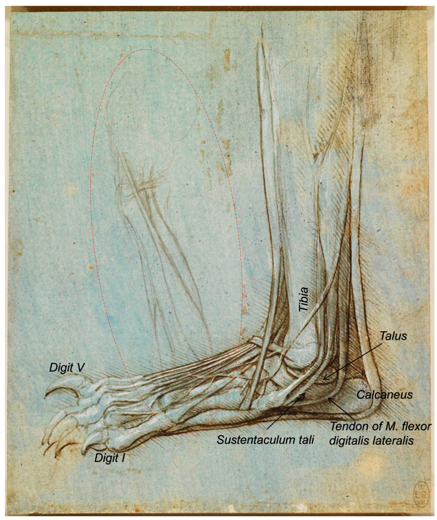
Figure 1. Bear’s foot series—Number 1. Bear distal right pelvic limb/pes, medial aspect. A bear’s foot c.1488–1490. Modified from www.rct.uk/collection/912372 (Royal Collection Trust[3]). This image is credited as Royal Collection Trust/© Her Majesty Queen Elizabeth II 2020.
This drawing shows with some accuracy the bones, muscles and tendons of a bear’s lower leg and foot, with the big toe, claw raised, away from the viewer”. The bear, as a plantigrade animal, walks like humans, with the whole plantar face of the pes (the sole with the heel) touching the ground. However, in contrast to humans, the shortest digit (toe) is not the fifth one (the lateral one), but the medial one, that is the first digit[20][21]. Hence, to the best of our knowledge, we support that the bear’s foot depicted by Leonardo corresponds to the right hind limb instead of the left one, as previously reported by O’Malley and Saunders [6] and Clayton and Philo[9] as well as at its description at the Royal Collection Trust website[3], maybe in resemblance to humans. In addition, the calcaneus bone of the tarsus is always in a lateral position to the talus, and the medial projection of the calcaneus bone to support the talus is quite visible, the so-called sustentaculum tali, with a groove to the tendon of the muscle
flexor digitalis lateralis
[
22]. F
]. Figure 1 also shows, on the left-centre, a preliminary sketch of some muscles that continues under the depicted bear’s foot. If observed upside-down, and once analyzed all the details, it seems to represent the forearm (antebrachium) and hand (manus) of a dog/wolf.
3. Anatomy of the Horse Trunk that Turned into a Dog’s Trunk
Later on, accordingure 1 also sh to Clayton and Philows,[10] “Writing oin the left-centre, a preliminary sketch of some musclesmid-sixteenth century, the biographer Giorgio Vasari stated that Leonardo compiled a treatise on the anatomy of the horse. One drawing of the viscera of a large quadruped, probably a horse, does survive from this period, suggesting that continues under the depicted bear’s foot. If observed upside-dLeonardo conducted full dissections to investigate the internal anatomy of the beast. But Vasari also stated that the treatise on the horse was lost when Milan was invaded by French forces in 1499. Ludovico Sforza was overthrown, and once analyzed allsoon afterwards, Leonardo left the city and returned to Florence” [10]. This text refers to the drawing RCIN 919097-retailscto, entitled ‘The viscera of a horse’ (1490–1492; Figure 6), and descrit seembed at the Royal Collection Trust[3] as: to represent the forearm“an anterior view of the arteries, veins and the genito-urinary system of an animal, probably a horse,” implying that Leonardo did not name (antebrachium)this drand hand (manus) wing. The drawing represents the ventral aspect of the trunk of an dog/wolf.
3. Anatomy of the Horse Trunk that Turned into a Dog’s Trunk
animal (supposedly, a horse) with the lungs and the
(esophagus, stomach and intestines) removed. The large vessels depicted at the centre, all the figure down, represent the
(on the right of the figure) and the vena
(on the left of the image). The way they ramify helps us to discard the idea that this drawing represents a human being. In humans, the
ends up dividing into two branches:
(
and
). In contrast, in animals, the end of the
(at the level of the pelvic limbs) produces two branches (
—
and
), well depicted, and subsequently continues and produces two more (
), well represented in the drawing, ending as the
, not depicted. Regarding the veins, the
is formed by the confluence of two
—
and
, each one resulting from the junction of the
and the
, following a similar pattern both in humans and domestic mammals.
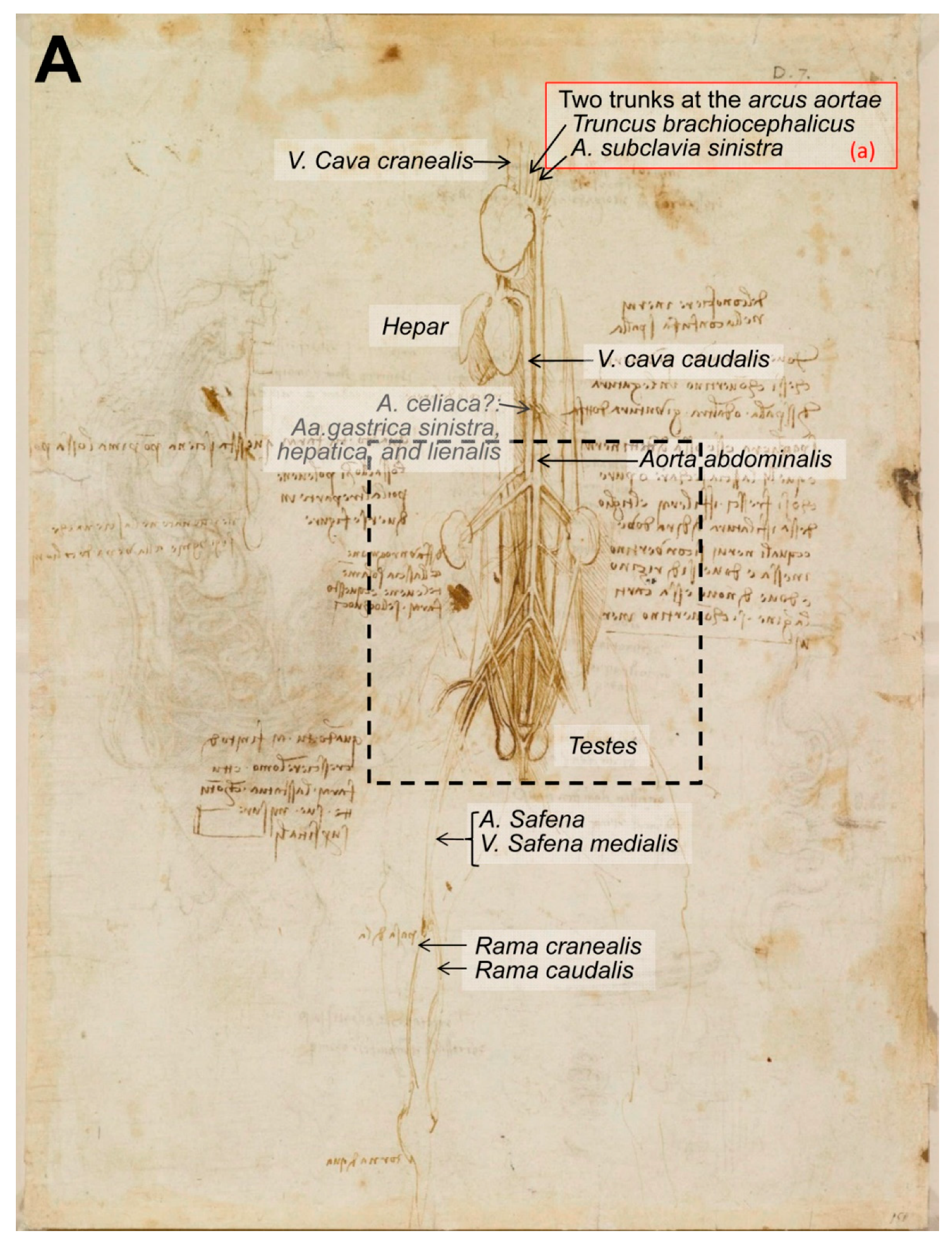
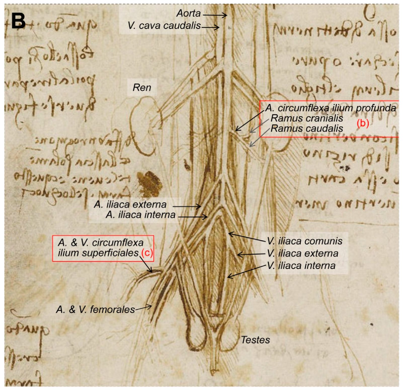
The viscera of a horse? (
A) Whole drawing representing the ventral aspect of the trunk of an animal with the
canalis alimentariusand lungs removed. (
B) Inset at higher magnification depicting the lumbar and pelvic regions. The viscera of a horse c.1490–1492. Modified from
www.rct.uk/collection/919097 recto (Royal Collection Trust [3]). This image is credited as Royal Collection Trust/© Her Majesty Queen Elizabeth II 2020.recto (Royal Collection Trust[3]). This image is credited as Royal Collection Trust/© Her Majesty Queen Elizabeth II 2020.
Regarding the blood vessels, the drawing (
) provides three key points: (a) The first huge vessel (on the left of the image), reaching the heart, could be the
, and the other curved vessel going down is the
and its
, with two big arteries leaving the aortic arch and some smaller ones (2–3) once the arch finishes and continues to the descendent aorta (
). Large domestic mammals (horses—Eq, and ruminants—Ru) only have one artery deriving from the aortic arch, the
, which is then divided into a
(could be absent in carnivores—dogs and cats) and two
. In contrast, carnivores and pigs (Su) have two arteries leaving the
: The
first and secondly the
. (b) On the other hand, at the kidney level, there are two arteries perfectly outlined in the drawing stemming from the
: The
(
and
), exclusive to carnivores [
] and dividing into the
and
. In contrast, the
derives from the
in Su, Ru and Eq [
], similar to humans[23], not stemming directly from the aorta. (c) The arteria and vena
, the first branches of the
and
, respectively, are exclusive to carnivores [
]. They leave their main vessels cranially oriented, at the medial and proximal part of the thigh. These vessels (a–c) in this drawing are the main clue to determine the species. Consequently, the horse representation/provenance of this drawing could be discarded. However, the horse is the unique domestic species in which the aorta does not end caudally as an
, which is not represented in the illustration.
Thanks given to the Royal Collection Trust for allowing free access to their digital collection of Leonardo da Vinci’s legacy, and for granting the copyright of these images free of charge.
References
- Forlani-Tempesti, A. The Robert Lehman Collection V: Italian Fifteenth- to Seventeenth-Century Drawings; The Metropolitan Museum of Art and Princeton University Press: New York, NY, USA; Princeton, NJ, USA, 1991; pp. 236–240.
- Pasipoularides, A. Historical continuity in the methodology of modern medical science: Leonardo leads the way. Int. J. Cardiol. 2014, 171, 103–115.
- Royal Collection Trust Website. Available online: www.royalcollection.org.uk/collection (accessed on 7 August 2020).
- Clark, K. The Drawings of Leonardo da Vinci in the Collection of Her Majesty the Queen at Windsor Castle, 2nd ed.; Blunt, A.F., Ed.; Phaidon Press Ltd.: London, UK, 1968; Volume 1.
- Clark, K. The Drawings of Leonardo da Vinci in the Collection of Her Majesty the Queen at Windsor Castle, 2nd ed.; Blunt, A.F., Ed.; Phaidon Press Ltd.: London, UK, 1969; Volume 3.
- O’Malley, C.D.; Saunders, J.B.D.C.M. Leonardo da Vinci on the Human Body: The Anatomical, Physiological, and Embryological Drawings of Leonardo da Vinci; Henry Schuman: New York, NY, USA, 1952.
- O’Malley, C.D.; Saunders, J.B.D.C.M. Leonardo da Vinci on the Human Body: The Anatomical, Physiological, and Embryological Drawings of Leonardo da Vinci; Crown Publications: Victoria, BC, Canada, 1983.
- O’Malley, C.D.; Saunders, J.B.D.C.M. Leonardo da Vinci on the Human Body: The Anatomical, Physiological, and Embryological Drawings of Leonardo da Vinci; Gramercy: New York, NY, USA, 2003.
- Clayton, M.; Philo, R. Leonardo da Vinci Anatomist; Royal Collection Publications: London, UK, 2012.
- Clayton, M.; Philo, R. Leonardo da Vinci. The Mechanics of Man; Royal Collection Trust: London, UK, 2013.
- Schultheiss, D.; Laurenza, D.; Götte, B.; Jonas, U. The Weimar anatomical sheet of Leonardo da Vinci (1452–1519): An illustration of the genitourinary tract. BJU Int. 1999, 84, 595–600.
- Jose, A.M. Anatomy and Leonardo da Vinci. Yale J. Biol. Med. 2001, 74, 185–195.
- Ganseman, Y.; Broos, P. Leonardo da Vinci and Andreas Vesalius; the shoulder girdle and the spine, a comparison. Acta Chir. Belg. 2008, 108, 477–483.
- Sterpetti, A.V. Anatomy and physiology by Leonardo: The hidden revolution? Surgery 2016, 159, 675–687.
- Bowen, G.; Gonzales, J.; Iwanaga, J.; Fisahn, C.; Loukas, M.; Oskouian, R.J.; Tubbs, R.S.; da Vinci, L. Leonardo da Vinci (1452–1519) and his depictions of the human spine. Childs Nerv. Syst. 2017, 33, 2067–2070.
- West, J.B. Leonardo da Vinci: Engineer, bioengineer, anatomist, and artist. Am. J. Physiol. Lung Cell. Mol. Physiol. 2017, 312, L392–L397.
- Keele, K.D. Leonardo da Vinci’s ‘Anatomia Naturale’ the inaugural John Fulton Lecture. Yale J. Biol. Med. 1979, 52, 369–409.
- Ochenkowski, H.;Wright, W. The quatercentenary of Leonardo da Vinci. Burlingt. Mag. Connoiss. 1919, 34, 186–203.
- Castiglioni, A. Leonardo da Vinci anatomo e fisiologo. In Il Volto di Ippocrate: Istorie di Medici e Medicine D’altri Tempi; Società Editrice Unitas: Milano, Italy, 1925; pp. 172–209.
- Sims, M.E. Comparison of Black Bear Paws to Human Hands and Feet; Identification Guides for Wildlife Law Enforcement No. USFWS; National Fish and Wildlife Forensics Laboratory: Ashland, OR, USA, 2007.
- Dogaroiul, C.; Dermengiu, D.; Viorel, V. Forensic comparison between bear hind paw and human feet. Case report and illustrated anatomical and radiological guide. Rom. J. Leg. Med. 2012, 20, 131–134.
- Schaller, O. Illustrated Veterinary Anatomical Nomenclature, 2nd ed.; Enke Verlag: Stuttgart, Germany, 2007.
- Feneis, H. Nomenclatura Anatómica Ilustrada; Salvat Editores: Barcelona, Spain, 1988.