HO-1 in Cancer Cell Survival: Comparison
Please note this is a comparison between Version 1 by Shuen-Ei Chen and Version 2 by Bruce Ren.

Heme oxygenases (HOs) act on heme degradation to produce carbon monoxide (CO), free iron, ferritin, and biliverdin. Upregulation of cellular HO-1 levels is signature of oxidative stress for its downstream effects particularly under pro-oxidative status. Subcellular traffics of HO-1 to different organelles constitute a network of interactions compromising a variety of effectors such as pro-oxidants, ROS, mitochondrial enzymes, and nucleic transcription factors. Some of the compartmentalized HO-1 have been demonstrated as functioning in the progression of cancer. Emerging data show the multiple roles of HO-1 in tumorigenesis from pathogenesis to the progression to malignancy, metastasis, and even resistance to therapy. However, the role of HO-1 in tumorigenesis has not been systematically addressed.

  • heme oxygenase-1
  • reactive oxygen species
  • cancers
  • subcellular localization
  • mitochondria
  • nuclei

Note: The entry will be online only after author check and submit it.

1. Heme Oxygenases (HOs) and Oxidative Stress

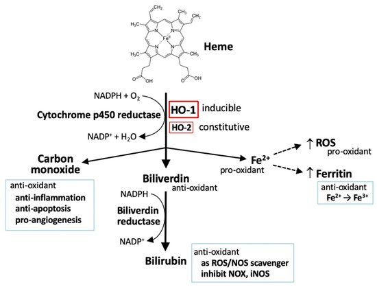
HOs are the key-limiting enzymes in heme degradation leading to carbon monoxide (CO), ferrous iron, and biliverdin products. Biliverdin is then rapidly converted to bilirubin by biliverdin reductase (Figure 1) [1][2][1,2]. HOs are expressed in a variety of cell types, rendering their broad contribution to cell functions. Currently, three mammalian HO isoforms are identified, namely HO-1, HO-2, and HO-3. HO-1 and HO-2 are identified in human and rats. HO-3 is only found in rats. HO-1 is inducible, synthetically enhanced by pro-oxidant stimuli. HO-1 is encoded by HMOX1 with a molecular weight of around 32 kDa (also called heat shock protein 32; HSP32) [3]. HMOX2 is constitutively expressed to encode a 36 kDa HO-2 protein, mainly functioning to maintain the basal heme metabolism and may also play a role in inflammatory responses [4]. HMOX3 is distinguished as a pseudogene and has no transcript form [5].
Figure 1. HO-1 and heme metabolism. Heme is degraded by heme oxygenases (HOs), generating biliverdin, carbon monoxide, and ferrous iron (Fe2+). Biliverdin is subsequently converted to bilirubin by biliverdin reductase. Both biliverdin and bilirubin act as anti-oxidants by scavenging or neutralizing reactive oxygen species (ROS). Carbon monoxide, a gaseous product, functions in signaling transduction, including the vasodilation of blood vessels, production of anti-inflammatory cytokines, upregulation of anti-apoptotic effectors, and promotion of angiogenesis. Ferrous iron (Fe2+) possesses pro-oxidant activity. However, activation of heme oxygenase-1 (HO-1) can upregulate ferritin expression, which binds to ferrous iron and detoxifies its pro-oxidant effect. Ferrous iron increases ROS generation via the Fenton reaction.
Oxidative stress is referred as a pro-oxidative circumstance, occurring at the imbalance between oxidants and antioxidants in favor of the oxidants, which has been implicated in governing normal physiological activities and pathological processes. Oxidative stress arises while the generation of reactive oxygen species (ROS) overload the neutralizing capacity of intrinsic antioxidants and antioxidative defenses. Intracellular ROS are mainly generated from mitochondrial electron transport chain, NADPH oxidases (NOXs), and xanthine oxidase [6], as well as, by exogenous stimuli such as electrophiles and ultraviolet light. They are also involved in the regulating of various cellular activities including growth, differentiation, inflammation, infection, ischemia, aging, and disease pathogenesis and progression [7]. Cellular enzymatic and non-enzymatic antioxidants function as an intrinsic defense to prevent oxidant attack and ameliorate oxidative stress. Enzymatic antioxidants include superoxide dismutases, catalase, peroxiredoxins, and glutathione peroxidase (GPX). The most abundant intracellular non-enzymatic antioxidant is glutathione (GSH) [6][7][6,7].
Despite the fact that ROS are produced as a byproduct in mitochondria biogenesis and intracellular metabolic activities, they are applied in the transduction of cellular signaling or triggering of intracellular defense. Moderately increased level of ROS promotes the systemic defense by inducing adaptive responses to support cell survival, whereas sustained oxidative stress is associated with many pathological conditions such as cancer, metabolic disorders, and neurodegenerative diseases [6][7][6,7]. An overload level of ROS can oxidize DNA, RNA, proteins, and lipids, causing irreversible damages and serious oxidative stress that eventually provoke cell death [6][7][6,7].
In addition to heme metabolism, HO-1 is induced by a broad range of incitements including oxidants, cytokines, growth factors and hormones, heavy metals, and physical cues (ischemia/reperfusion injury and hypoxia/hyperoxia), especially being highly sensitive to pro-oxidant stimuli, such as ultraviolet, heavy metals, inflammatory cytokines, and iron-containing molecules, that contribute to a regulatory network of cell functions [8]. Thus, HO-1 is regarded as a pro-oxidant indicator. Several in vivo studies with HO-1-deficiency have demonstrated a cytoprotective effect of HO-1 in human diseases including systemic inflammation, hemolysis, nephritis, asplenia, nephropathy, and vascular endothelial injury [9][10][9,10]. Results from clinical studies further confirmed that HO-1 protects cells by diminishing oxidative stress and inflammation, and maintaining mitochondrial integrity, thereby promoting cell survival [11]. Due to the manner in regulation of iron metabolism [12], HO-1 also play a role in mediating ferroptosis, a newly identified iron-dependent cell death [13][14][15][16][13,14,15,16]. Since heme degradation generates distinctive metabolites including pro-oxidant ferrous iron and anti-oxidant biliverdin [1][2][1,2], HO-1 apparently possesses a dual role either to protect or deteriorate cancer-cell death [8][17][18][8,17,18]. The mechanisms of HO-1 in redox homeostasis and how HO-1 interplays with oxidative stress to regulate tumor progression are addressed in the later sections.

2. The Metabolism of Heme

2.1. Heme

Circulating irons (Fe) are imported into cells in two different forms, namely free iron and heme-containing iron. More than 80% of bioavailable Fe in mammals is contained within heme, a coordinated complex of Fe with porphyrin called ferriprotoporphyrin. Heme is utilized as a gradient for hemeproteins such as hemoglobin (in red blood cells), myoglobin (in muscle cells), and cytochromes. Extracellular free Fe exists in the plasma mainly by binding to transferrin. Non-heme forms of Fe can transit into the heme along the de novo heme synthesis that transfers Fe to the protoporphyrin IX ring [19]. The entry of free heme or non-binding heme, for example, from hemoglobin release is achieved through several mechanisms. Free heme is captured in plasma by hemopexin or albumin to form heme–hemopexin or heme–albumin complexes. Circulating hemoglobin–haptoglobin complexes, as well as heme–hemopexin complexes, are recognized by the transmembrane protein CD163 and receptor CD91, respectively, for the internalization into cells. Internalization of heme–albumin and free heme is mediated by heme transporters including feline leukemia virus C receptor 2 (FLVCR2), heme responsive gene-1 (HRG-1), and heme carrier protein 1 (HCP1). Intercellular heme transferred from endosomes to the cytoplasma is mediated by HRG-1 [19][20][19,20] (Figure 2). The Fe–transferrin complex can interact with transferrin receptor 1, transferrin receptor 2, or cubilin for the internalization through endocytosis. Most of the endosomal ferrous iron (Fe2+) is reduced into ferric iron (Fe3+) by metalloreductase and transported into the cytoplasm by divalent metal transporter1 (DMT1), followed by the reduction of duodenal cytochrome b (DCYTB). Intracellular iron either binds to ferritin for storage or is oxidized to ferric iron by hephasestin and thereafter exported to the circulating transferrin by ferroportin [19][20][21][19,20,21] (Figure 2).
Figure 2. Metabolism of heme. Heme–hemopexin and hemoglobin–haptoglobin complexes are recognized by CD91 and CD163 receptors, receptively, and internalized through endocytosis. Extracellular heme–albumin or free heme is internalized via heme transporters, FLVCR2 (feline leukemia virus C receptor 2), HRG-1 (heme responsive gene-1), and HCP1 (heme carrier protein 1). Internalized heme is then released to transit from the endosome to the cytoplasm via HRG-1 and is further catabolized by HO-1 and HO-2, generating Fe2+. The uptake of extracellular Fe3+–transferrin is mediated by TFR1 (transferrin receptor 1) through the endocytic process into endosomes, during which they undergo acidification to release Fe3+. Free Fe3+ is further reduced by metalloreductase enzymes such STEAP3 and reduced Fe2+ is transported into the cytoplasma via DMT1 (divalent metal transporter 1). Endosomes are recruited to and fuse with the plasma membranes to release both unbound transferrin and TFRs. Cytosolic Fe2+ is either utilized directly as a cofactor of enzymatic proteins or transported to the mitochondria for the synthesis of heme, for Fe–sulfur proteins, or is stored by binding with ferritin. Under the catalysis by ferroxidase, ferritin converts Fe2+ to Fe3+. Ferritin then is transported to the autophagosome to release Fe3+, during which Fe3+ is reduced to Fe2+ and transferred into the cytoplasmic labile iron pool (LIP). Fe2+ can be mobilized as described above or further pumped out the cell via the iron exporter ferroportin. Extracellular free Fe2+ may enter cells directly through DMT1. Extracellular Fe2+ can be oxidized into less reactive Fe3+ by ferroxidase ceruloplasmin (CP) and hephaestin (HEPH).

2.2. Iron

Cytoplasmic heme is catabolized by HOs to release free iron. Intracellular iron either binds to ferritin for storage or is oxidized to ferric iron (Fe3+) by hephaestin, which is exported to circulating transferrin by ferroportin [19][20][21][19,20,21]. Production of ferrous iron (Fe2+) can accelerate the Fenton reaction to facilitate the generation of ROS and following both stress and damages. Ferritin is the major iron storage protein, which processes ferroxidase activity to oxidize Fe2+ to Fe3+ [22]. Heme thus exerts a pro-oxidative and cytotoxic effect due to its iron function group to provoke ROS production and lipid peroxidation [23].
Intracellular iron is primarily utilized in the mitochondria for heme synthesis and iron–sulfur clustering, and also in the cytosol and other organelles. Lysosomal iron is recycled from the mitochondria and cytosolic ferritin through the selective autophagic process and through both mitophagy and ferritinophagy, respectively [24]. Increased ferritin degradation elevates cellular Fe2+ levels and provokes both ROS production and lipid peroxidation, leading to ferroptosis [24].

2.3. Carbon Monoxide (CO)

The HO system contributes to approximately 85% of CO production and accounts for the main source of endogenous CO. CO is a small gas molecule acting as a gasotransmitter in signaling pathways. For example, CO induces soluble guanylyl cyclase to generate cyclin GMP [25], which controls several critical physiological processes such as vasodilation, redox control, and intracellular signaling [26]. CO can render endothelial cells resistant to endoplasmic reticulum (ER) stress by downregulating CCAAT/enhancement-binding protein homologous protein (CHOP) expression and upregulating the Nrf2/HO-1 pathway [27]. CO also mediates the anti-apoptotic effect of HO-1 via p38 MAPK, PI3K–Akt activation, and K+ channel inhibition [28][29][30][28,29,30]. Both in vivo and in vitro studies showed that a low dose of CO selectively inhibited the expression of lipopolysaccharide-induced pro-inflammatory cytokine production including tumor necrosis factor (TNF-α), interleukin (IL)-1β, and macrophage inflammatory protein 1β (MIP1β), whereas it increased anti-inflammatory cytokine IL-10 [31]. Additionally, CO regulates the physiological activities through the implication with cytochrome oxidase, cytochrome p450 reductase, inducible nitric oxide synthase, NADPH oxidases, and mitochondrial cytochromes [32][33][34][32,33,34]. CO also exerts a cytoprotective effect by regulating mitochondrial biogenesis and maintaining mitochondrial integrity for normal membrane potential, permeabilization, and the inhibition of mitochondrial pro-apoptotic pathways [35].

2.4. Bilirubin and Biliverdin

Bilirubin possesses a potent antioxidant and a quick oxidation by H2O2 back to biliverdin, forming a catalytic antioxidant cycle driven by NADPH and biliverdin reductase [36]. Bilirubin and biliverdin can directly scavenge ROS including singlet oxygen, O2•−, ONOO, and RO2 radicals, inhibiting the activity of NADPH oxidase and inducible nitric oxide synthase, and thereby exerting a cytoprotective function [37][38][39][37,38,39].

3. The Contradictory Role of HO-1 in Tumorigenesis

Tumorigenesis is a complicated process, characterized by several stages including mutation, cell transformation hyperproliferation, genome instability, immortalization, angiogenesis, epithelial–mesenchymal transition, and metastasis. ROS is proposed to act as a major regulator in tumorigenesis [40][79]. As an oxidative stress response, not surprisingly, HO-1 is expressed in a broad range of cancer types such as lymphosarcoma, adenocarcinoma, hepatoma, glioblastoma, melanoma, prostate cancer, and pancreatic cancer [41][42][64,80]. The correlational relationship between ROS/HO-1 and tumorigenesis has been discussed in several reviews [41][42][43][64,80,81].

3.1. HO-1 Deficiency or Mutation in Tumorigenesis

In normal cells, HO-1 is critical in maintaining cellular redox homeostasis by scavenging ROS to prevent DNA damage. Naive HMOX1–/– mice exhibit an excessively dysfunctional γ-H2AX foci [44][82]. The stimuli of genotoxic stressors or irradiation in HO-1-deficient cells caused a loss of ataxia-telangiectasia-mutated (ATM)/ataxia telangiectasia Rad3-related (ATR) proteins and breast cancer 1 proteins (BRCA1), leading to a significant increase of dysfunctional γ-H2AX foci and DNA damage. HO-1 induction or exposure to CO induced the homologous recombination-mediated DNA repair through ATM/ATR in HMOX1–/– mice, suggesting the role of HO-1 in DNA-repair signaling [44][82]. Moreover, in Mdr2−/− mice for chronic liver inflammation and inflammation-induced tumor development, administration of HO-1-inducer, CoPP, increased CD8+ T cell numbers, reduced DNA damage in liver macrophages of aged mice, and moreover delayed and suppressed tumor growth [45][83].
Pharmacological inhibition and genetic knockdown of HO-1 was shown to potentiate hemin-triggered ROS generation and oxidative DNA damage, and the results were more profound in human colonocyte epithelial cells than those observed in the colorectal cancer cell line [46][84]. The cytoprotective role of HO-1 also acts at the mitochondrion as observed in skin cells under radiation exposure [47][85]. HO-1 with the G143H mutant was shown to enhance diethylnitrosamine-induced liver injury and accelerate the tumorigenesis and progression of tumor growth, accompanied with an enhancement of ROS production, hepatocyte damages, and inflammatory IL-6 production [48][86]. Under hypoxia, induction of HO-2 expression in endothelial cells increased the association with polysomes to enhance the translation of transcripts, allowing cells to maintain a steady level of HO-2 against apoptosis [49][87].

3.2. HO-1-Regulated Proliferation and Development of Cancer Cells

In human primary head and neck squamous cell carcinoma (HNSCC) specimens, HO-1 was found with a high level of expression, mostly localized in the nuclei in cancerous tissues than non-tumor tissues. In a mouse model of squamous cell carcinoma and HNSCC, cytoplasmic HO-1 expression was observed in pre-neoplastic lesions, whereas nuclear HO-1 expression was identified in tumor tissues, suggesting the role of nuclear HO-1 in promoting tumor growth [50][75]. Moreover, nuclear localization of HO-1 is associated with malignant performance in colorectal, prostate, and breast cancer [51][52][53][88,89,90]. However, in some human astrocytoma and oligodendroglioma subtypes, tumor malignancy is paralleled with total cellular HO-1 levels not compartmentalized HO-1 in the nuclei [54][91]. In fact, HO-1 are involved in substantial mechanisms to support the proliferation and invasiveness of the tumor. HO-1 can act as a BCR/ABL-dependent survival factor in chronic myeloid leukemia [55][92]. It also participates in the hepatocyte growth factor-induced c-Met–Ras signaling-enhanced proliferation of renal cell carcinoma [56][93]. In human colon cancer cells, namely HT-29, HO-1 mediates EGFR–Src–NF-κB signaling to promote cell proliferation [57][94].
In tissue-associated leukocytes, HO-1 is highly expressed in monocytic cells in the microenvironments surrounding the tumor, rendering the cells differentiated into tumor-associated macrophages (TAMs) [58][95]. Iron metabolism plays a pivotal role in the microenvironments for tumor cell growth, especially by TAMs [59][96]. TAMs are the main population of immune cells in tumor microenvironments, in which they acquire diverse phenotypes and functional profiles to differentiate into pro-inflammatory (M1) or anti-inflammatory (M2) states. M2-like TAMs are found in the hypoxic and necrotic areas of tumor microenvironments, which are characterized by high levels of ferroportin and low levels of ferritin, presenting an enhanced phenotype of iron-release. Accordingly, M2-like TAMs are capable of supporting tumor cell proliferation, angiogenesis, and metastasis via promoting vascularization in the tumor microenvironments [59][96]. In the prostate cancer xenograft mouse model, deletion of HO-1 in macrophages suppressed tumor growth, in which HO-1-derived CO from TAMs’ downregulated E-cadherin expression to mediate tumor pathogenesis and progression [60][97].

3.3. HO-1-Regulated Angiogenesis of Cancer Cells

Angiogenesis is necessary for continued growth, invasion, and metastasis of solid tumors [61][98]. HO-1 overexpression in pancreatic cancer cells markedly promoted tumor angiogenesis and accelerated the occurrence of metastasis in a lung colonization model [62][99]. Angiogenesis by HO-1 is likely mediated by the upregulation or activation of proangiogenic factors such as VEGF and stroma cell-derived factor-1 (SDF-1) [63][64][100,101]. Nuclear translocalization of HO-1 increased VEGF expression and secretion in prostate cancer cells [65][102]. Treatment of ZnPP, a HO-1 inhibitor, suppressed HIF-1α expression and VEGF production, accompanied by the enhanced proliferation of HCT-15 cells, suggesting that the angiogenesis for tumor growth is mediated by HIF-1α and VEGF [66][103]. VEGF-enhanced angiogenesis by HO-1 was further shown as operating at the upregulation of cyclin A1, cyclin E1, and cyclin-dependent kinase 2 activity, as well as vimentin to enhance the proliferation of human endothelial cells [67][104]. In addition, induction of HO-1 expression attenuated high glucose-mediated ER stress and downstream events in endothelial cells, including oxidative stress, activation of inflammatory responses, and apoptosis, as well as enhanced VEGF-A expression [68][105]. In addition, CO, the metabolite by HO-1, can promote VEGF expression by increasing HIF-1α content at the translational level and post-translational stabilization of the HIF-1α protein [69][106].
The lung metastasis resulting from subcutaneous tumors or circulating tumor cells was significantly repressed in mice bearing bone marrow HO-1+/– as compared to those in wild type mice [70][107], suggesting that HO-1 expression in hematopoietic cells impacts tumor colonization at the metastatic site. The mechanism was further attributed to chemoattractant-induced myeloid cell migration through p38 kinase signaling and to tumor cell transendothelial migration through the vascular endothelial growth factor, IL-10, and STAT3 activation [70][107]. In a similar manner, mice intravenously injected with HO-1-overexpressed melanoma cells, namely B16-HO1, were characterized by augmented vascularization and a higher level of vascular endothelial growth factors in the tumor, whereas a lower level of serum TNF-α but a higher level of soluble receptor TNF-RI were observed. HO-1 overexpression apparently accelerated B16 melanoma cell metastasis in the lungs and resulted in a low survival rate [71][108].
Despite the numerous reports regarding the pro-tumor effects of HO-1, overexpression of HO-1 in non-small cell lung carcinoma upregulates the tumor-suppressive factors, miR-378 and p53 expression; downregulates angiopoietin-1 and mucin-5AC (MUC5AC); suppresses cell proliferation and migration; and rather unexpectedly diminishes angiogenic potential CO to act as a mediator of HO-1 effects [72][52]. Conversely, miR-378 overexpression downregulated HO-1 and p53 expression but increased VEGF and MUC5AC expression, cell proliferation, and migration [72][52].

3.4. HO-1-Regulated Metastasis of Cancer Cells

The epithelial to mesenchymal transition plays an important role in cancer progression from initiation, primary tumor growth, invasion, dissemination, and metastasis to colonization as well as resistance to therapy [73][109]. The analysis of the microarray dataset from clinical biopsies showed that HMOX1 expression levels significantly increase in glioma grade IV brain biopsies when compared to grade I, II, and III. Additionally, the expression level of HO-1 in glioma grade IV brain biopsies was correlated to the chemotaxis gene expression [74][110]. In A2780 and SKOV-3 ovarian cancer cells, ROS scavengers, namely N-acetyl-L-cysteine and HO-1 inhibitor ZnPP, were shown to relieve ROS production and autophagy, and ameliorate cell migration and invasion by reversing the epithelial–mesenchymal transition [75][111]. Genetic silence of GRP78, an ER stress response protein, enhanced the metastasis by promoting vimentin and decreasing E-cadherin expression through the Nrf2/HO-1 pathway in HT-29 colon cancer cells [76][112]. Additionally, the deficiency of GRIM-19, an essential subunit of the mitochondrial MRC complex I, accelerated gastric cancer metastasis through the ROS–Nrf2–HO-1 axis [77][113]. The role of HO-1 in cancer progression involves cell cycle regulation. Mice treated with HO-1 inhibitor ZnPP had a reduced thyroid cancer xenograft growth and diminished cyclin D1 and Ki-67 expression [78][114]. The results were further confirmed in vitro, showing that ZnPP induced a G0/G1 arrest of cell cycle, accompanied by decreased cyclin D1 and CDK4, and an increase of p21 and p27 expression [78][114]. Moreover, ATF4 and Nrf2 can work together to transcriptionally activate HO-1 to ameliorate oxidative stress and prevent both anoikis and lung metastasis [79][41]. These results reveal that both HO-1 and ROS crosstalk with each other in coordinating subcellular compartmentalization, related effectors, and cascadings to contribute the epithelial–mesenchymal transition.
The anti-tumor effects by mitigating metastasis were also observed in NCI-H292 lung mucoepidermoid carcinoma cells. Nrf2 overexpression-derived HO-1 inhibited NCI-H292 cell proliferation and migration, and downregulated oncogenic miR-378, multiple matrix metalloproteinases (MMP-1 and MMP-9), and inflammatory IL-1β expression [80][115]. Furthermore, the Notch1/Slug pathway was found to mediate the antitumor role of HO-1 in mouse mammary carcinoma [81][116].
In summary, in normal cells, ROS or chemicals/radiation-increased ROS can cause DNA damage and mutation, which may further lead to the cell transformation to cancer cells. In response to increased ROS, HO-1 is thereby raised to neutralize ROS and eliminate DNA damage that reduce the chances of acquired mutation. Once tumorigenesis is initiated, more fuels are required to support cancer cell proliferation. In the proliferation from initiated cells, the increase of both ROS and HO-1 regulates the mitochondrial biogenesis, which may co-work with autophagy and redistribute to the metabolic system, allowing for the adaption of enhanced fuel requirements for the fast growth of cancer cells. For high-fuel demand following the fast growth, angiogenesis is necessary to establish the transportation network. In this stage, ROS and HO-1 communicate with the nuclei to upregulate pro-angiogenesis factors such as VEGF and promote the proliferation of vascular epithelial cells. For metastatic colonization, ROS and HO-1 co-work to regulate the protein expression responsible for the epithelial–mesenchymal transition and cell cycle. Based on clinical observations, ROS and HO-1 levels increase gradually along the malignancy. It is reasonable to propose that ROS and HO-1 assist each other to contribute to tumorigenesis via serving as the communicators in linking with the ER, mitochondria, and nuclei to set up an optimal environment for cancer cells (Figure 3).
Figure 3. HO-1, ROS, and tumorigenesis. HO-1 can suppress the transformation of cancer cells via preventing ROS-induced mutation. Once the tumorigenesis process is triggered, ROS and HO-1 may serve as the mediator roles to support the proliferation, angiogenesis, invasion, and metastasis of cancer cells. Abbreviation: EMT, epithelial–mesenchymal transition.